Chapter 20: Heart and Neck Vessels Exam 2022
Document Content and Description Below
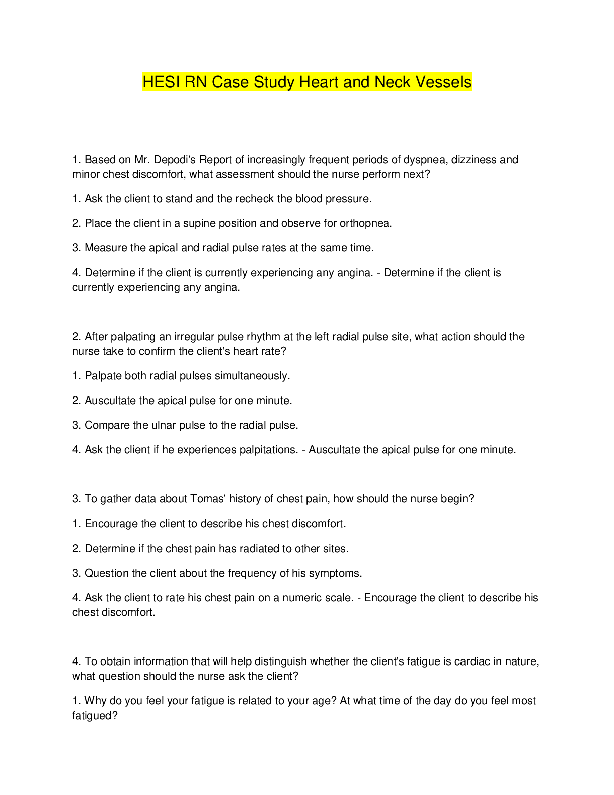
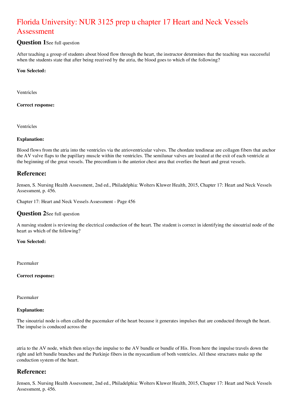
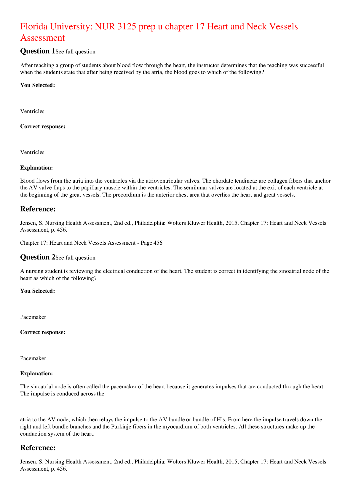
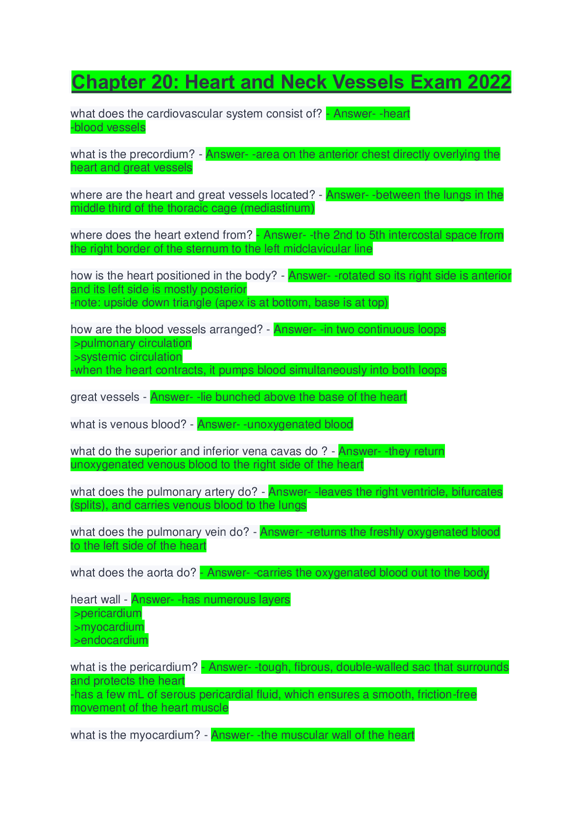
Chapter 20: Heart and Neck Vessels Exam 2022 what does the cardiovascular system consist of? - Answer- -heart -blood vessels what is the precordium? - Answer- -area on the anterior chest directly o... verlying the heart and great vessels where are the heart and great vessels located? - Answer- -between the lungs in the middle third of the thoracic cage (mediastinum) where does the heart extend from? - Answer- -the 2nd to 5th intercostal space from the right border of the sternum to the left midclavicular line how is the heart positioned in the body? - Answer- -rotated so its right side is anterior and its left side is mostly posterior -note: upside down triangle (apex is at bottom, base is at top) how are the blood vessels arranged? - Answer- -in two continuous loops >pulmonary circulation >systemic circulation -when the heart contracts, it pumps blood simultaneously into both loops great vessels - Answer- -lie bunched above the base of the heart what is venous blood? - Answer- -unoxygenated blood what do the superior and inferior vena cavas do ? - Answer- -they return unoxygenated venous blood to the right side of the heart what does the pulmonary artery do? - Answer- -leaves the right ventricle, bifurcates (splits), and carries venous blood to the lungs what does the pulmonary vein do? - Answer- -returns the freshly oxygenated blood to the left side of the heart what does the aorta do? - Answer- -carries the oxygenated blood out to the body heart wall - Answer- -has numerous layers >pericardium >myocardium >endocardium what is the pericardium? - Answer- -tough, fibrous, double-walled sac that surrounds and protects the heart -has a few mL of serous pericardial fluid, which ensures a smooth, friction-free movement of the heart muscle what is the myocardium? - Answer- -the muscular wall of the heart -it does the pumping what is the endocardium? - Answer- -thin layer of endothelial tissue that lines the inner surface of the heart chamber and the valves what are the two pumps of the heart? - Answer- -right side: pumps blood into lungs -left side: pumps blood into the body what is a septum? - Answer- -wall that separates the right and left side of the heart what is an atrium? - Answer- -upper chamber of heart -right and left atria >RA right atrium >LA left atrium what is a ventricle? - Answer- -lower chamber of heart -right and left ventricles >RV right ventricle >LV left ventricle how are the four chambers of the heart separated? what is the purpose? - Answer- -valves -purpose is to prevent backflow of blood -note: valves are unidirectional and can only open one way, they open and close passively in response to the pressure gradients in the moving blood how many valves are there in the heart? what are the called? - Answer- -four valves -2 atrioventricular valves(AV) -2 semilunar valves (SL) atrioventricular valves (AV) - Answer- -separate the atria and the ventricles -RIGHT AV VALVE IS TRICUSPID -LEFT AV VALVE IS BICUSPID (MITRAL) what is the chordae tendinae? - Answer- -collagenous fibers -they anchor the valves thin leaflets to papillary muscle embedded in the ventricle floor when do the AV valves OPEN? - Answer- -during the heart's filling phase (diastole) to allow the ventricles to fill with blood when do the AV valves CLOSE? - Answer- -during the pumping phase (systole) to prevent regurgitation of blood back into atria semilunar valves (SL) - Answer- -set between ventricles and arteries -each valve has 3 cusps that look like half moons (hence semilunar) -the SL valves are the pulmonic and aortic where is the pulmonic valve located? - Answer- -in the right side of the heart where is the aortic valve located? - Answer- -in the left side of the heart when do the semilunar (SL) valves open? - Answer- -during pumping (systole), when blood ejects from the heart where are there no valves? - Answer- -in between the vena cava and the right atrium or between the pulmonary veins and the left atrium -note: due to this, abnormally high blood pressure in the left side of the heart gives a person symptoms of pulmonary congestion-- abnormally high blood pressure in the right side of the heart shows in the distended neck veins and abdomen direction of blood flow - Answer- (1) from liver to right atrium (RA) through inferior vena cava >superior vena cava drains venous blood from the head and upper extremities >from RA venous blood travels through tricuspid valve to right ventricle (RV) (2) from RV venous blood flows through pulmonic valve to pulmonary artery >pulmonary artery delivers unoxygenated blood to lungs (3) lungs oxygenate blood >pulmonary veins return fresh blood back to left atrium (LA) (4) from LA arterial blood travels through mitral valve to left ventricle (LV) >LV ejects blood through aortic valve into aorta (5) aorta delivers oxygenated blood to body -NOTE: circulation is a continuous loop, the blood is kept moving by continually shifting pressure gradients. it flows from an area of high pressure to lower pressure which chamber of the heart is the strongest? why? - Answer- -left ventricle -it has to pump blood a greater distance, the walls become thicker and stronger to withstand a greater force cardiac cycle - Answer- -rhythmic movement of blood through heart -two phases: diastole and systole what happens during diastole? - Answer- -ventricles relax and fill with blood -AV valves (tricuspid and mitral) open -pressure in atria is higher than that in the ventricles; so blood pours rapidly into the ventricles (protodiastolic filling) -toward the end of diastole, atria contract and push the last amount of blood (ab 25% of stroke volume) into the ventricles (presystolic or atrial systole) -takes up two-thirds of the cardiac cycle what happens during systole? - Answer- -heart contraction -blood is pumped from the ventricles and fills the pulmonary and systemic arteries -since so much blood is pumped into ventricles, ventricular pressure is higher than in atria, so tricuspid and mitral valves swing shut -after ventricles contents are rejected, the pressure falls -when pressure falls below pressure of aorta, some blood flows backward toward ventricle, causing aortic valve to swing shut -one-third of the cardiac cycle what is protodiastolic filling? - Answer- -first passive filling phase -aka early filling what is presystole? - Answer- -active filling phase -causes a small rise in left ventricle pressure -aka atrial systole or atrial kick what is S1? - Answer- -heart sound 1 -closure of the AV valves and signals systole what is S2? - Answer- -heart sound 2 -closure of the SL valves and signals end of systole events in the right and left sides - Answer- -the same events are happening at the same time in the right side of the hear, but pressures in the right side of the heart are much lower than those of the left side because less energy is needed to pump blood to its destination, the pulmonary circulation heart sounds - Answer- -S1: closure of AV, can hear all over precordium, but usually loudest at apex -S2: closure of SL, can hear all over precordium, but usually loudest at base effect of respiration - Answer- -moRe to the Right heart, Less to the Left -during inspiration, intrathoracic pressure is decreased -this pushes more blood into the vena cava, increasing venous return to the right side of the heart, which increases right ventricular stroke volume -when aortic valve closes significantly earlier than the pulmonic valve, you can hear the two components separately (split S2) what is S3? - Answer- -extra heart sound -normally diastole is silent, but in some conditions ventricular filling creates vibrations that can be heard all over the chest -S3 occurs when ventricles are resistant to filling during the early rapid filling phase (protodiastole) -occurs immediately after S2 when AV open murmurs - Answer- -some conditions create turbulent blood flow and collision currents -gentle, blowing, swooshing sound that can be heard on chest wall what are the conditions resulting in a murmur? - Answer- -velocity of blood increases (exercise;flow murmur) -viscosity of blood decrease (anemia) -structural defects in valves (a stenotic or narrowed valve, an incompetent or regurgitant valve) or unusual openings occur in the chambers (dilated chamber, septal defect) characteristics of sound - Answer- (1) frequency (pitch): heart sounds described as high pitches or low pitched (2) intensity (loudness): loud or soft (3) duration: very short for heart sounds; silent periods are longer (4) timing: systole or diastole conduction - Answer- -heart can contract by itself, independent of any signals or stimulation from the body -contracts in response to an electrical current conveyed by a conduction system -sinoatrial node (SA node): initiates electrical impulse -SA is termed the pacemaker of heart current flow in heart - Answer- -current flows in an orderly sequence: (1) across atria to AV node low in atrial septum (2) then its delayed slightly so atria can contract before ventricles are stimulated (3) impulse travels to the bundle of His, right and left bundle branches (4) through ventricles ECG wave - Answer- -PQRST -P wave: depolarization of atria -PR interval: from the beginning of P wave to the beginning of QRS complex (the time is necessary for atrial depolarization plus time for the impulse to travel through AV node to the ventricles) -QRS complex: depolarization of the ventricles -T wave: repolarization of the ventricles pumping ability of heart - Answer- -normally pumps between 4 and 6 L per minute throughout body cardiac output - Answer- CO = SV x R -stroke volume times beats per minute -heart can alter cardiac output to adapt to the metabolic needs of body what is preload? - Answer- -volume -venous return that builds during diastole -length to which the ventricular muscle is stretched at the end of diastole just before contraction what is afterload? - Answer- -pressure -opposing pressure the ventricle must generate to open the aortic valve against the higher aortic pressure -is is the resistance against which the ventricle must pump its blood -once the ventricle is filled with blood, the ventricular end diastolic pressure is 5-10 mmHg, whereas the aorta is 70-80 mmHg -ventricular muscle tenses to overcome this difference -rapid ejection occurs neck vessels - Answer- -carotid artery and jugular veins -these vessels reflect the efficiency of cardiac function carotid artery - Answer- -located in the groove between the trachea and the sternomastoid muscle, medial to and alongside that muscle CONTINUES..... [Show More]
Last updated: 2 years ago
Preview 1 out of 13 pages
Buy this document to get the full access instantly
Instant Download Access after purchase
Buy NowInstant download
We Accept:

Reviews( 0 )
$10.50
Can't find what you want? Try our AI powered Search
Document information
Connected school, study & course
About the document
Uploaded On
Sep 04, 2022
Number of pages
13
Written in
Additional information
This document has been written for:
Uploaded
Sep 04, 2022
Downloads
0
Views
88


