NR 224 Exam 2 Study Outline
The following is a list of concepts for you to understand to be successful on Exam 2
Skin
Pressure ulcers
o Stages – describe, identify
Category/Stage I: Nonblanchable Redness.
Intact
...
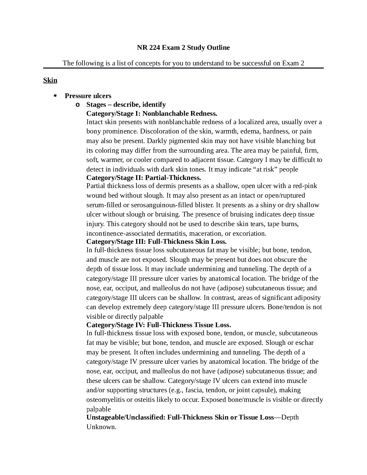
NR 224 Exam 2 Study Outline
The following is a list of concepts for you to understand to be successful on Exam 2
Skin
Pressure ulcers
o Stages – describe, identify
Category/Stage I: Nonblanchable Redness.
Intact skin presents with nonblanchable redness of a localized area, usually over a
bony prominence. Discoloration of the skin, warmth, edema, hardness, or pain
may also be present. Darkly pigmented skin may not have visible blanching but
its coloring may differ from the surrounding area. The area may be painful, firm,
soft, warmer, or cooler compared to adjacent tissue. Category I may be difficult to
detect in individuals with dark skin tones. It may indicate “at risk” people
Category/Stage II: Partial-Thickness.
Partial thickness loss of dermis presents as a shallow, open ulcer with a red-pink
wound bed without slough. It may also present as an intact or open/ruptured
serum-filled or serosanguinous-filled blister. It presents as a shiny or dry shallow
ulcer without slough or bruising. The presence of bruising indicates deep tissue
injury. This category should not be used to describe skin tears, tape burns,
incontinence-associated dermatitis, maceration, or excoriation.
Category/Stage III: Full-Thickness Skin Loss.
In full-thickness tissue loss subcutaneous fat may be visible; but bone, tendon,
and muscle are not exposed. Slough may be present but does not obscure the
depth of tissue loss. It may include undermining and tunneling. The depth of a
category/stage III pressure ulcer varies by anatomical location. The bridge of the
nose, ear, occiput, and malleolus do not have (adipose) subcutaneous tissue; and
category/stage III ulcers can be shallow. In contrast, areas of significant adiposity
can develop extremely deep category/stage III pressure ulcers. Bone/tendon is not
visible or directly palpable
Category/Stage IV: Full-Thickness Tissue Loss.
In full-thickness tissue loss with exposed bone, tendon, or muscle, subcutaneous
fat may be visible; but bone, tendon, and muscle are exposed. Slough or eschar
may be present. It often includes undermining and tunneling. The depth of a
category/stage IV pressure ulcer varies by anatomical location. The bridge of the
nose, ear, occiput, and malleolus do not have (adipose) subcutaneous tissue; and
these ulcers can be shallow. Category/stage IV ulcers can extend into muscle
and/or supporting structures (e.g., fascia, tendon, or joint capsule), making
osteomyelitis or osteitis likely to occur. Exposed bone/muscle is visible or directly
palpable
Unstageable/Unclassified: Full-Thickness Skin or Tissue Loss—Depth
Unknown.Full-thickness tissue loss in which actual depth of an ulcer is completely obscured
by slough (yellow, tan, gray, green or brown) and/or eschar (tan, brown or black)
in the wound bed is unstageable. Until enough slough and/or eschar are removed
to expose the base of a wound, the true depth cannot be determined; but it will be
either a category/stage III or IV. Stable (dry, adherent, intact without erythema or
fluctuance) eschar on the heels serves as “the natural (biological) cover of the
body” and should not be removed
Suspected Deep-Tissue Injury—Depth Unknown.
Suspected deep-tissue injury is a purple or maroon localized area of discolored
intact skin or a blood-filled blister caused by damage of underlying soft tissue
from pressure and/or shear. The area may be preceded by tissue that is painful,
firm, mushy, boggy, warmer, or cooler compared 1188to adjacent tissue. Deeptissue injury may be difficult to detect in individuals with dark skin tones. It may
begin as a thin blister over a dark wound bed. The wound may further evolve and
become covered by thin eschar. Evolution may be rapid, exposing additional
layers of tissue even with optimal treatment.
o Prevention – specific interventions
Quick Guide to Pressure Ulcer PreventionRisk Factor Nursing Interventions
Decreased sensory
perception
Provide pressure-redistribution surface. Be sure to include protection
for pressure points from medical devices such as oxygen tubing,
feeding tubes, and casts (Black et al., 2015 ; Fletcher, 2012).
Moisture
Following each incontinent episode, clean area with no-rinse perineal
cleaner and protect skin with moisture-barrier ointment (Rolstad
et al., 2016 ).
Keep skin dry and free of maceration (Gray et al., 2011 ; Colwell
et al., 2011 ). Turn patient off of at-risk areas often.
Friction and shear
Reposition patient using drawsheet or a transfer board surface.
Provide trapeze to facilitate movement in bed.
Position patient at a 30-degree lateral turn and limit head elevation to
30 degrees (see Figure 48-15).
Decreased
activity/mobility
Establish and post individualized turning schedule.
Poor nutrition
Provide adequate nutritional and fluid intake; help with intake as
necessary.
Consult dietitian for nutritional assessment and recommended
nutrients
[Show More]