NURSING > STUDY GUIDE > N5315 Advanced Pathophysiology Gastrointestinal Best Study Guide Ever Already Graded A+ (All)
N5315 Advanced Pathophysiology Gastrointestinal Best Study Guide Ever Already Graded A+
Document Content and Description Below
Last updated: 3 years ago
Preview 1 out of 12 pages
Instant download
Buy this Document to get the Full Access Instantly
Provided by Students Who Aced it
We Verify Document Content to Gurantee Accuracy
Reviews( 0 )
Document information
Connected school, study & course
About the document
Uploaded On
Sep 04, 2021
Number of pages
12
Written in
All

Additional information
This document has been written for:
Uploaded
Sep 04, 2021
Downloads
0
Views
153
Document Keyword Tags
Recommended For You
Get more on STUDY GUIDE »
N5315 / N5315 Advanced Pathophysiology Module 7 Study Guide (L...
N5315 / N5315 Advanced Pathophysiology Module 6 Study Guide (L...
N5315 / N5315 Advanced Pathophysiology Module 5 Study Guide (L...
.png)
N5315 Advanced Pathophysiology Gastrointestinal Latest Updated...

N5315 Advanced Pathophysiology Renal and Urologic System Core...

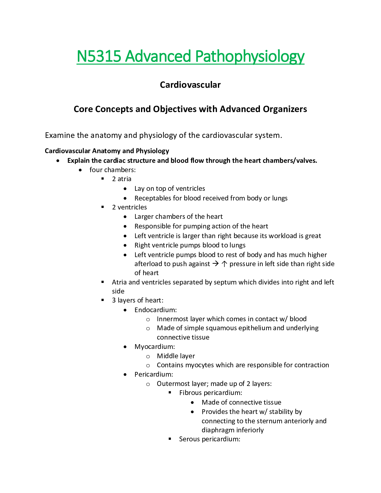
N5315 Advanced Pathophysiology Cardiovascular Core Concepts an...

N5315 Advanced Pathophysiology Gastrointestinal (M 9) Core Con...
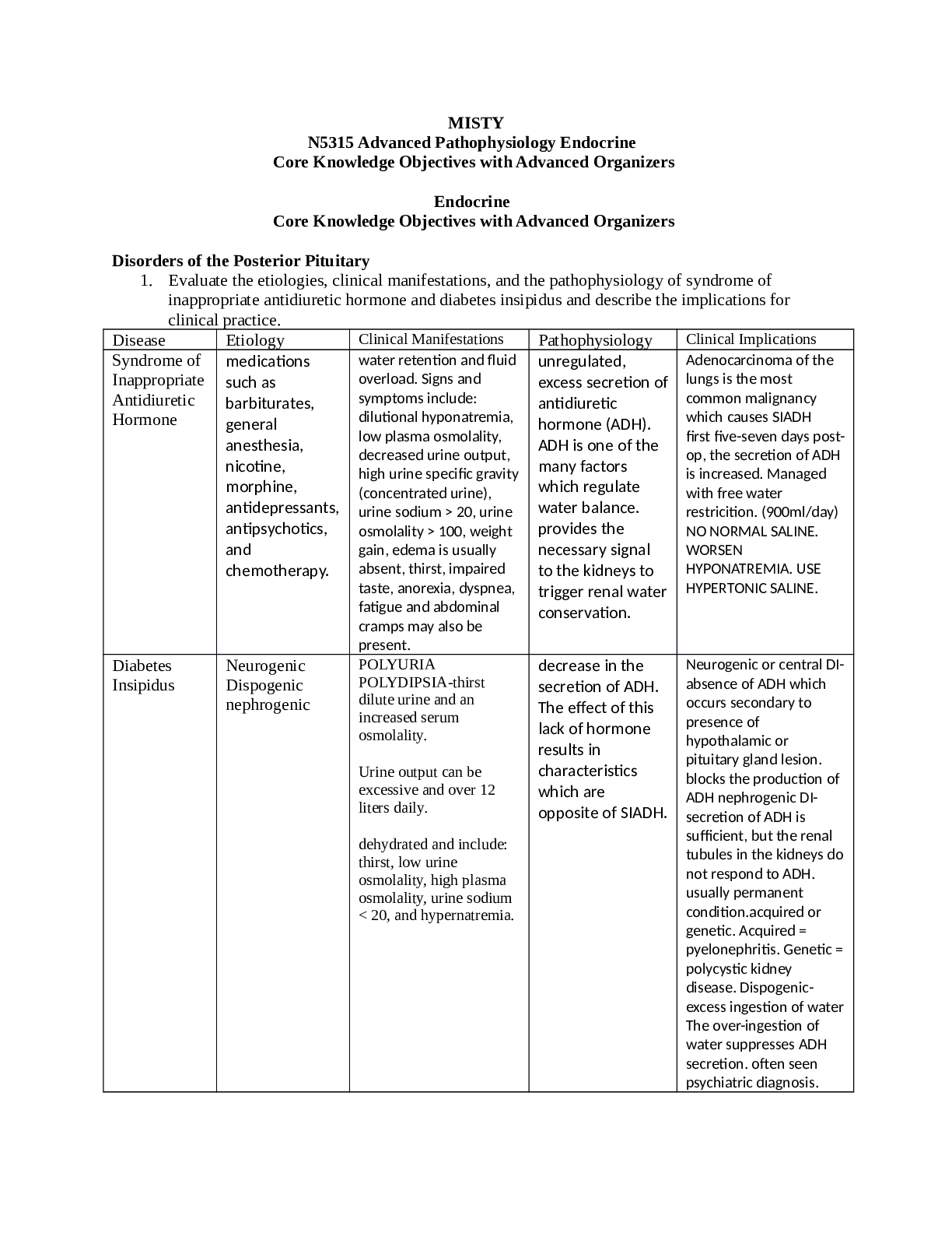
N5315 Advanced Pathophysiology Endocrine Core Knowledge Objec...

N5315 Advanced Pathophysiology Cardiovascular Core Concepts an...

N5315 Advanced Pathophysiology Inflammation, Altered Immunity...

CHIM NCE Health Information Management Practice Exam 1 – Ver...
Essentials of Psychiatric Nursing 2nd Edition Boyd TEST BANK
Test Bank for Essentials of Psychiatric Nursing 2nd Edition by...

Test Bank for Essentials of Psychiatric Nursing 2nd Edition B...