NUR2063 Exam 3 Focused Review
Ch. 2 Homeostasis, Allostasis, and Adaptive Responses to Stressors
• Electrolyte pools- bones act as an electrolyte pool and store calcium magnesium, and phosphate
ions; shift of electrol
...
NUR2063 Exam 3 Focused Review
Ch. 2 Homeostasis, Allostasis, and Adaptive Responses to Stressors
• Electrolyte pools- bones act as an electrolyte pool and store calcium magnesium, and phosphate
ions; shift of electrolytes into electrolyte pools will decrease plasma electrolyte concentration
and shift of electrolytes from an electrolyte pool into the extracellular fluid will increase the
plasma electrolyte concentration
Ch. 7 Neoplasia
• Metastasis- process by which cancer cells escape their tissue of origin and initiate new colonies
of cancer in distant sites
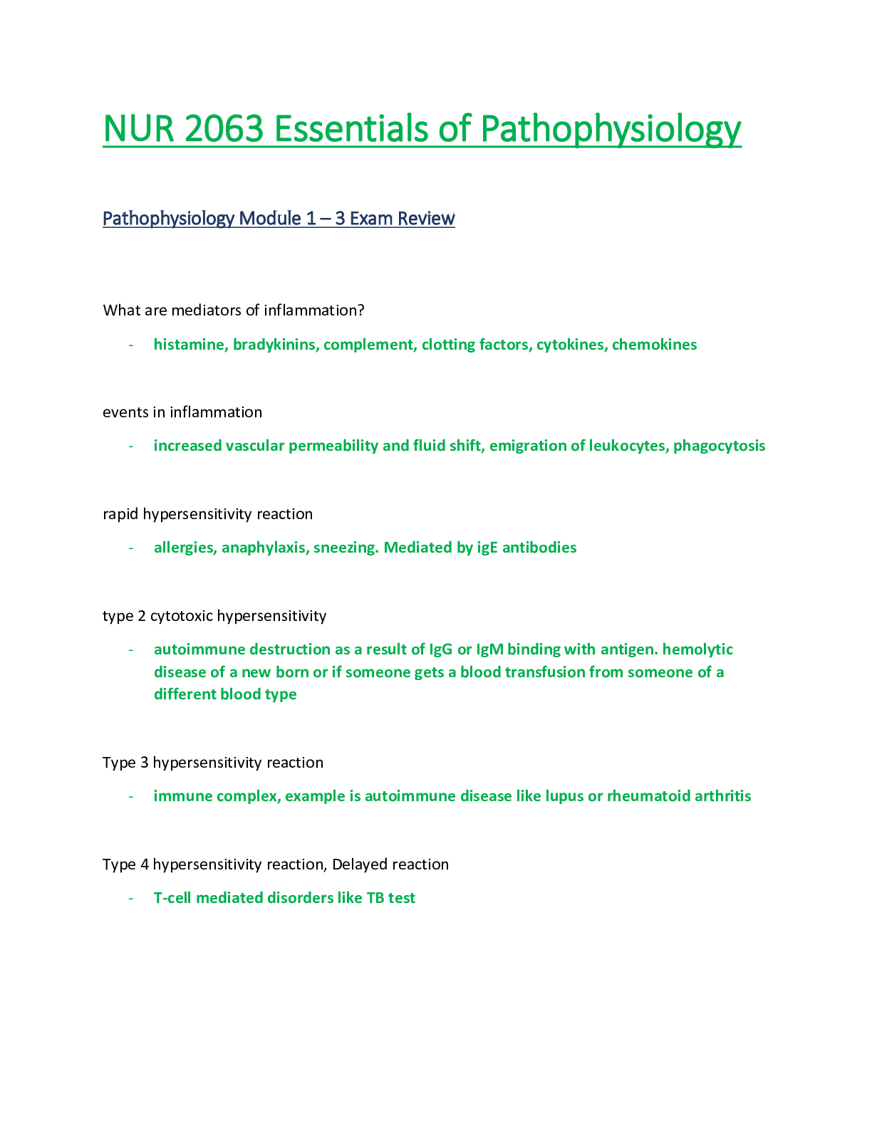
Ch. 9 Inflammation and Immunity
• Passive vs active immunity- Passive: transfer of preformed antibodies against specific antigen
from a protected or immunized individual to an unprotected or nonimmunized individual; types:
mother to fetus: IgG- can cross placenta, mother to infant: IgA- from breast milk, serotherapy-
direct injections of antibodies (human or animal); active: a protected state owing to the body’s
immune response as a result of active infection or immunization, types: immunizations- vaccines
• Immunoglobulins- antibodies; two identical light polypeptide chains joined to identical heavy
polypeptide chains by disulfide bonds; five different classes: IgG, IgM, IgA, IgD, & IgE
•
Ch. 11 Malignant Disorders of White Blood Cells
• Chronic myeloid leukemia (CML)- characterized by malignant granulocytes that carry the
Philadelphia chromosome (Ph+); does not respond well to chemotherapy: poor overallsurvival
time; Blast stage CML- poor prognosis: 3 to 4 months; treatment- bone marrow transplant
• Age of onset for multiple myeloma- median age 65 years
• Malignant Disorders of WBCs- may be nonspecific signs & symptoms
•
Ch. 13 Alterationsin Oxygen Transport
• Anemia- occursfrom a manifestation of diseases; a deficit of RBCs; leads to hypoxia
• Polycythemia- excess of RBCs; increases blood viscosity & volume; leadsto hypertension
• Erythropoietin- is stimulated from the kidney secretion hormone to stimulates RBC production
• Acute blood loss- type of anemia; may be from trauma or secondary to a disease process;
symptoms develop with activity at 20% loss of blood volume (tachycardia and postural drop in
BP), and increase in severity with continued blood loss; shock and death can occur with 50% loss
of circulating volume; treatment: blood volume replacement therapy with crystalloids, colloids,
and fresh whole blood; prognosis: excellent with treatment unless blood loss is severe
•
Ch. 14 Alterationsin Hemostasis and Blood Coagulation
• Thrombocytopenia causes- 1) bone marrow suppression from chemotherapy, 2) recent
immunizations, 3) alcohol ingestion, 4) decreased platelet production, 5) decreased platelet
survival, 6) splenic sequestration, & 7) intravascular dilution of circulating platelets from
transfusion
• DIC (Disseminated Intravascular Coagulation)- acquired hemorrhagic syndrome in which
clotting, and bleeding occursimultaneously. Widespread clot formation in small vessels; clotting
and platelets consumed resulting in bleeding. Causes: trauma, malignancy, burns, shock, and
abruptio placentae. Clinical manifestations: fibrinogen level and platelet count decreased,
increased bleeding time, elevated PT/INR/aPTT, elevated D-dimer/fibrin split products.
Treatment: Removal/correction of underlying cause,support major organs, fresh frozen plasma,
packed red blood cells, platelets, or cryoprecipitate, heparin used to minimize further
consumption of clotting factors (controversial).
•
Ch. 15 Alterations in Blood Flow
• Atherosclerosisis the leading cause of coronary artery/heart disease
Ch. 16 Alterationsin Blood Pressure
• Treatment of hypertension- (lifestyle modifications) weight loss, exercise, DASH diet, alcohol
moderation, decreased sodium intake; drug therapy affects HR, SVR, and/or stroke volume
• Long term problems from hypertension- Renal failure, stroke, heart disease, Damage to arterial
system and acceleration of atherosclerosis lead to cardiovascular disease, Increased myocardial
work results in heart failure, Glomerular damage results in kidney failure, Affects
microcirculation of the eyes, Increased pressure in cerebral vasculature can result in hemorrhage
• Modifiable vs nonmodifiable risk factors for hypertension- modifiable: dietary factors,
sedentary lifestyle, obesity/weight gain, metabolic syndrome, elevated blood glucose
levels/diabetes, elevated total cholesterol, alcohol & smoking; nonmodifiable: family history,
age, ethnicity/genetics
•
Ch. 18 Alterationsin Cardiac Function
• Coronary artery disease: characterized by insufficient delivery of oxygenated blood to the
myocardium caused by atherosclerotic coronary arteries; known risk factor: atherosclerosis
• How to determine causes of acute coronary syndrome: associated with acute changes in plaque
morphology and thrombosis; evidence of acute ischemia on ECG- STEMI-candidates for acute
reperfusion therapy; no ST elevation on ECG- non-STEMI- candidates for antiplatelet drugs;
treatment: reperfusion
• diagnosis and prognosis: diagnosis: Signs and symptoms, Electrocardiographic changes,
Elevations ofspecific marker proteinsin the blood, Severe crushing, excruciating chest pain that
may radiate to the arm, shoulder, jaw, or back, Accompanied by nausea, vomiting, diaphoresis
(sweating), shortness of breath, prognosis: difficult to determine b/c of many different factors;
After 18 to 24 hours: area of infarction becomes paler than surrounding tissues, 5 to 7 days:
turns yellowish and soft with a rim of red vascular connective tissue, At 1 to 2 weeks: necrotic
tissue progressively degraded and cleared away; infarcted myocardium weakened and
susceptible to rupture, By 6 weeks: necrotic tissue replaced by tough fibrousscar tissue
• Unstable angina pectoris vs stable angina- angina pectoris: chest pain associated with
intermittent myocardial ischemia;stable angina: most common; also called classic; chronic
occlusion of a coronary vessel; chronic syndromes with slow progression because of chronic
obstruction from stable atherosclerotic plaques; unstable/crescendo angina pectoris:
acute/partial occlusion; plaque distribution and thrombus formation and results in unstable
angina or MI; acute coronary syndrome (ACS) associated with acute changes in plaque
morphology and thrombosis; abrupt and can be life-threatening (non-STEMI)
• Myocardial ischemia- acute occlusion: plaque distribution and thrombus formation and results
in unstable angina or MI; may be caused by conditions that reduce coronary blood flow or
increase myocardial demandsfor oxygen; may by uncommonly caused by: coronary vasospasm,
hypoxemia, & low perfusion pressure from volume depletion or shock
•
Ch. 19 Heart Failure & Dysrhythmias: Common Sequelae of Cardiac Diseases
• Left and rightsided heart failure including treatment: left-sided: backward effects, which result
in accumulation of blood within the pulmonary circulation, pulmonary congestion, and edema;
forward effects, which result in insufficient CO with diminished delivery of oxygen & nutrientsto
peripheral tissues & organs; right-sided: backward effects, caused by congestion in the systemic
venous system; forward effects cause low output to left ventricle leading to low CO; treatment:
Aimed at improving CO while minimizing congestive symptoms and cardiac workload, Obtained
by manipulating preload, afterload, and contractility, Reduce preload—reduces intravascular
volume with diuretics and ACE inhibitors, modify fluid and salt intake, Improve CO—digitalis,
Inhibit SNS effects—β-blockers, Improve contractility—digitalis or other cardiac glycoside, β
agonists(not for long-term use), Reduce effects of Ang II—ACE inhibitors and ARBs, Pacemakers:
synchronize ventricular contraction
• Treatment of myocardial infarction- Aimed at improving CO while minimizing congestive
symptoms and cardiac workload, Obtained by manipulating preload, afterload, and contractility,
Reduce preload—reduces intravascular volume with diuretics and ACE inhibitors, modify fluid
and salt intake, Improve CO—digitalis, Inhibit SNS effects—β-blockers, Improve contractility—
digitalis or other cardiac glycoside, β agonists (not for long-term use), Reduce effects of Ang II—
ACE inhibitors and ARBs, Pacemakers: synchronize ventricular contraction
•
Ch. 20 Shock
• Shock- imbalance between oxygen supply & oxygen requirements at the cellular level; common
factor among all types is hypoperfusion and impaired cellular oxygen utilization (impaired tissue
perfusion & inadequate cellular oxygenation) s/s: decreased cardiac output, increased HR,
increased RR, cool, clammy skin, altered LOC/confusion, decreased capillary refill, decreased
kidney perfusion, decreased urinary output; impaired tissue oxygenation leads to cellular
dysfunction & cellular hypoxia; GOAL is to treat the underlying cause of the shock
• Cardiogenic shock- inadequate cardiac output despite sufficient vascular volume; usually the
result of severe ventricular dysfunction associated with MI
• Obstructive shock- circulatory blockage,such as a large pulmonary embolus, cardiac tamponade,
and tension pneumothorax; disrupts cardiac output; results from mechanical obstructions that
prevent effective cardiac filling and stroke volume
• Hypovolemic shock- loss of blood volume as a result of hemorrhage or excessive loss of
extracellular fluids; results from inadequate circulation blood volume precipitated by
hemorrhage, burns, or leakage of fluid into interstitialspaces; most common cause: external
hemorrhage
• Anaphylactic shock- result of excessive mast cell degranulation mediated by IgE antibodiesin
response to antigen
• Septic shock including usual cause- response from severe inflammatory response to infection;
characterized by release of immune mediators resulting in widespread inflammation; common
causes: gram-negative & gram-positive bacteria, fungal infections
• Reperfusion injury- ischemic cells may produce oxygen-free radicals when oxygen supplies are
restored
Ch. 21 Respiratory Function & Alterationsin Gas Exchange
• Hypoventilation vs hyperventilation in relation to carbon dioxide- hypoventilation: air delivered
to alveoli is insufficient to provide O2 and remove CO2 (increased amount of CO2 in the blood);
hypoxemia; hyperventilation: increase of air entering the alveoli leads to hypocapnia (decreased
amount of CO2 in the blood)
[Show More]