Health Care > TEST BANKS > Test Bank Illustrated Dental Embryology Histology and Anatomy- 4th Edition- Fehrenbach Chapter 02: O (All)
Test Bank Illustrated Dental Embryology Histology and Anatomy- 4th Edition- Fehrenbach Chapter 02: Oral Cavity and Pharynx
Document Content and Description Below
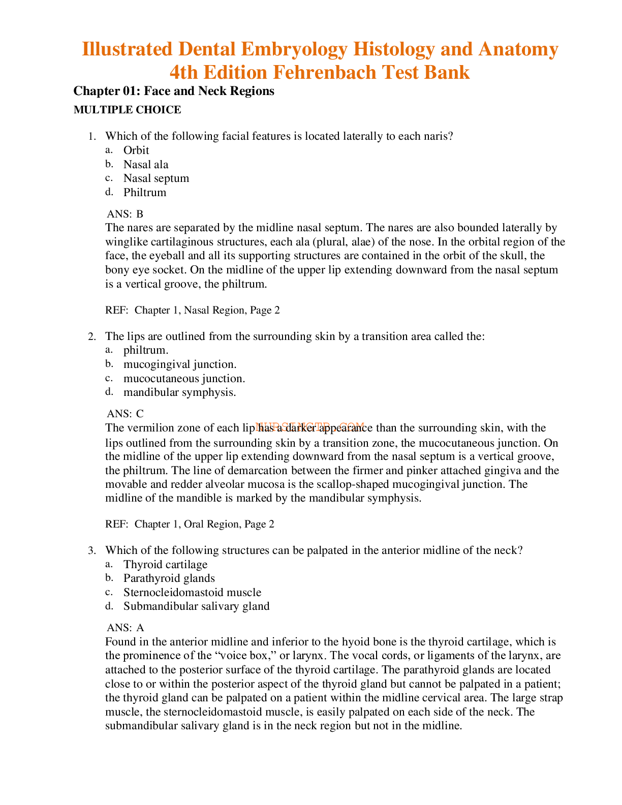
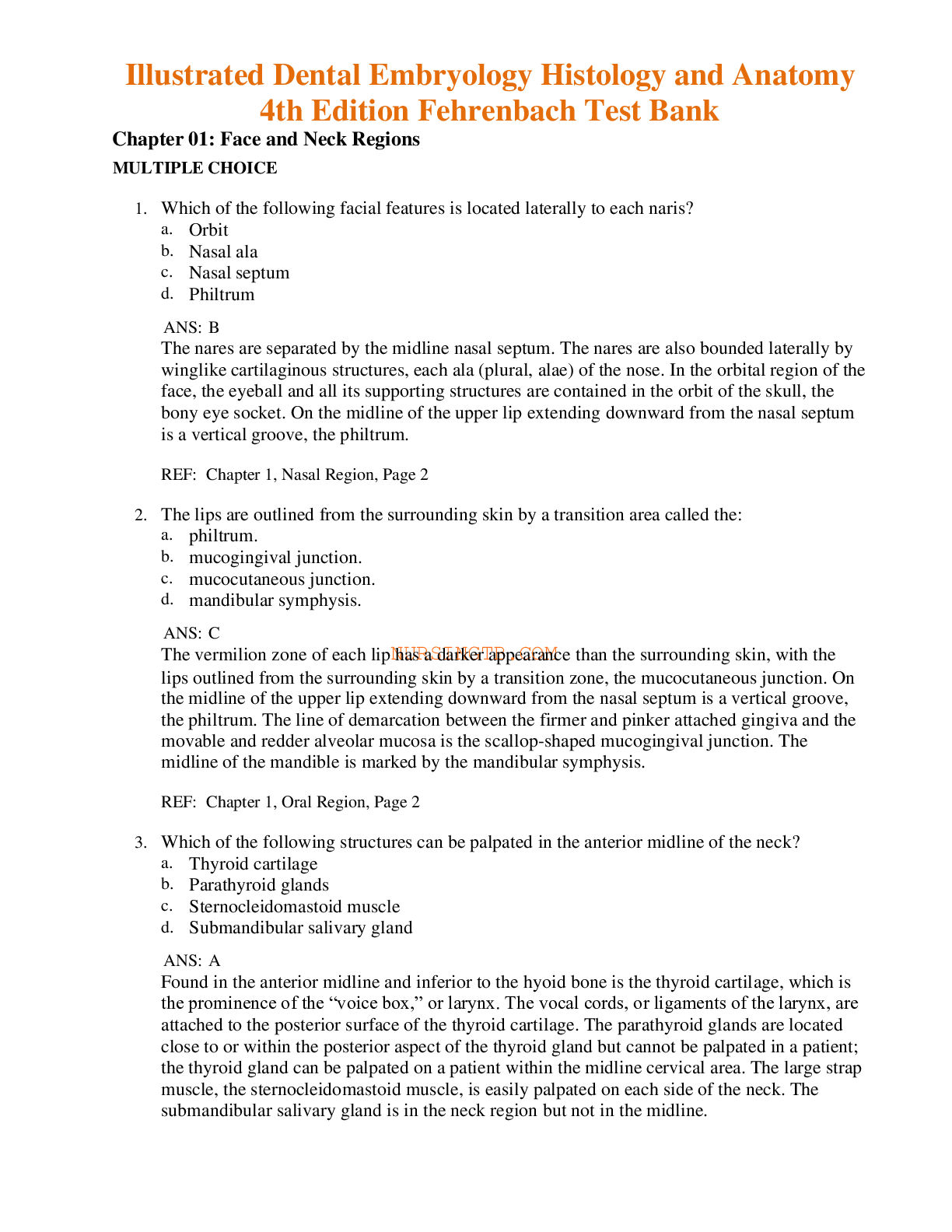
Test Bank IllusChapter 02: Oral Cavity and Pharynx MULTIPLE CHOICE 1. Which of the following oral landmarks may be noted on the soft palate? a. Incisive papilla b. Palatine rugae c. Median palati... ne raphe d. Uvula ANS: D A midline muscular structure, the uvula of the palate, hangs down from the posterior margin of the soft palate. The incisive papilla is a small bulge of tissue at the most anterior part of the hard palate. The median palatine raphe is a midline ridge of tissue on the hard palate. The palatine rugae are firm, irregular ridges of tissue on the hard palate. REF: Chapter 2, Palate, Page 15 2. Which of the following oral landmarks separates the base from the body of the tongue? a. Sublingual fold b. Lingual tonsil c. Plica fimbriatae d. Sulcus terminalis ANS: D Posteriorly on the dorsal surface of the tongue is an inverted V-shaped groove, the sulcus terminalis; it separates the base from the body of the tongue, demarcating a line of fusion of tissue during the tongue’s development. A ridge of tissue on each side of the floor of the mouth, the sublingual fold, joins in a V-shaped configuration extending from the lingual frenum to the base of the tongue; it contains openings of the sublingual duct from the sublingual salivary gland. Even farther posteriorly on the dorsal surface of the base of the tongue is an irregular mass of tissue, the lingual tonsil. Lateral to each deep lingual vein on the ventral surface of the tongue is the plica fimbriata (plural, plicae fimbriatae) with fringelike projections. REF: Chapter 2, Tongue, Page 15 3. Which of the following statements concerning Fordyce spots is correct? a. Composed of salivary gland tissue b. Located on the attached gingiva c. Composed of sebum from sebaceous tissue d. Indicate a disease state in the tissue ANS: C https://www.coursehero.com/file/81804525/ILLUSTRATED-DENTAL-EMBRYOLOGY-HISTOLOGY-AND-ANATOMY-4TH-EDITION-BY-FEHRENBACH-10pdf/ This study resource was shared via CourseHero.com TEST BANK FOR ILLUSTRATED DENTAL EMBRYOLOGY HISTOLOGY AND ANATOMY 4TH EDITION BY FEHRENBACH On the surface of the labial and buccal mucosa is a common variation, Fordyce spots. These are visible as small, yellowish elevations on the oral mucosa. They represent deeper deposits of sebum from trapped or misplaced sebaceous gland tissue, usually associated with hair follicles. REF: Chapter 2, Clinical Considerations with Oral Mucosa, Page 10 4. The line of demarcation between the attached gingiva and the alveolar mucosa is the: a. mucogingival junction. b. interdental gingiva. c. mucobuccal fold. d. marginal gingiva. ANS: A The line of demarcation between the firmer and pinker attached gingiva and the movable and redder alveolar mucosa is the scallop-shaped mucogingival junction. The interdental gingiva is the gingival tissue between adjacent teeth adjoining attached gingiva. Deep within each vestibule is the vestibular fornix, where the pink labial mucosa or buccal mucosa meets the redder alveolar mucosa at the mucobuccal fold. At the gingival margin of each tooth is the marginal gingiva, which forms a cuff above the neck of the tooth. REF: Chapter 2, Gingival Tissue, Page 13 5. The root of the mature and fully erupted tooth is composed of: a. enamel, dentin, and pulp. b. dentin and pulp. c. dentin, pulp, and cementum. d. pulp, cementum, and periodontal ligament. ANS: C The crown of the tooth is composed of the extremely hard outer enamel layer and the moderately hard inner dentin layer overlying the pulp of the tooth. The pulp is the soft innermost layer in the tooth. The moderately hard dentin continues to cover the soft tissue of the pulp of the tooth in the root(s), but the outermost layer of the root(s) is composed of cementum. The bonelike cementum is the part of the tooth that attaches to the periodontal ligament, which then attaches to the alveolus of bone, holding the tooth in its socket. REF: Chapter 2, Jaws, Alveolar Processes, and Teeth, Page 12 6. Which of the following lingual papillae are located on the lateral surface of the tongue? a. Circumvallate papillae b. Filiform papillae c. Fungiform papillae d. Foliate papillae ANS: D Certain surfaces of the tongue have small, elevated structures of specialized mucosa, the lingual papillae, some of which are associated with taste buds. The side or lateral surface of the This study source was downloaded by 100000808701186 from CourseHero.com on 11-03-2021 16:46:42 GMT -05:00 https://www.coursehero.com/file/81804525/ILLUSTRATED-DENTAL-EMBRYOLOGY-HISTOLOGY-AND-ANATOMY-4TH-EDITION-BY-FEHRENBACH-10pdf/ This study restrated Dental Embryology Histology and Anatomy- 4th Edition- Fehrenbach Chapter 02: Oral Cavity and Pharynx [Show More]
Last updated: 2 years ago
Preview 1 out of 9 pages

Buy this document to get the full access instantly
Instant Download Access after purchase
Buy NowInstant download
We Accept:

Reviews( 0 )
$10.00
Can't find what you want? Try our AI powered Search
Document information
Connected school, study & course
About the document
Uploaded On
Nov 04, 2021
Number of pages
9
Written in
Additional information
This document has been written for:
Uploaded
Nov 04, 2021
Downloads
0
Views
165



 (1).png)
