MICROSCOPY LAB REPORT WITH ANSWERS
1. Give a description of the part of the microscope named
arm
• connects the body tube to the ba
microscope
body tube • connects the eyepiece to the obj
coarse focus adjustment •
...
MICROSCOPY LAB REPORT WITH ANSWERS
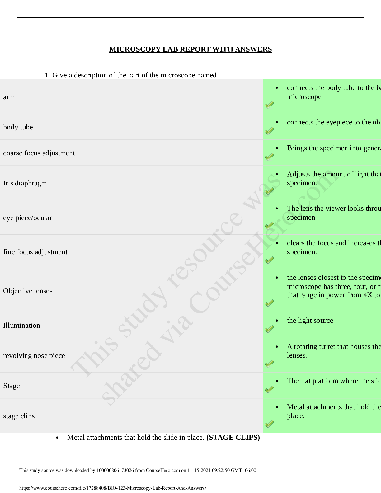
1. Give a description of the part of the microscope named
arm
• connects the body tube to the ba
microscope
body tube • connects the eyepiece to the obj
coarse focus adjustment • Brings the specimen into genera
Iris diaphragm
• Adjusts the amount of light that
specimen.
eye piece/ocular
• The lens the viewer looks throu
specimen
fine focus adjustment
• clears the focus and increases th
specimen.
Objective lenses
• the lenses closest to the specime
microscope has three, four, or fi
that range in power from 4X to
Illumination • the light source
revolving nose piece
• A rotating turret that houses the
lenses.
Stage • The flat platform where the slid
stage clips
• Metal attachments that hold the
place.
• Metal attachments that hold the slide in place. (STAGE CLIPS)
• connects the body tube to the base of the microscope (ARM)
• the lenses closest to the specimen. A standard microscope has three, four, or five of these
that range in power from 4X to 100X. (OBJECTIVE LENS)
• Adjusts the amount of light that reaches the specimen. (IRIS DIAPHRAGM)
• A rotating turret that houses the objective lenses. (REVOLVING NOSE PIECE)
• connects the eyepiece to the objective lenses (BODY TUBE)
• The flat platform where the slide is placed. (STAGE)
• The lens the viewer looks through to see the specimen
• the light source (EYEPIECE/OCULAR)
• Brings the specimen into general focus. (COURSE FOCUS ADJUSTMENT)
• clears the focus and increases the detail of the specimen. (FINE FOCUS
ADJUSTMENT)
2. Calculate the magnification options of the compound microscope
Ocular Lens x Objective Lens = Total Magnification Color of the Objective Lens
x10 4x 40x red
x10 10x 100x yellow
x10 40x 400x blue
x10 100x 1000x white
Feedback
Comments
Comment:
the colors are not correct
3. Describe how the letter e appears under the microscope. Based on what you know about
inversion, what do you think the letter e appears like when you are looking at it with the naked
eye (i.e. not under the microscope).
4. Zoom in to 400x on the onion root tip. You should be able to see square-like shapes with dark
areas in the center. Based on what you have learned about cells and cell structure, what are the
dark areas in the middle of each square? Define its function.
5. Because you are looking at a plant cell, there is possibility you could identify other organelles
under the microscope as well. List at least 3 organelles you (in theory) could identify in the plant
cell that would not be in the animal cell. Include their functions
6. Zoom in to the highest power on the bacterial capsule. How can you tell you are looking at a
prokaryote, and not a eukaryote?
8. Describe the cell theory.
9. Describe the differences between prokaryotic and eukaryotic cells.
10. Eukaryotic cells contain many different organelles that constantly keep our bodies working at
a normal and healthy state. One particular set we learn about is the endomembrane system which
includes 5 different organelles. Describe this system more in detail. Include how it differs
between plants and animals.
11. Zoom in to 400X on the human cheek smear. Are you looking at a prokaryotic or eukaryotic
cell?
Select one:
A. Prokaryotic
Powered by TCPDF (www.tcpdf.org)
[Show More]