Portage Learning A&P 1 101 Answers of All Modules- Final
Pre Exam Questions
1. Describe the position of the epigastric region relative to the umbilical region in anatomical position terms. Use a full sen- tence for
...
Portage Learning A&P 1 101 Answers of All Modules- Final
Pre Exam Questions
1. Describe the position of the epigastric region relative to the umbilical region in anatomical position terms. Use a full sen- tence for your description
• The epigastric region is superior to the umbilical region. (OR) The umbilical region is inferior to the epigastric region.
2. Which anatomical action is occurring at the elbow joint, in the direction of the arrow?
• (Elbow) Flexion
3. Look at the figure below. The right femur is moved in the direction of the arrow. What anatomical action term best describes this movement?
• Abduction (The femur is moving away from midline)
Matching:
A. Emphysema
B. Pleurisy
C. Pulmonary edema
D. Cystic Fibrosis
A. Emphysema- Smoking is often the cause of this disease.
B. Pleurisy This is inflammation of the pleurae.
D. Cystic Fibrosis- With this disease, mucus clogs the respiratory tract.
C. Pulmonary edema- This condition results in fluid-filled alveoli.
Answer the following two questions:
1. Explain why a patient with liver disease would have intolerance to fatty foods.
• The liver produces bile which breaks down fats. If it is not producing bile (or less bile), fats will not be broken down effectively.
2. Explain in detail how the stomach contents enter the small intestine.
• The pyloric sphincter (valve), located at the base of the stomach, relaxes causing a small quantity of chyme to pass through the opening into the first part of the small intestine.
This initiates a reflex that causes the muscles of the sphincter to contract and close the opening temporarily. Then the sphincter relaxes again and allows more chyme to enter.
Label the diagram below, including left or right. Discuss the purpose of tooth (B).
A: Mandible
B:Right Molar
C:Right Premolar (bicuspid)
F:Right Central Incisor
Purpose of tooth type B: Flat molars for crushing food
1. Label the following bone landmarks:
B:Supraspinatus fossa
D:Acromion E:Neck H:Lateral border I:Inferior angle
1. Yellow bone marrow:
A. is found primarily in long bones.
B. is found primarily in short and flat bones.
C. is found primarily in newborns, not adults.
D. produces red blood cells.
2. The diaphysis of a bone:
A. is found at the ends of long bones.
B. contains the articular cartilage at joint articulations.
C. contains the proximal epiphysis.
D. is the center length of a bone.
E. both A. and C.
3. Compact bone:
A. forms the exterior of bones.
B. forms the interior of bones.
C. is lighter than spongy bone.
D. contains numerous bars and plates with irregular spaces.
E. both B. and D.
4. Intramembranous ossification is the formation of from :
A. a growth plate; the center of a bone.
B. long bones; hyaline cartilage.
C. flat bones; connective tissue.
D. a primary ossification center; a cartilaginous disc.
E. both A. and C.
5. What term best describes the type of fracture pictured below?
Greenstick (the bone is broken, but not all the way across)
Answer the following five short-answer questions:
1. Tendons connect which types of tissue? Connect muscle to bone
2. Acetylcholine is what type of substance? Neurotransmitter
3. During a muscle contraction which protein myofilament contains cross-bridges? Myosin
4. What is the name of the thick myofilament? Myosin
5. What must occur for a muscle contraction to stop? Calcium ions must be pumped back into the sarcoplasmic reticulum
Answer the following essay question:
Describe how acetylcholine, sodium ions and calcium ions work together to enable a muscle contraction.
• Acetylcholine (a special chemical called a neurotransmitter) is released from a motor nerve at the neuromuscular junction (or NMJ).
Acetylcholine binds to receptors on the muscle fiber that cause sodium channels to open. Sodium ions rush in- side the cell, triggering an action potential which eventually reaches the sarcoplasmic reticulum.
Calcium ions are released from the sarcoplasmic reticulum of the muscle cell, causing a muscle contraction to oc- cur.
1. Explain the concept of negative feedback and how it helps to maintain homeostasis.
• The effect or increased level of the hormone acts to shut down the continued release of the hormone.
• The brain is constantly monitoring hormone levels to keep levels within a certain range or set-point (homeosta- sis). Negative feedback is a way of “turning off” hormone production when the desired level is achieved
Matching each of the following conditions with the one best explanation (1-5):
1. Jet Lag F. Melatonin production is produced according to body’s normal rhythm
2. Pituitary dwarf A. Underproduction of GH as a child
3. Acromegaly C. Overproduction of GH as an adult
4. Inflammation E. Cortisol helps to counteract this condition
5. Tetany D. Results if PTH is not produced in response to low blood calcium
1. Identify the region and the layer of skin that is highlighted in blue. (Also indicated by the arrow, below) *NOTE: To receive full credit you must label two regions.
• Stratum granulosum; Epidermis
2. A- Identify the sensory cell receptor highlighted in blue, below, also indicated by the arrow. B- What sensory information is detected by this type of receptor?
• Meisner’s corpuscles;
• Detects light touch and vibration (mechanoreceptor)
1. All of the following are functions of skin except:
A. Absorb UV rays
B. Produce vitamin C
C. Thermoregulation
D. Protection for underlying tissues
2. The thickest region of the dermis is the:
A. Reticular
B. Papillary
C. Basement membrane
D. Subcutaneous
E. None of the above are layers of the dermis
3. The hypodermis is comprised mainly of:
A. Adipocytes
B. Reticular cells
C. Dermatomes
D. Sebaceous glands
4. What is a contusion?
A. A third degree burn
B. An infection in the epidermis
C. Damage to a broken blood vessel
D. Subcutaneous injection
5. Information from a dermatome travels to:
A. The brain
B. A hair follicle
C. A gland
D. The skin
Module 1 Answers
All multiple choice questions have one answer unless otherwise specified. Choose the best response to the question with the information provided.
1. What is the study of the structure of the human body?
A. Anatomy
B. Physiology
C. Anabolism
D. Catabolism
E. Metabolism
2. The liver is an example of the organ level of organization in the human body
3. What is the breakdown of nutrients?
A. Anatomy
B. Physiology
C. Anabolism
D. Catabolism
E. Metabolism
1. List the four basic types of tissues.
• Epithelium, muscle, connective, and nervous tissue
2. List the three layers of the skin in order from deep to superficial. The deepest layer of the skin should be at the top of your list. To receive credit the layers must be in this order.
• hypodermis (inner, also called subcutaneous tissue layer)
• dermis (middle layer)
• epidermis (outer layer)
*MUST be in this order (top to bottom) to receive credit
3. Testosterone contributes to muscle strength and bone mass. Based upon your learning of metabolism, would it be consid- ered an anabolic or catabolic hormone? Explain why.
• Anabolic because it is building new tissues from smaller cells and using energy to do so.
Note: Essay answers must clearly be in your own words. All multiple choice questions have one answer unless otherwise specified. Choose the best response to the question with the information provided.
1. What anatomical orientation term is used to indicate "toward the front of the body"?
A. Superior
B. Inferior
C. Ventral
D. Dorsal
E. Medial
2. What anatomical orientation term is used to indicate "toward the upper part of a structure"?
A. Superior
B. Inferior
C. Ventral
D. Dorsal
E. Medial
3. What anatomical orientation term is used to indicate "away from the head end"?
• Inferior/Caudal
4. What anatomical orientation term is used to indicate "further from the origin of the body part"?
• Distal
5. You are looking at a diagram of a patient standing in anatomical position. On the sheet of paper containing the dia- gram, which thumb points to the left side of the paper, right or left?
• Right
Note: Essay answers must clearly be in your own words. All multiple choice questions have one answer unless otherwise specified. Choose the best response to the question with the information provided.
1. Are the arms part of the axial or appendicular region of the body?
• Appendicular
2. A patient is standing in anatomical position. The patient moves his hand to scratch his nose. What anatomical action term is best used to indicate the motion occurring at his elbow?
A. Flexion
B. Extension
C. Elevation
D. Depression
E. Abduction
3. What anatomical action term is used to indicate "movement away from midline"?
A. Flexion
B. Extension
C. Elevation
D. Depression
E. Abduction
Note: Essay answers must clearly be in your own words. All multiple choice questions have one answer unless otherwise specified. Choose the best response to the question with the information provided.
1. Describe the position of the diaphragm in relation to the thoracic cavity in anatomical position terms. Use a full sen- tence for your description.
• The diaphragm is inferior to the thoracic cavity.
(OR) The thoracic cavity is superior to the diaphragm.
2. Which anatomical action is occurring at the elbow joint, in the direction of the arrow?
• (Elbow) Extension
3. Look at the figure below. The right femur is moved in the direction of the arrow. What anatomical action term best describes this movement?
• Adduction (The femur is moving towards midline)
Note: Essay answers must clearly be in your own words. All multiple choice questions have one answer unless otherwise specified. Choose the best response to the question with the information provided.
1. List two organs contained in the pelvic cavity.
• The pelvic cavity contains the bladder, reproductive organs and the rectum
2. True or False: The ventral body cavity contains the cranial cavity, the thoracic cavity, and the abdominal cavity. If your answer is false, rephrase the statement to make it a true statement.
False. The ventral cavity contains the thoracic cavity and the abdominal cavity. (It does not contain the cranial cavity)
1. The body is divided into superior and inferior sections by what type of plane?
• Transverse or horizontal plane
2. A sagittal plane that lies exactly in the midline is referred to as what type of plane?
• Median or midsagittal plane.
3. Look at the diagram below. What type of plane is shown?
• Frontal (coronal)
1. Using the letters given, match the cell with the type of solution it has been placed in:
• A cell beginning to shrink- hypertonic
• A red blood cell placed in distilled (purified) water- hypotonic
• A cell neither shrinking or swelling - isotonic
2. Using the letters given, match each molecule/item with its typical means of entering a cell. You will need to use some means of entry more than once.
• Lipid insoluble molecules Facilitated diffusion
• Other cells Endocytosis
• Water Diffusion
• Carbon Dioxide Diffusion
3. Which one of the following is not true of active transport?
A. The sodium-potassium pump is an example
B. Additional energy is needed to drive this process
C. Active transport moves particles with the concentration gradient
D. Active transport requires carrier proteins.
4. List the three organelles, in order, involved in the synthesis, transport and shipping of proteins.
A. ribosomes, rough ER, Golgi complex
B. rough ER, Golgi complex, lysosomes
C. ribosomes, smooth ER, Golgi complex
D. rough ER, Golgi complex, vacuole
1. Label the 5 organelles listed below:
3: Nucleolus
4: Nuclear Envelope
5: Mitochondria
7: Lysosome
8: Endoplasmic Reticulum (ER)
2. True or False?
a. The purpose of cellular respiration is to produce ADP. (T or F) a. FALSE (ATP is produced, not ADP)
b. Peripheral proteins are found on the outer surface of a cell membrane. (T or F) b. TRUE (Peripheral proteins can be found on the outer or inner surface, but do not span across the entire membrane).
c. Exocytosis is the process that occurs to bring biomacromolecules inside the cell. (T or F) c. FALSE (This process is called endocytosis)
d. Receptor-mediated endocytosis uses a signaling molecule from another cell, binding to the cell membrane to bring about changes within the cell proteins. (T or F) d. TRUE
e. Endocytosis is a passive process. It does not require cellular energy expenditure.(T or F) e. FALSE (Endocytosis is an ac- tive process and requires cellular energy)
1. In your own words, discuss Tay-Sachs disease. What organelle within the cell is impacted? What are the symptoms of the disease and why do they occur?
(1) Lysosomes
(2) buildup of toxic lipids inside the cell (biomacromolecules)
(3) disability and death
In the genetic disease known as Tay-Sachs, one of the normally present digestive enzymes inside lysosomes is lacking. Thus, a toxic lipid in the brain cells cannot be broken down. The resulting buildup of lipids in these cells can cause intel- lectual disability and death.
2. You are observing two cells under the microscope. They are the same type of eukaryotic cell but one appears much larger. Based on appearance alone, which one would you expect to be carrying out respiration at a more active rate, the larger or smaller cell? Explain why.
The smaller cell. Cells need to remain relatively small because as a cell expands the amount of surface area relative to the volume of the cell decreases. The smaller cell is more active because relative to its volume, its surface area is larger than a bigger cell. With a larger surface area (relative to its volume) this allows the metabolic processes to occur faster.
Module 2 Answers
1. How many lobes does the right human lung have?
Three Lobes
2. Air (and not food) pass in which of the following areas:
A. Esophagus
B. Nasopharynx
C. Oropharynx
D. Both A. and C.
3. Rings of cartilage line much of the respiratory tract. In which of one the following would cartilage NOT be found?
A. Trachea
B. Larynx
C. Bronchioles
D. Alveoli
Label the following five items from the diagram:
Label A: Nasal Cavity
Label C: Soft Palate (Uvula also acceptable answer) Label E: Glottis (not epiglottis)
Label F: Trachea Label H: Tongue
1. Explain what happens to the soft palate during swallowing. Why?
The upward movement of the soft palate prevents food or liquid from entering the nasal passages during swallowing.
2. Explain at least two differences between Type I and Type II alveolar cells.
a. Type I form the very thin simple squamous epithelium of the alveoli in junction with capillaries. Make up roughly 95% of alveolar epithelial cells.
b. Type II produce and secrete pulmonary surfactant which is needed throughout the alveolar surface to keep the alveoli open. In addition, Type II cells can divide to replace damaged Type I cells. Make up roughly 5% of alveolar epithelial cells.
1. Positive pressure is used to move air into the lungs. False (Negative pressure)
2. During inhalation the diaphragm contracts to pull the lungs open. True
3. During inhalation the rib cage is lowered pull the lungs open. False (rib cage is lifted upward during inhalation)
4. As the thoracic cavity expands and lung volume increases, the density of the gases filling the lungs increases. False
5. As the rib cage expands during inhalation, air will naturally flow into the lungs. True
Answer the following five multiple choice questions:
1. According to Charles's Law gas volume is:
A. Indirectly proportional to its temperature
B. Directly proportional to its pressure
C. Directly proportional to its temperature
D. Indirectly proportional to its pressure
2. Which one of the following tracheal cartilages are paired?
A. Thyroid
B. Cricoid
C. Arytenoid
D. Epiglottal
E. Hyaline
3. Which one of the following is not true of the pleurae?
A. The pleurae are membranes that cover surfaces
B. Parietal pleurae cover the surfaces surrounding the lungs
C. Visceral pleurae cover the diaphragm
D. A healthy pleural cavity is nearly empty of air or fluid
4. Which one of the following is not true of the bronchioles?
A. Bronchioles can contain respiratory epithelium
B. Bronchioles can contain simple squamous cells
C. Bronchioles can contain fossae and mucus
D. Bronchioles contain cartilage and goblet cells
E. Bronchioles can contain cilia and mucus
5. Which one of the following is true of the respiratory physiology calculations? (You may find it helpful to draw the respi- ratory physiology diagram on a piece of scratch paper to help you answer this question.)
A. Vital Capacity (VC) =TLC + RV
B. Total Lung Capacity (TLC) =IRV + TV
C. Inspiratory Capacity (IC) = TV- IRV
D. Functional Residual Capacity (FRC) = RV+ERV
1. The gas-exchange region in the lungs must be dry, thin and large. False - requires moisture
2. During inhalation, negative pressure draws air into the lungs. True
3. During exhalation, the diaphragm rises. True
4. Because of diffusion, oxygen naturally moves out of the blood and into the lungs. False
5. Each red blood cell contains from 10-100 molecules of hemoglobin. False Matching:
• Smoking is often the cause of this disease. -Emphysema
• With this disease, mucus clogs the respiratory tract. -Cystic Fibrosis
• This condition results in fluid-filled alveoli. -Pulmonary edema
• This is inflammation of the pleurae. -Pleurisy
1. The pressure of three gases equals 1 atmosphere. What is the partial pressure of oxygen (in mmHG) if nitrogen is 300 mmHg and carbon dioxide is 350 mmHg? To receive full credit you must show your work.
2. Why would warming air be beneficial to gas exchange?
It would expand the air to make it more available for gas exchange.
3. Explain why incomplete ventilation is important for effective gas exchange.
Incomplete ventilation helps to maintain a constant temperature within the lungs. The increased temperature allows for greater gas exchange.
Answer the following three questions:
1. From widest to narrowest, the branches of the bronchial tree are:
A. Primary bronchi, secondary bronchi, tertiary bronchi, bronchioles
B. Bronchioles, primary bronchi, secondary bronchi, tertiary bronchi
C. Tertiary bronchi, secondary bronchi, primary bronchi, bronchioles
D. Secondary bronchi, tertiary bronchi, primary bronchi, bronchioles
2. In gas exchange (external respiration):
A. Carbon dioxide diffuses from alveoli into capillaries, oxygen diffuses from capillaries into alveoli
B. Oxygen diffuses from alveoli into capillaries, carbon dioxide diffuses from capillaries into alveoli
C. Oxygen and carbon dioxide is carried from alveoli into the bronchioles
D. Oxygen is chemically transformed into carbon dioxide within the alveoli
3. The purpose of these cells are the primary immune defense of the alveoli:
A. Type I alveolar cells
B. Type II alveolar cells
C. Surfactant
D. Alveolar macrophages
1. Emphysema is a disease which causes air to become trapped in the lungs upon exhalation. If a patient is diagnosed with em- physema:
1) Which of the following pulmonary function values would be abnormally high?
2) Explain your answer.
A. Inspiratory Reserve Volume
B. Expiratory Reserve Volume increases
C. Residual Volume decreases
C. Residual Volume as it is the amount of air remaining in the lungs after exhalation that cannot be exhaled
Module 3 Answers
1. Explain why someone who has hypergastrinemia (excessive secretion of gastrin) might develop gastric/stomach ulcers.
Gastrin stimulates the secretions of pepsinogens and hydrochloric acid. Excess amounts would lead to erosion of the stomach lining.
2. Fill in the four blanks:
Food enters the stomach from the esophagus via the cardioesophageal sphincter. Food exits the stomach via the pyloric sphincter into the small intestine.
Look carefully at the diagram below. Label the following 5 organs of the digestive system. To receive credit for the intestines you must label the specific region.
3: stomach
4: gall bladder
7: jejunum
1. A patient is experiencing vitamin toxicity. What type of vitamin would be causing this toxicity? Explain your answer.
B. Fat soluble vitamin. Fat soluble vitamins are stored within the body’s fat stores making it harder for the body to rid itself of them.
2. Explain which digestive system functions are carried out by the small intestine.
• Nutrient absorption & chemical digestion
1. Describe parietal cells and chief cells: name their location, secretions and purposes.
1. Match the digestive organ with the one substance it produces: (4 points):
Pancreas - Lipase
Liver -Urea
Mouth- Salivary amylase
2. What is the purpose of the hormone Ghrelin?
A. Increase hunger, decrease satiety
B. Increase hunger, increase satiety
C. Decrease hunger, increase satiety
D. Decrease hunger, decrease satiety
3. Which of the following statements is false?
A. Taste buds are located on the surface of the mouth and the wall of the pharynx.
B. The ileocecal valve controls the entrance of chyme into to the small intestine.
C. The gall bladder, teeth, and tongue are considered accessory organs to the digestive system.
D. The alimentary canal is a continuous muscular tube, open at both ends.
1. Lipids can be divided into two categories: complete or incomplete. False
2. Catabolism combines smaller molecules to make larger molecules. False (Anabolism)
3. The oropharynx is the most superior region of the pharynx. False (The nasopharynx is the most superior region)
4. The pharynx has two types of skeletal muscle: circumferential and oblique. False (It does not include an oblique layer)
5. The oblique layer of the stomach churning food into smaller pieces is an example of mechanical digestion. True
1. Label the following three types of raised bumps on the tongue. Describe the purpose of type C.
A: Filiform papillae
B: Circumvallate papillae
C: Fungiform papillae
Describe C: It helps with gripping food and contains taste buds. Answer the following three questions:
1. Which of the following statements is true concerning glycolysis?
A. Pyruvic acid are broken down into glucose.
B. Five ATP molecules are formed.
C. Glucose is broken down into two pyruvate molecules.
D. Glucose is broken down into two ADP molecules.
2. Which of the following statements is false concerning the second stage of glucose breakdown?
A. This second stage is also known as the TCA cycle.
B. Pyruvic acid is converted to carbon dioxide.
C. Additional ATP is formed.
D. This is also known as the electron transport system.
3. Which of the following statements is true concerning the Electron Transport System?
A. Can proceed can proceed only aerobically
B. Approximately a total of 2 ATP are formed.
C. Protons are pumped out of the inner mitochondrial membrane.
D. The proton motive force forms ADP.
Label the diagram below, including left or right. Describe tooth (A).
A: left central incisor
C: left canine
E: molar
F: maxilla/hard palate
Describe tooth type A:
Incisors are for biting
1. Pancreas:
A. Storage site for bile
B. Attached to the soft palate
C. Secretes juices which digest all types of foods
D. Stores glycogen and produces urea
E. Forms food into a bolus
2. Stomach:
A. Hangs from the cecum
B. Connects mouth to esophagus
C. Attaches to the greater omentum
D. Contains the hepatic flexure
E. Site of the ileocecal valve
3. Submucosal (layer of tissue):
A. Made of two layers of muscle
B. Innermost layer
C. Secretes mucous
D. Contains blood and lymph vessels
E. Begins in the mouth
4. Enamel:
A. Lies below the gum line
B. Innermost layer
C. Largest portion of each tooth
D. Contains nerves and vessels
E. Located on the surface of the crown
5. Common Bile Duct
A. Contains secretions from the pancreas
B. Joins with the pancreatic duct
C. Enters the jejunum
D. Contains secretions from the salivary glands
E. Contains lymph vessels and nerves
Module 4 Answers
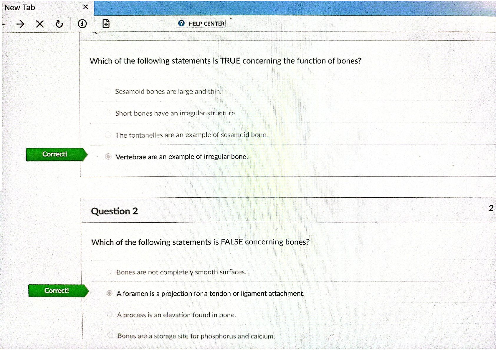
1. Which of the following statements is TRUE concerning the function bones?
A. Sesamoid bones are large and thin.
B. Short bones have an irregular structure
C. The fontanelles are an example of sesamoid bone.
D. Vertebrae are an example of irregular bone.
2. Which of the following statements is FALSE concerning bones?
A. Bones are a storage site for phosphorus and calcium.
B. A tuberosity is a projection for a tendon or ligament attachment.
C. A process is a depression found in bone.
D. Bones are completely smooth surfaces.
E. C. and D. are false
3. Which of the following statements is FALSE about the skeletal system?
A. The two main divisions of the skeletal system are: axial and appendicular.
B. The two main divisions of the skeletal system are: vertebral and thoracic.
C. The axial skeleton lies along the midline of the body.
D. The vertebral division of the skeleton is included in the appendicular skeleton.
E. B. and D. are false
F. A. and C are false
4. The skull is formed by bones; the facial skeleton contains bones.
A. 30; 10
B. 22; 14
C. 10; 5
D. 15; 7
5. What bone is highlighted in blue in the figure below? (superior/internal view)
Sphenoid bone
1. Label the following bones of the skeleton from the figure below:
1:Frontal bone (or frontal sinuses)
3:Maxilla
5:Vomer
7:Maxilla
8:Mandible
1. Label the following bone landmarks:
A: head
C: lesser tubercle
F: deltoid tuberosity I: capitulum
K: medial epicondyle
1. Which of the following statements is FALSE about the scapula?
A. The clavicle connects the coracoid process of the scapula.
B. The lateral border of the scapula connects to the neck of the scapula.
C. The supraspinatus fossa is located on the posterior side of the scapula.
D. The clavicle articulates with the scapula.
2. Which of the following statements is TRUE about the humerus?
A. The lateral epicondyle of the humerus articulates with the ulna.
B. The capitulum articulates with the ulna.
C. The lateral epicondyle is the prominent bone landmark of the medial side of the elbow (in anatomical position).
D. The head of the humerus articulates with the scapula.
E. B. and D. are true
3. The glenohumeral joint is prone to dislocation. In your own words, (1) Describe dislocation and (2) What happens in disloca- tion of the glenohumeral joint?
Dislocation of a joint means that the bone is removed from its socket. Dislocation of the shoulder occurs when the head of the humerus is removed from the glenoid cavity in any direction.
Label the two figures:
1. Label the bones in the figure below: A:Trapezium
B:Scaphoid
C:Lunate
D:Triquetral
E:5th metacarpal (must include both parts for full credit)
2. Label the following bones in the figure below: A:
1st Metatarsal (optional: Hallux)
C:Lateral Cuneiform E:Medial Cuneiform F:Navicular
G:Talus
1. Red bone marrow:
A. is found primarily in long bones.
B. is found primarily in short and flat bones.
C. is only found in adult bones.
D. produces fat storage.
2. The epiphysis of a bone:
A. is found at the ends of long bones.
B. contains the articular cartilage at joint articulations.
C. contains the diaphysis.
D. is the center length of a bone.
E. both A. and B.
3. Spongy bone:
A. forms the interior of bones.
B. forms the exterior of bones.
C. is heavier than spongy bone.
D. contains numerous bars and plates with irregular spaces.
E. both A. and D.
4. Endochondral ossification:
A. begins at the primary ossification center at end of long bones
B. begins at the primary ossification center at the center of long bones.
C. is the formation of long bones from hyaline cartilage.
D. is the formation of long bones from connective tissue.
E. both A. and D.
F. both B. and C.
5. What term best describes the type of fracture pictured below?
Open compound fracture.
1. A patient has a diagnosis of osteoporosis. (1) In your own words, describe this diagnosis and (2) What type of bone cell would they be lacking? Explain your answer.
Osteoporosis is a bone disease that causes bone tissue to degenerate faster than it can be replaced. This causes paid and is more likely to fracture. The bone cell that would be lacking is osteoblasts.
2. Your patient has back pain due to a herniated disc. (1) In your own words explain what it means to have a herniated disc. (2) As reviewed in the module, discuss one treatment option to address your patient’s pain.
(1) A herniated disc is an injury to the intervertebral disc, where the center portion of the disc bulges into the vertebral fora- men, causing pain.
(2) Explanation of
1- Physical therapy for strengthening to support back ligaments. OR 2- Surgery to fuse two vertebrae together.
1. Matching: Match the joint with the correct joint classification (A-F).
*NOTE: Some joints may fall into more than one category. Mark all that apply.
A= Fibrous B= Cartilaginous C= Synovial D= Hinge E= Ball-and-Socket F=Saddle
1. Knee joint- C, D (Synovial, Hinge) 2. Thumb joint- C, F (Synovial, Saddle)
3. Shoulder joint C, E (Synovial, Ball and socket) 4. Vertebral joint B (Cartilaginous)
5. Cranial joints A (Fibrous)
1. Name the ligament highlighted in blue in the figure below:
Iliolumbar ligament
2. Name the ligament highlighted in blue in the figure below:
PCL (Posterior cruciate ligament)
3. Name the ligament highlighted in blue in the figure below:
Coracohumeral ligament
Module 5
Answer the following five true or false questions:
1. There are three types of muscle tissue found in the body. True
2. Smooth and cardiac muscle tissue are both under voluntary control. False
3. The lumbar plexus is part of the central nervous system. False- this is a peripheral nerve
4. A sensory signal is a signal that is sent from a muscle to the central nervous system. True
5. The lumbar plexus contains nerves that innervate the thigh. True
1. Ligaments connect which types of tissue? Connect bone to bone
2. Acetylcholine is what type of substance? Neurotransmitter
3. During a muscle contraction which protein myofilament is pulled along? Actin
4. What is the name of the thin myofilament? Actin
5. The return of calcium ions into the sarcoplasmic reticulum causes what to occur? Muscle relaxation (Muscle contraction stops)
Label the following five muscles:
B: Left Temporalis C: orbicularis oculi E: zygomaticus major G: buccinator (deep)
J: depressor labii inferioris
Fill in the following muscle chart (10 blank spaces):
Muscle Origin Insertion Action Innervation
Flexor digitorum
superficialis ------- ---------- 1. 2.
Pronator Teres ------- 3. 4. 5.
Vastus Medialis 6. ---------- 7. --------------
Peroneus Brevis 8. 9. ----------- 10.
1. Flexion of the wrist & digits 205 (middle & proximal phalanx) 2. Median nerve
3. mid lateral surface of radius 4. pronation
5. median nerve
6. intertrochanteric line of femur 7. knee extension
8. lateral, distal fibula
9. lateral side of 5th metatarsal 10. superficial peroneal nerve
1. Your patient sustained an injury to their facial nerve (CN VII). Which actions would be impaired?
A. Eye closure
B. Raising eyebrows
C. Jaw closure
D. Neck flexion
E. A&B
F. C&D
2. Your patient is having difficulty when asked to bring his chin to his chest. He is also having difficulty turning his face from side to side. What muscle is most likely impaired?
A. Thyrohyoid
B. Semispinalis capitis
C. Splenius Capitis
D. Sternocleidomastoid
E. Scalenes
3. A person sustained a back injury to the muscle group closest to the spine. What muscle group was injured?
A. Longissimus
B. Spinalis
C. Sternocleidomastoid
D. Iliocostalis
E. Rectus abdominis
4. True or False: When a muscle contracts, the Z lines within a sarcomere come closer together towards the M line. True
5. True or False: The I bands contain thick filaments. False (I bands contain thin filaments)
1. Which muscle contracts to enable the main effort required to stand on your toes?
A. Biceps femoris
B. Quadriceps
C. Gastrocnemius
D. Tibialis anterior
2. When gluteus maximus contracts, which bone is pulled posteriorly by this muscle?
A. Tibia
B. Ilium
C. Ulna
D. Femur
3. When flexor digitorum profundus contracts, what action(s) occurs?
A. Wrist flexion
B. Elbow flexion
C. Flexion of digits 2-5
D. Flexion of digit 1
E. A&D
F. B&C
4. Contraction of results in ?
A. obturator externus, spine flexion
B. obturator externus, spine extension
C. iliacus, hip flexion-
D. iliacus, hip extension
5. When piriformis contracts, what action(s) will not occur at the hip?
A. Internal rotation
B. Lateral rotation
C. Adduction
D. Flexion
E. Extension
F. A&D-
G. B&E
Use the figure answer the following five questions:
1. Which muscle is the prime mover of adduction, flexion and inward rotation of the humerus? (specify letter on diagram with the name of the muscle for full credit)
B (Pectoralis Major)
2. Which muscle is innervated by the musculocutaneous nerve? (specify letter on diagram with the name of the muscle for full credit)
F or G (Coracobrachialis)
3. What is the name of muscle C? (specify letter on diagram with the name of the muscle for full credit) Supraspinatus
4. What is the innervation of muscle C? (specify letter on diagram with the name of the muscle for full credit) Suprascapular nerve
5. What is the origin of muscle E? (specify letter on diagram with the name of the muscle for full credit)
Inferior angle of scapula (Teres major)
Use the figure to label the following six muscles: (Viewed anteriorly)
A: flexor carpi radialis
B: flexor digitorum superficialis C: palmaris longus
D: flexor carpi ulnaris E: flexor pollicis longus
F: flexor digitorum profundus
Use the figure to label the following six muscles:
A: gastrocnemius B: tibialis anterior
C: extensor digitorum longus G: tibialis posterior
H: flexor digitorum longus I: flexor hallucis longus
Describe what is meant by muscle fiber “cross-bridges”. What occurs to enable movement of the cross-bridges? Cross bridges occur when calcium ions are present. This causes portions of the myosin filaments to bend backwards & attach to actin filaments. Once this attachment occurs, the cross- bridges bend forward and the actin filament is pulled along.
The cross-bridges attach and detach between 50-100x as the thin filaments are pulled to the center of a sar- comere. ADP is need on a cellular level for the myosin cross-bridges to pull the actin filaments.
Module 6
1. Explain two reasons why a woman with low levels of LH would not be able to become pregnant.
A woman would not be able to become pregnant with low levels of LH because:
1. LH acts on the ovary to cause ovulation to occur
2. LH causes progesterone release to facilitate the potential fertilization of the effort and pregnancy. Progesterone is a key hormone for the maintenance of pregnancy.
2. Would you expect a female to have testosterone in their bloodstream? Explain why or why not.
Yes because males and females produce some levels of this hormone but females do not produce enough to affect the gonads/ the ovaries the way that it affects the testes.
Label the endocrine glands (A-C)
A:pineal gland B:hypothalamus C: pituitary gland
1. Explain the concept of negative feedback and how it helps to maintain homeostasis.
Negative feedback one way the body maintains homeostasis. It is like a thermostat in which the body acts to maintain all its levels for function.
1. This type of hormone is derived from cholesterol:
A. Peptide hormones
B. Catecholamines
C. Steroid hormones
D. Tyrosine
2. Growth hormone
A. is most active during childhood/adolescence.
B. can cause disease if levels are not correct.
C. cannot affect an adult.
D. A &B
3. Neurons in the hypothalamus are called
A. Neurosecretory
B. Vasopressin
C. Melanocytes
D. Somatotropic
4. Which hormone production is increased with sunlight?
A. MSH –melanocyte stimulating hormone
B. Melatonin
C. Cortisol
D. GH -Growth hormone
5. The posterior pituitary stores these two hormones:
A. ADH and LH
B. ADH and Oxytocin
C. LH and FSH
D. TSH and Prolactin
1. List the hormone that best describes each of the four statements below:
A. I promote the reabsorption of water at the collecting ducts of the kidneys. ADH
B. I am secreted by the pituitary to stimulate the thyroid. TSH
C. I am secreted by the beta cells of the pancreas. Insulin
D. I cause blood glucose to rise and breakdown stored nutrients. Glucagon
1. Explain in detail how PTH and calcitonin work to maintain calcium balance.
Calcitonin is a hormone that lowers the level of calcium in the blood and deposits calcium into the bone. It also opposes the action of the PTH produced by the parathyroid glands.
PTH also retain calcium through excreting phosphate at the kidneys. In the bones, pTH promotes the activity of osteo- clasts to demineralize to demineralize the bone, increasing the amount of calcium in the blood. When the blood calcium level reaches appropriate level through all these means, the parathyroid glands stop producing PTH through negative feedback.
1. Which hormone/s are involved in milk production?
A. Thyroxin
B. Oxytocin
C. Prolactin
D. B &C
E. A&B
2. Which cells mature in the thymus?
A. B cells
B. T cells
C. Red blood cells
D. the islets of Langerhans
E. A & B
3. Iodine is needed to produce which hormone/s?
A. T3/T4
B. Gonadotropic
C. ACTH
D. TSH
E. A & D
4. Which of the following is NOT a function of oxytocin?
A. Uterine contraction
B. Bone growth
C. Milk letdown
D. Given to aide in childbirth process
5. Which hormones are produced in the anterior pituitary?
A. TSH, MSH, and calcitonin
B. ADH, GH, and prolactin
C. PTH, TSH, and FH
D. ACTH, GH, and prolactin
1. Aldosterone is involved in the regulation of sodium and potassium in the body. Explain how too much aldosterone could contribute to high blood pressure.
• Aldosterone’s primary target organ is the kidney, where it promotes renal absorption of sodium and renal excretion of potassium. The blood sodium level is particularly important to the maintenance of blood pressure. Too much sodium causes retention of fluid and increases blood pressure. Therefore, too much aldosterone will cause increased and po- tentially high blood pressure.
Matching each of the following conditions with the one best explanation (1-5):
1. Congenital hypothyroidism
C. Low thyroxin production since birth
2. Pituitary giant
A. Overproduction of GH as a child
3. Acromegaly
H. Overproduction of GH as an adult
4. Tetany
F. Results if PTH is not produced in response to low blood calcium
5. Anemia
E. Lack of erythropoietin to act on the bone marrow
1. In addition to the endocrine system, the pancreas is also part of which system?
A. Excretory
B. Nervous
C. Circulatory
D. Digestive
2. Which part of the adrenal glands is vital to life?
A. Cortex
B. Medulla
C. The entire gland
D. None of adrenal gland is vital to life
3. Which of the following is false regarding aldosterone?
A. It is a regulated by the concentration of sodium.
B. It causes the kidneys to excrete potassium and retain sodium.
C. It causes the kidneys to retain potassium and excrete sodium.
D. It is a mineralocorticoid.
E. It increases blood pressure
4. If you were calcium deficient, which hormone would most likely need to increase in your body in order to compen- sate?
A. PTH
B. Calcitonin
C. Erythropietin
D. TSH
5. Which of the following is true regarding estrogen?
A. Production is highest in childhood.
B. It causes growth of the uterus and vagina.
C. It decreases fat storage.
D. It causes females to have a narrower pelvis (compared to males).
Module 7
1. Identify the region and the layer of skin that is highlighted in blue. (Also indicated by the arrow, below) *NOTE: To re- ceive full credit you must label two regions.
Stratum granulosum; Epidermis (Must have both for full credit)
2. A- Identify the sensory cell receptor highlighted in blue, below, also indicated by the arrow. B- What sensory information is detected by this type of receptor?
A) Meisner’s corpuscles;
B) Detects light touch and vibration (mechanoreceptor)
1. The layers of the epidermis, from deep to superficial are:
A. Stratum corneum, stratum granulosum, stratum spinosum, stratum basale.
B. Stratum basale, stratum spinosum, stratum granulosum, stratum corneum.
C. Stratum basale, stratum granulosum, stratum corneum, Stratum spinosum.
D. Stratum corneum, stratum spinosum, stratum granulosum, stratum basale.
2. Exocrine glands secrete ; Endocrine glands secrete .
A. for body-wide distribution, into an epithelial lining
B. into an epithelial lining, for body-wide distribution
C. through a duct, into the bloodstream
D. into the bloodstream, through a duct
E. A&D
F. B&C
3. Which of the following is false regarding the epidermis:
A. It is avascular.
B. As cells divide they are pushed towards the basement membrane.
• False, as cells divide they are pushed towards the external surface of the stratum corneum
C. The cells of the stratum basale cells have a high rate of cell division.
D. Epidermal ridges form a person’s fingerprints.
1. Match the cell (#1-4) with its single best description (A-F), using each letter only once.
1. Keratinocytes
E. Produces a protein to protect the skin
2. Fibroblasts
B. Produces collagen
3. Melanocytes
F. Produces a pigment that absorbs UV rays
4. Langerhans cells
A. Assists in immune responses
1. A person touches a hot cup of coffee. What type of sensory organ detects this information?
Thermoreceptors
2. Merkel cells are embedded in what layer of the skin? epidermis
3. In your own words, how can the skin be used to determine if a person is dehydrated?
A person's skin can be tested for dehydration by pinching the skin on the back of the hand. If this skin stay pinched in in- stead of going back to its normal shape it is an indicator of dehydration.
Answer the following two questions:
1. What is the name and function of the structure below? (Highlighted in blue, also indicated by the arrow)
This is the arrector pili muscle and its function is to contract when the body is cold or experience emotional responses.
2. What is the name and function of the structure below? (Highlighted in blue, also indicated by the arrow) Sweat gland; its function is to produce sweat in almost every part of the skin.
1. All of the following are functions of skin except:
A. Absorb UV rays
B. Thermoregulation
C. Perspiration
D. Produce vitamin K
2. The most superficial region of the dermis is the:
A. Reticular
B. Papillary
C. Stratum corneum
D. Subcutaneous
E. None of the above are layers of the dermis
3. A subcutaneous injection is to be given in what region of the skin?
A. Epidermis
B. Dermis
C. Hypodermis
D. Muscle tissue
4. What is a contusion?
A. A third degree burn
B. An infection in the epidermis
C. Subcutaneous injection
D. Damage to a broken blood vessel
5. Information from a dermatome carries information from:
A. The brain
B. The spinal cord
C. A gland
D. The skin
1. This injury, seen below, is best described as what degree of burn? Explain what layer/s of skin are damaged in this type of burn.
Second degree burn. The layers of the skin that are damaged are the epidermis and part of the epidermis.
1. Your patient was injured in a cooking accident. The burn damaged the entire epidermis, dermis and slightly into the subcutaneous layer.
1- What degree of burn best describes your patient’s injury?
2- Would your patient experience pain in the injured area? Why or why not?
1. Third degree burn
2. The patient would experience a high level or pain because of the extensive damage that has been done to the dermis layer of the skin.
1. This part of a hair contains blood and nervous supply:
A. Bulb
B. Root
C. Shaft
D. Keratin
2. Hair is made up of:
A. Living epidermal cells converted to collagen
B. Dead epidermal cells converted to collagen
C. Living epidermal cells converted to keratin
D. Dead epidermal cells converted to keratin
3. Nails are hard, dead cells which have been converted to
A. Keratin
B. Collagen
C. Calcium
D. Corneum
4. The nail is a portion of the nail not visible.
A. cuticle
B. body
C. root
D. lunula
1. Why would a person receive a skin graft?
A skin graft may be necessary if the skin has been too severely damaged or if the burn covers a large surface area.
2. What substance from the blood accumulates to form a clot?
A. White blood cells
B. Red blood cells
C. Macrophages
D. Platelets
3. Mast cells secrete what chemical?
A. Collagen
B. Histamine
C. Platelets
D. Heparin
4. Macrophages
A. secrete histamine.
B. remove fibroblasts.
C. engulf bacteria.
D. replace old tissue matrix.
5. A scar .
A. is called clotting.
B. maintains full function of the original tissue.
C. is an overgrowth of fibrous connective tissue.
D. supports the tissue matrix until the old cells are developed.
[Show More]