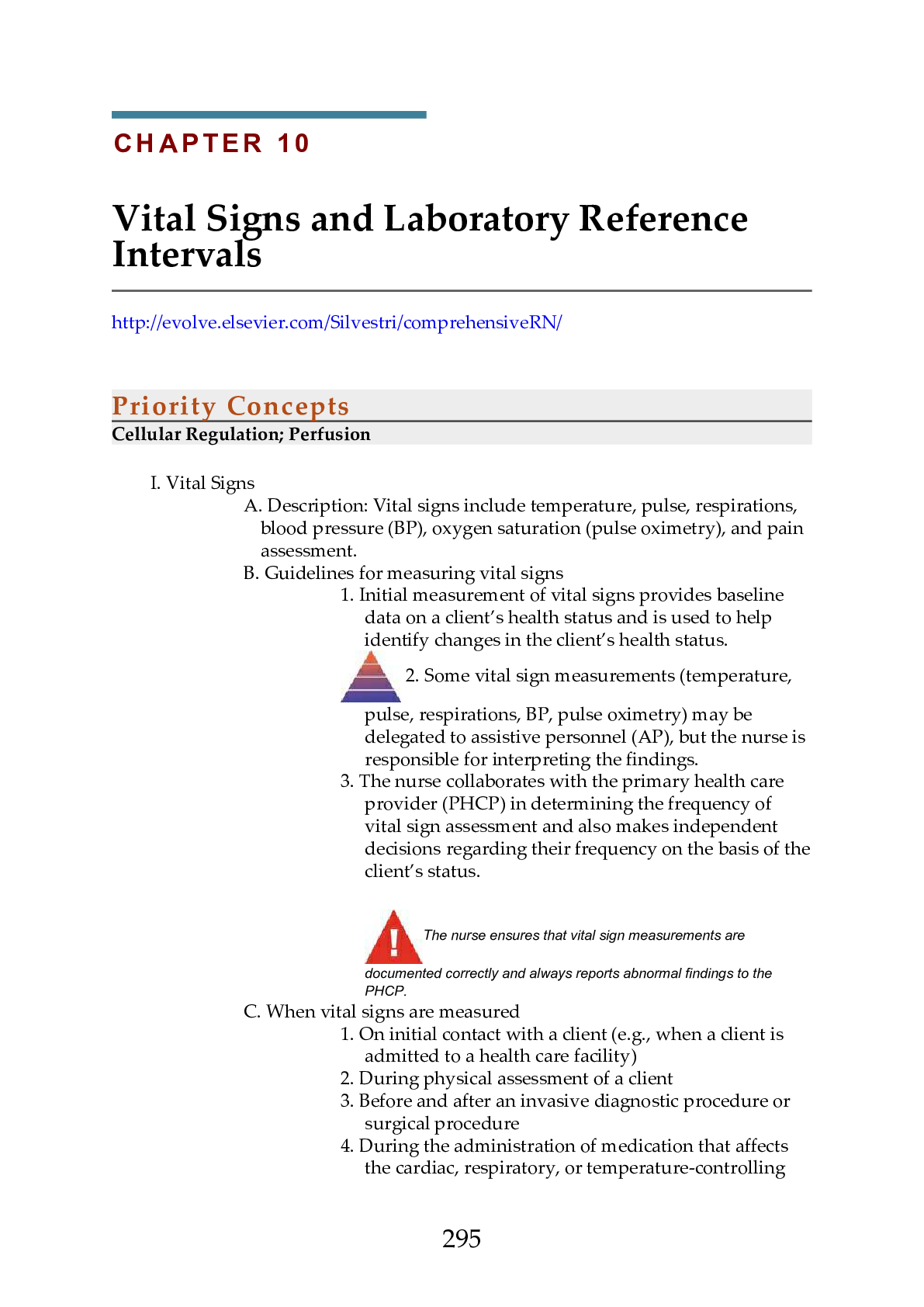
Priority Concepts
Cellular Regulation; Perfusion
I. Vital Signs
A. Description: Vital signs include temperature, pulse, respirations,
blood pressure (BP), oxygen saturation (pulse oximetry), and pain
assessment.
B.
...
Priority Concepts
Cellular Regulation; Perfusion
I. Vital Signs
A. Description: Vital signs include temperature, pulse, respirations,
blood pressure (BP), oxygen saturation (pulse oximetry), and pain
assessment.
B. Guidelines for measuring vital signs
1. Initial measurement of vital signs provides baseline
data on a client’s health status and is used to help
identify changes in the client’s health status.
2. Some vital sign measurements (temperature,
pulse, respirations, BP, pulse oximetry) may be
delegated to assistive personnel (AP), but the nurse is
responsible for interpreting the findings.
3. The nurse collaborates with the primary health care
provider (PHCP) in determining the frequency of
vital sign assessment and also makes independent
decisions regarding their frequency on the basis of the
client’s status.
The nurse ensures that vital sign measurements are
documented correctly and always reports abnormal findings to the
PHCP.
C. When vital signs are measured
1. On initial contact with a client (e.g., when a client is
admitted to a health care facility)
2. During physical assessment of a client
3. Before and after an invasive diagnostic procedure or
surgical procedure
4. During the administration of medication that affects
the cardiac, respiratory, or temperature-controlling
295functions (e.g., in a client who has a fever); may be
required before, during, and after administration of
the medication
5. Before, during, and after a blood transfusion
6. Whenever a client’s condition changes or the client
verbalizes unusual feelings such as nonspecific
symptoms of physical distress (i.e., feeling funny or
different)
7. Whenever an intervention (e.g., ambulation) may
affect a client’s condition
8. When a fever or known infection is present (check
vital signs every 2 to 4 hours)
II. Temperature
A. Description
1. Normal body temperature ranges from 36.4° to 37.5°
Celsius (C) (97.5° to 99.5° Fahrenheit [F]); the average
in a healthy young adult is 37.0° C (98.6° F).
2. Common measurement sites are the mouth, rectum
(unless contraindicated), axilla, ear, and across the
forehead (temporal artery site); various types of
electronic measuring devices are commonly used to
measure temperature.
3. Rectal temperatures are usually 1° F (0.5° C) higher
and tympanic and axillary temperatures about 1° F
(0.5° C) lower than the normal oral temperature.
4. Know how to convert a temperature to a Fahrenheit or
Celsius value (Box 10-1).
B. Nursing considerations
1. Time of day
a. Temperature is generally in the lownormal range at the time of awakening
as a result of muscle inactivity.
b. Afternoon body temperature may be
high-normal as a result of the
metabolic process, activity, and
environmental temperature.
2. Environmental temperature: Body temperature is
lower in cold weather and higher in warm weather.
3. Age: Temperature may fluctuate during the first year
of life because the infant’s heat-regulating mechanism
is not fully developed.
4. Physical exercise: Use of the large muscles creates
heat, causing an increase in body temperature.
5. Menstrual cycle: Temperature decreases slightly just
before ovulation but may increase to 1° F above
normal during ovulation.
6. Pregnancy: Body temperature may consistently stay at
high-normal because of an increase in the woman’s
296metabolic rate.
7. Stress: Emotions increase hormonal secretion, leading
to increased heat production and a higher
temperature.
8. Illness: Infective agents and the inflammatory
response may cause an increase in temperature.
9. The inability to obtain a temperature should not be
ignored, because it could represent a condition of
hypothermia, a life-threatening condition in very
young and older clients.
C. Methods of measurement
1. Oral
a. If the client has recently consumed hot
or cold foods or liquids or has smoked
or chewed gum, the nurse must wait
15 to 30 minutes before taking the
temperature orally.
b. The thermometer is placed under the
tongue in one of the posterior
sublingual pockets; ask the client to
keep the tongue down and the lips
closed and to not bite down on the
thermometer.
2. Rectal
a. Place the client in the Sims’ position.
b. The temperature is taken rectally when
an accurate temperature cannot be
obtained orally or via other methods
including by an electronic method, or
when the client has nasal congestion,
has undergone nasal or oral surgery or
had the jaws wired, has a nasogastric
tube in place, is unable to keep the
mouth closed, or is at risk for seizures.
c. The thermometer is lubricated and
inserted into the rectum, toward the
umbilicus, about 1.5 inches (3.8 cm)
(no more than 0.5 inch [1.25 cm] in an
infant).
The temperature is not taken rectally in
cardiac clients; the client who has undergone rectal
surgery; or the client with diarrhea, fecal impaction,
or rectal bleeding or who is at risk for bleeding.
3. Axillary
a. This method of taking the temperature
297is used when the oral or other methods
of temperature measurement are
contraindicated.
b. Axillary measurement is not as accurate
as the oral, rectal, tympanic, or
temporal artery method but is used
when other methods of measurement
are not possible.
c. The thermometer is placed in the
client’s dry axilla, and the client is
asked to hold the arm tightly against
the chest, resting the arm on the chest;
follow the instructions accompanying
the measurement device for the
amount of time the thermometer
should remain in the axillary area.
4. Tympanic
a. The auditory canal is checked
for the presence of redness, swelling,
discharge, or a foreign body before the
probe is inserted; the probe should not
be inserted if the client has an
inflammatory condition of the auditory
canal or if there is discharge from the
ear.
b. The reading may be affected by an ear
infection or excessive wax blocking the
ear canal.
5. Temporal artery
a. Ensure that the client’s forehead is dry.
b. The thermometer probe is placed flush
against the skin and slid across the
forehead or placed in the area of the
temporal artery and held in place.
c. If the client is diaphoretic, the temporal
artery thermometer probe may be
placed on the neck, just behind the
earlobe.
III. Pulse
A. Description
1. Pulse is a palpable bounding of blood flow in a
peripheral artery; it is an indirect indicator of
circulatory status.
2. The average adult pulse (heart) rate is 60 to 100 beats
per minute.
3. Changes in pulse rate are used to evaluate the client’s
tolerance of interventions such as ambulation,
298bathing, dressing, and exercise.
4. Pedal pulses are checked to determine whether the
circulation is blocked in the artery up to that pulse
point.
5. When the pedal pulse is difficult to locate, a Doppler
ultrasound stethoscope (ultrasonic stethoscope) may
be needed to amplify the sounds of pulse waves.
B. Nursing considerations
1. The heart rate slows with age.
2. Exercise increases the heart rate.
3. Emotions stimulate the sympathetic nervous system,
increasing the heart rate.
4. Pain increases the heart rate.
5. Increased body temperature causes the heart rate to
increase.
6. Stimulant medications increase the heart rate;
depressants and medications affecting the cardiac
system slow it.
7. When the BP is low, the heart rate is usually increased.
8. Hemorrhage increases the heart rate.
C. Assessing pulse qualities
1. When the pulse is being counted, note the rate,
rhythm, strength (force or amplitude), and equality.
2. Once you have checked these parameters, use the
grading scale for pulses to assess the information you
have elicited (Box 10-2).
D. Pulse points and locations
1. The temporal artery can be palpated anterior to or in
the front of the ear.
2. The carotid artery is located in the groove between the
trachea and the sternocleidomastoid muscle, medial
to and alongside the muscle.
3. The apical pulse may be detected at the left
midclavicular, fifth intercostal space.
4. The brachial pulse is located above the elbow at the
antecubital fossa, between the biceps and triceps
muscles.
5. The radial pulse is located in the groove along the
radial or thumb side of the client’s inner wrist.
6. The ulnar pulse is located on the medial side of the
wrist (little finger side of the forearm at the wrist).
7. The femoral pulse is located below the inguinal
ligament, midway between the symphysis pubis and
the anterosuperior iliac spine.
8. The popliteal pulse is located behind the knee.
9. The posterior tibial pulse is located on the inner side of
the ankle, behind and below the medial malleolus
299(ankle bone).
10. The dorsalis pedis pulse is located on the top of the
foot, in line with the groove between the extensor
tendons of the great and first toes.
The apical pulse is counted for 1 full minute and is assessed in
clients with an irregular radial pulse or a heart condition, before the
administration of cardiac medications such as digoxin and beta
blockers, and in children younger than 2 years.
E. Pulse deficit
1. In this condition, the peripheral pulse rate (radial
pulse) is less than the ventricular contraction rate
(apical pulse).
2. A pulse deficit indicates a lack of peripheral perfusion;
it can be an indication of cardiac dysrhythmias.
3. One-examiner technique: Auscultate and count the
apical pulse first and then immediately count the
radial pulse.
4. Two-examiner technique: One person counts the
apical pulse and the other counts the radial pulse
simultaneously.
5. A pulse deficit indicates that cardiac contractions are
ineffective, failing to send pulse waves to the
periphery.
6. If a difference in pulse rate is noted, the PHCP is
notified.
IV. Respirations
A. Description
1. Respiration is a mechanism the body uses to exchange
gases between the atmosphere and the blood and
between the blood and the cells.
2. Respiratory rates vary with age.
3. The normal adult respiratory rate is 12 to 20 breaths
per minute.
B. Nursing considerations
1. Many of the factors that affect the pulse rate also affect
the respiratory rate.
2. An increased level of carbon dioxide or a lower level
of oxygen in the blood results in an increase in
respiratory rate.
3. Head injury or increased intracranial pressure will
depress the respiratory center in the brain, resulting
in shallow respirations or slowed breathing.
4. Medications such as opioid analgesics depress
respirations.
5. Additional factors that can affect the respiratory rate
300include exercise, pain, anxiety, smoking, and body
position.
C. Assessing respiratory rate
1. Count the client’s respirations after measuring the
radial pulse. (Continue holding the client’s wrist
while counting the respirations or position the hand
on the client's chest.)
2. One respiration includes both inspiration and
expiration.
3. The rate, depth, pattern, and sounds are assessed.
The respiratory rate may be counted for 30 seconds and
multiplied by 2, except in a client who is known to be very ill or is
exhibiting irregular respirations, in which case respirations are counted
for 1 full minute.
V. Blood Pressure
A. Description
1. Blood pressure (BP) is the force on the walls of an
artery exerted by the pulsating blood under pressure
from the heart.
2. The heart’s contraction forces blood under high
pressure into the aorta; the peak of maximum
pressure when ejection occurs is the systolic pressure;
the blood remaining in the arteries when the
ventricles relax exerts a force known as the diastolic
pressure.
3. The difference between the systolic and diastolic
pressures is called the pulse pressure.
4. For an adult (age 18 years and older), a normal BP is a
systolic pressure below 120 mm Hg and a diastolic
pressure below 80 mm Hg.
5. Categories of hypertension (Box 10-3).
6. In postural (orthostatic) hypotension, a normotensive
client exhibits symptoms and low BP on rising to an
upright position.
7. To obtain orthostatic vital sign measurements,
check the BP and pulse with the client supine, sitting,
and standing; readings are obtained 1 to 3 minutes
after the client changes position.
B. Nursing considerations
1. Factors affecting BP
a. BP tends to increase as the aging
process progresses.
b. Stress results in sympathetic
301
Practice Questions
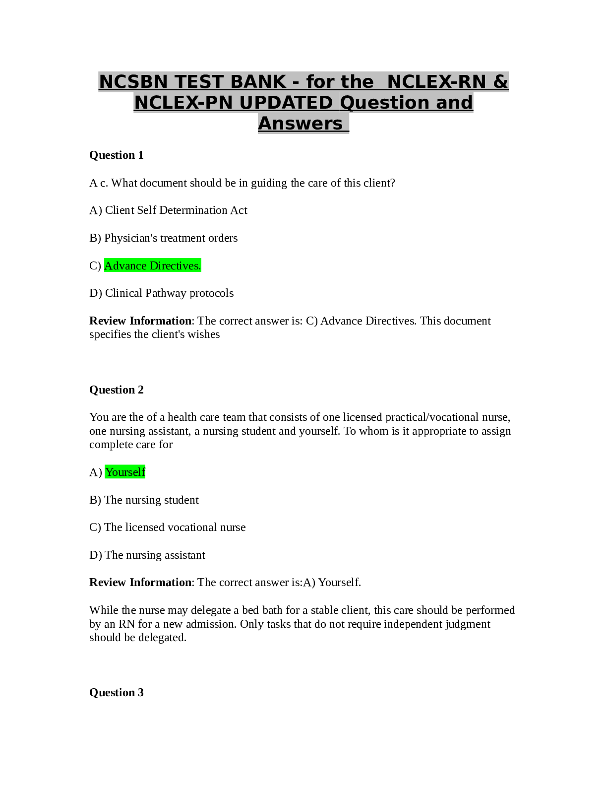
64. A client with atrial fibrillation who is receiving maintenance therapy of
warfarin sodium has a prothrombin time (PT) of 35 seconds. On the basis of
these laboratory values, the nurse anticipates which prescription?
1. Adding a dose of heparin sodium
2. Holding the next dose of warfarin
3. Increasing the next dose of warfarin
4. Administering the next dose of warfarin
65. A staff nurse is precepting a new graduate nurse and the new graduate is
assigned to care for a client with chronic pain. Which statement, if made by
323the new graduate nurse, indicates the need for further teaching regarding
pain management?
1. “I will be sure to ask my client what his pain level is on a scale of 0
to 10.”
2. “I know that I should follow up after giving medication to make
sure it is effective.”
3. “I will be sure to cue in to any indicators that the client may be
exaggerating their pain.”
4. “I know that pain in the older client might manifest as sleep
disturbances or depression.”
66. A client has been admitted to the hospital for gastroenteritis and
dehydration. The nurse determines that the client has received adequate
volume replacement if the blood urea nitrogen (BUN) level drops to which
value?
1. 3 mg/dL (1.08 mmol/L)
2. 15 mg/dL (5.4 mmol/L)
3. 29 mg/dL (10.44 mmol/L)
4. 35 mg/dL (12.6 mmol/L)
67. The nurse is explaining the appropriate methods for measuring an accurate
temperature to an assistive personnel (AP). Which method, if noted by the
UAP as being an appropriate method, indicates the need for further
teaching?
1. Taking a rectal temperature for a client who has undergone nasal
surgery
2. Taking an oral temperature for a client with a cough and nasal
congestion
3. Taking an axillary temperature for a client who has just consumed
hot coffee
4. Taking a temperature on the neck behind the ear using an
electronic device for a client who is diaphoretic
68. A client is receiving a continuous intravenous infusion of heparin sodium to
treat deep vein thrombosis. The client’s activated partial thromboplastin time
(aPTT) is 65 seconds. The nurse anticipates that which action is needed?
1. Discontinuing the heparin infusion
2. Increasing the rate of the heparin infusion
3. Decreasing the rate of the heparin infusion
4. Leaving the rate of the heparin infusion as is
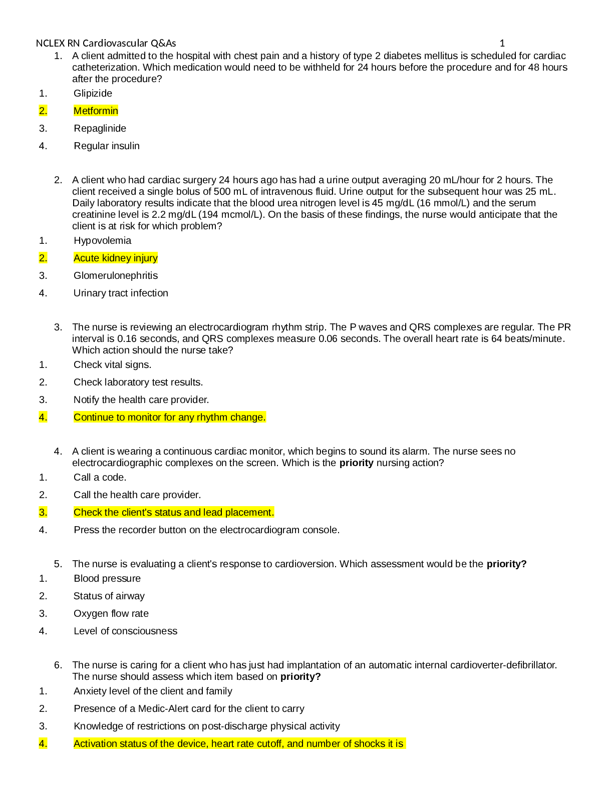
69. 69. A client with a history of heart failure is due for a morning dose of
furosemide. Which serum potassium level, if noted in the client’s laboratory
report, should be reported before administering the dose of furosemide?
1. 3.2 mEq/L (3.2 mmol/L)
2. 3.8 mEq/L (3.8 mmol/L)
3. 2 mEq/L (4.2 mmol/L)
4. 8 mEq/L (4.8 mmol/L)
70. Several laboratory tests are prescribed for a client, and the nurse reviews the
results of the tests. Which laboratory test results should the nurse report?
Select all that apply.
3241. Platelets 35,000 mm3 (35 × 109/L)
2. Sodium 150 mEq/L (150 mmol/L)
3. Potassium 5.0 mEq/L (5.0 mmol/L)
4. Segmented neutrophils 40% (0.40)
5. Serum creatinine, 1 mg/dL (88.3 mcmol/L)
6. White blood cells, 3000 mm3 (3.0 × 10⁹/L)
71. The nurse is caring for a client who takes ibuprofen for pain. The nurse is
gathering information on the client’s medication history and determines it is
necessary to contact the primary health care provider (PHCP) if the client is
also taking which medications? Select all that apply.
1. Warfarin
2. Glimepiride
3. Amlodipine
4. Simvastatin
5. Atorvastatin
72. A client with diabetes mellitus has a glycosylated hemoglobin A1c level of
8%. On the basis of this test result, the nurse plans to teach the client about
the need for which measure?
1. Avoiding infection
2. Taking in adequate fluids
3. Preventing and recognizing hypoglycemia
4. Preventing and recognizing hyperglycemia
73. The nurse is caring for a client with a diagnosis of breast cancer who is
immunosuppressed. The nurse would consider implementing neutropenic
precautions if the client’s white blood cell count was which value?
1. 2000 mm3 (2.0 × 10⁹/L)
2. 5800 mm3 (5.8 × 10⁹/L)
3. 8400 mm3 (8.4 × 10⁹/L)
4. 11,500 mm3 (11.5 × 10⁹/L)
74. A client brought to the emergency department states that he has accidentally
been taking 2 times his prescribed dose of warfarin for the past week. After
noting that the client has no evidence of obvious bleeding, the nurse plans to
take which action?
1. Prepare to administer an antidote.
2. Draw a sample for type and crossmatch and transfuse the client.
3. Draw a sample for an activated partial thromboplastin time
(aPTT) level.
4. Draw a sample for prothrombin time (PT) and international
normalized ratio (INR).
75. The nurse is caring for a postoperative client who is receiving demand-dose
hydromorphone via a patient-controlled analgesia (PCA) pump for pain
325control. The nurse enters the client’s room and finds the client drowsy and
records the following vital signs: temperature 97.2° F (36.2° C) orally, pulse
52 beats per minute, blood pressure 101/58 mm Hg, respiratory rate 11
breaths per minute, and SpO2 of 93% on 3 liters of oxygen via nasal cannula.
Which action should the nurse take next?
1. Document the findings.
2. Attempt to arouse the client.
3. Contact the primary health care provider (PHCP) immediately.
4. Check the medication administration history on the PCA pump.
76. An adult female client has a hemoglobin level of 10.8 g/dL (108 mmol/L). The
nurse interprets that this result is most likely caused by which condition
noted in the client’s history?
1. Dehydration
2. Heart failure
3. Iron deficiency anemia
4. Chronic obstructive pulmonary disease
77. A client with a history of upper gastrointestinal bleeding has a platelet count
of 300,000 mm3 (300 × 109/L). The nurse should take which action after seeing
the laboratory results?
1. Report the abnormally low count.
2. Report the abnormally high count.
3. Place the client on bleeding precautions.
4. Place the normal report in the client’s medical record.
[Show More]