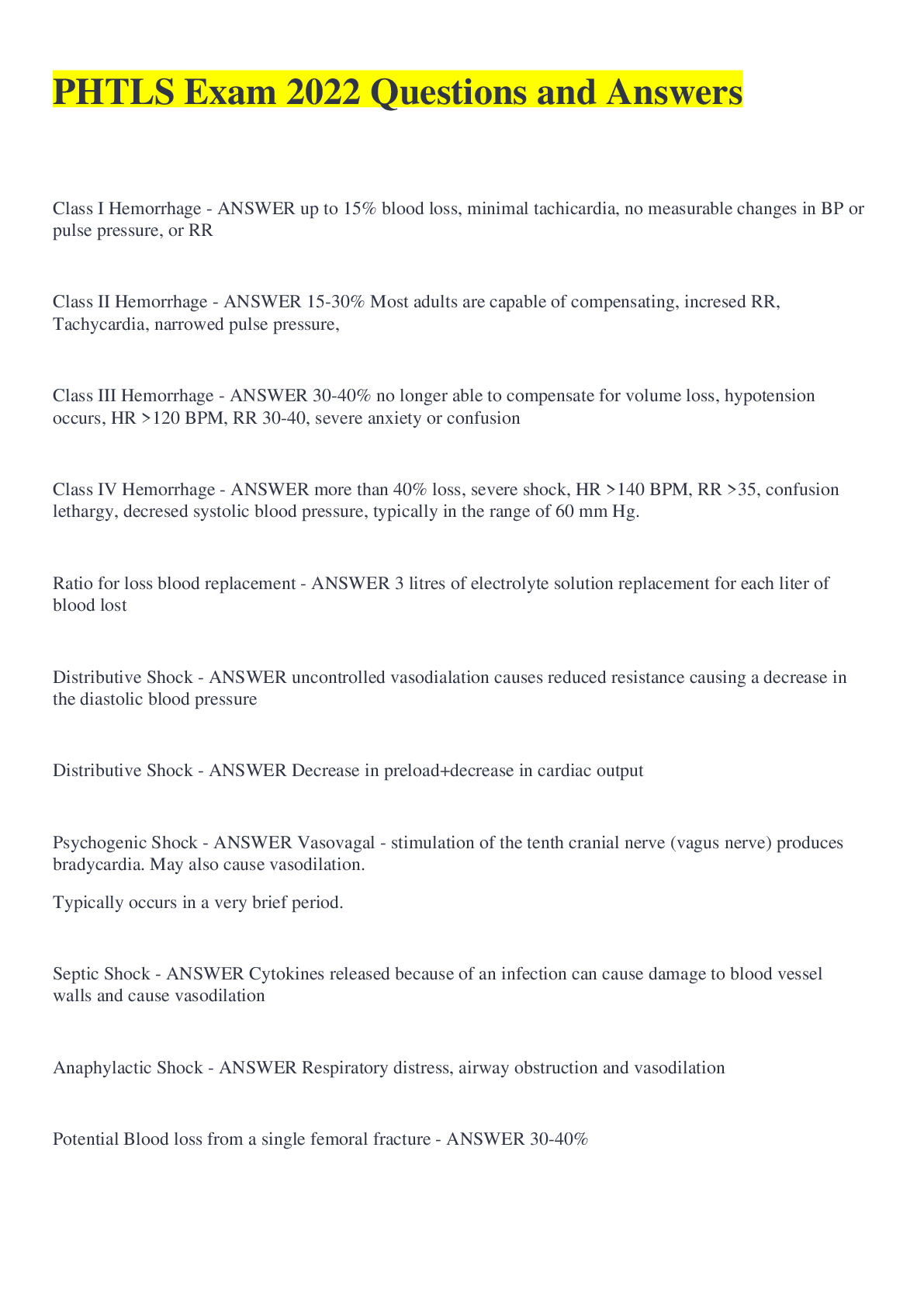
Class I Hemorrhage - ANSWER up to 15% blood loss, minimal tachicardia, no measurable changes in BP or pulse pressure, or RR
Class II Hemorrhage - ANSWER 15-30% Most adults are capable of compensating, incresed RR, Tac
...
Class I Hemorrhage - ANSWER up to 15% blood loss, minimal tachicardia, no measurable changes in BP or pulse pressure, or RR
Class II Hemorrhage - ANSWER 15-30% Most adults are capable of compensating, incresed RR, Tachycardia, narrowed pulse pressure,
Class III Hemorrhage - ANSWER 30-40% no longer able to compensate for volume loss, hypotension occurs, HR >120 BPM, RR 30-40, severe anxiety or confusion
Class IV Hemorrhage - ANSWER more than 40% loss, severe shock, HR >140 BPM, RR >35, confusion lethargy, decresed systolic blood pressure, typically in the range of 60 mm Hg.
Ratio for loss blood replacement - ANSWER 3 litres of electrolyte solution replacement for each liter of blood lost
Distributive Shock - ANSWER uncontrolled vasodialation causes reduced resistance causing a decrease in the diastolic blood pressure
Distributive Shock - ANSWER Decrease in preload+decrease in cardiac output
Psychogenic Shock - ANSWER Vasovagal - stimulation of the tenth cranial nerve (vagus nerve) produces bradycardia. May also cause vasodilation.
Typically occurs in a very brief period.
Septic Shock - ANSWER Cytokines released because of an infection can cause damage to blood vessel walls and cause vasodilation
Anaphylactic Shock - ANSWER Respiratory distress, airway obstruction and vasodilation
Potential Blood loss from a single femoral fracture - ANSWER 30-40%
Amount of blood loss that can happen before signs of compensatory mechanisms fail and BP become <90 mmHq - ANSWER Greater than 30%
Hypertension medications that may prevent compensatory tachycardia to maintain BP - ANSWER Beta Adrenergic blocking agents and calcium channel blockers
Managing Volume Resuscitation
Uncontrolled hemorrhage-suspected chest, abdomen, retroperitonium - ANSWER Maintain a systolic bp 80-90 or MAP 60-65
Managing Volume Resuscitation
CNS injuries or TBI - ANSWER maintaining the systolic BP (SBP) above 90 or MAP 85-90
Managing Volume Resuscitation
Controlled Hemorrhage
large scalp or extremity controlled with a tourniquet, PT falls into class II,III,IV shock - ANSWER rapid bolus of 1-2 litres
Three responses of initial fluid bolus
Rapid Response - ANSWER vital signs return indicating that PT lost less than 20%
PT is best managed at SBP between 80-90, IV fluid should be titrated
Three responses of initial fluid bolus
Transient Response - ANSWER Vital signs improve (pulse slows, BP increases) however PT shows deterioration. PT has typically lost 20-40% of blood volume
Three responses of initial fluid bolus
Minimal or no response - ANSWER No change in PT after 1-2 liter Bolus
Leading cause of TBI - ANSWER MVC
Three seperate membranes that surround the brain - ANSWER meninges
Outer most meninges - ANSWER Dura mater- composed of tough fibrous tissue
The inner most meninges that is directly in contact with the brain - ANSWER Pia Mater-
Vessels that lay on top of the Pia Mater - ANSWER Cerebral Blood Vessels
The meninge that lays in the middle between the dura and pia matter - ANSWER Arachnoid membrane-loosely covers the brain and its blood vessels
MAP-Mean Arterial Pressure - ANSWER The average pressure for the entire cardiac cycle
MAP=Diastolic Pressure + 1/3 of the Pulse Pressure
PP- Pulse Pressure - ANSWER The difference between the systolic and diastolic
PP= Systolic (SBP) - Distolic (DBP)
CPP- Cerebral Perfusion Pressure - ANSWER The amount of pressure it takes to push blood through the cerebral circulation. CPP=Mean Arterial pressure (MAP) - Intercranial pressure (ICP)
CPP- is normally below ? mmHg - ANSWER CPP is normally 15 mmHg or below
Autoregulation - ANSWER The brain changes the cerebral blood vessel resistance (CVR) to compensate for changes in the cerebral pulse pressure (CPP)
For neurological injured PT's the systolic blood pressure is best if maintained at or above - ANSWER 90 mmHg
Hyperventilation and TBI - ANSWER Hyperventilation can reduced CBF by causing vasoconstriction due to hypocapnia, PaCO2 >35 increases risk of cerebral ischemia
Primary Brain injury - ANSWER The direct trauma or mechanical injury, includes contusions, hemorrhages and lacerations
Secondary brain injury - ANSWER pathologic mechanisms that related to intercranial mass effect, elevated ICP, and herniation, hemotoma, hypoxia and hypotension
Mass effect - ANSWER The effect of a growing mass or tumor
Uncal Herniation - ANSWER temporal lobe epidural hematoma forces pressure on the medial portion of the temporal lobe (uncus) which in turn puts pressure on the 3rd cranial nerve causing dilation or blown pupil on the side of the herniation. May also cause loss of function of the motor track on the same side.
Final stage of uncal herniation - ANSWER The RAS is affected patient lapses into coma
decorticate posturing - ANSWER flexion of the upper extremities with rigidity and extension of the lower extremities.
decerebrate posturing - ANSWER all extrem become flaccid and motor activity is absent
Cheyne strokes ventilation - ANSWER repeating cycle of slow hallow breaths that become deeper and more rapid and then return to slow
Central neurogenic hyperventilation - ANSWER rapid deep breaths
Ataxic breathing - ANSWER erratic ventilatory efforts that lack any discernible pattern
Cushings phenomenon - ANSWER greatly increased arterial blood pressure and the resultant bradycardia (increase BP + decreased HR) may occur with severely increasing ICP
Hypercapnia (high CO2) - ANSWER causes vasodilation
Hypocapnia (low CO2) - ANSWER causes vasoconstriction
When people have 20 mmHg CO2 in their blood (half of the official norm), - ANSWER they have about 40% less blood supply to the brain in comparison with normal conditions
Indication of TBI
Hemiparesis - ANSWER One sided weakness
Indication of TBI
Hemiplegia - ANSWER One sided paralysis
Indications for a nose fracture - ANSWER ecchymosis, edema, nasal deformity, swelling, epistaxis
Epidural Hematoma
1-brief Loss of consciousness
2-Lucid Interval
3-Rapid decline in level of consciousness - ANSWER The period of time after a brief loss of consciousness where the PT may be oriented, lethargic or confused followed by a rapid decline of consciousness
Subdural Hematoma - ANSWER Account for 30% of TBI-Generally results from venous blood from bridging viens that are torn during a violent blow to the head.
Epidural Hematoma - ANSWER Account for 2% of TBI-Arterial blood starts to dissect or peal the dura off of the inner table of the skull creating an epidural space filled with blood
Epidural Hematoma Signs - ANSWER Altered LOC, dilated and sluggish non reactive pupil on the side of herniation, hemiparesis or hemiplegia on opposite side of injury.
Subdural Hematoma - ANSWER Can be chronic or acute. Pt on coumadin are at high risk of un identified subdural hematomas that may be chronic. Often may be confused with stroke, infection or general decline of PT
Cerebral Contusions - ANSWER actual bleeding into the substance of the brain, CC's often take 12-24 hrs to appear on CT scans, only indicator is a depressed GCS of 9-13
Subarachnoid Hemorrhage - ANSWER rarely causes mass effect and does not require surgery, it does increase the risk of cerebral contusion by 63-73%
Normal ventilatory rates
Adults= - ANSWER 10 BPM
Normal ventilatory rates
Children= - ANSWER 20 BPM
Normal ventilatory rates
Infants= - ANSWER 25BPM
Warning signs of possible ICP - ANSWER Decline in GCS
sluggish or non reactive pupils
Hemiplegia or hemiparesis
Cushings phenomenon
Osmotherapy - ANSWER the use of osmotically active agents that may assist in the treatment of intracranial hypertension`
Number of cervical vertebrae - ANSWER 7
Number of thoracic vertebrae - ANSWER 12
Number of lumbar vertebrae - ANSWER 5
Number of coccygeal vertebrae - ANSWER 4
Number of pounds the human head averages - ANSWER 16-22 pounds
Space between the spinal cord and the spinal canal wall - ANSWER 3mm
Name of the 1st cervical vertebrae - ANSWER Atlas
Name of the 2nd cervical vertebrae - ANSWER Axis
Function of the ascending nervee tracts - ANSWER Sensory impulses from body parts through the cord up to the brain
Function of the descending nerve tracts - ANSWER impulses from the brain through the cord down to the body
Dorsal root= - ANSWER sensory
Ventral root= - ANSWER motor
Dermatome - ANSWER The sensory area on the body for which a nerve root is responsible
The phrenic nerve originates from - ANSWER the cervical vertebrae c2-c5
Skeletal Injuries
Compression - ANSWER Compression fractures that produce wedge compression or total flattening of the body of the vertebra
Skeletal Injuries
Fractures - ANSWER Produce small fragments of bone that may lie in the spinal canal near the cord
Skeletal Injuries
Sublaxation - ANSWER a partial dislocation of the vertebra from its normal alignment in the spinal column
Skeletal Injuries
Overstretching - ANSWER Tearing of the ligaments and muscles producing instability between the vertebrae
Skeletal Injuries
Percentage of falls from a height greater than 15 feet involve an associated lumbar spine fracture - ANSWER 20%
Cause and Symptoms
Anterior Cord syndrome - ANSWER Results from bony fragments or pressure on spinal arteries
Symptoms-loss of motor function and pain, temperature and light touch PT's will have some light touch sensations such as vibration.
Cause and Symptoms
Central cord syndrome - ANSWER usually occurs with hyperextension
Symptoms include weakness or paresthesia (tickling, tingling, burning, pricking, or numbness) in the upper extremities but normal strength in the lower extremities
Cause and Symptoms
Brown Sequard syndrome - ANSWER Caused by penetrating injury and involves hemi-transection of the cord.
Symptoms include complete cord damage and loss of function of the affected side with loss of pain, temp and sensation on the opposite side of the injury
Conditions that should mandate spinal immobilization - ANSWER Altered LOC
Spinal Pain or tenderness
Neurologic deficit or complaint
Anatomic deformity of the spine
Pulmonary Volume Terminology
Dead Space - ANSWER Amount of air brought into the lungs that does not have the opportunity to exchange oxygen and carbon dioxide
Pulmonary Volume Terminology
Minute Volume (Ve) - ANSWER Total volume of air moved into and out of during a 1 minute interval.
Pulmonary Volume Terminology
Tidal Volume (Vo) - ANSWER Amount of air that is inhaled then exhaled druing a normal breath (.4-.5 litres)
Pulmonary Volume Terminology
Total Lung Capacity (TLC) - ANSWER Total volume the lungs contain when maximally inflated. This volume declines with age from 6 L. in young adults to 4 L. in elderly.
Pulmonary Contusion - ANSWER Blunt force can tear lung tissue resulting in bleeding into the alveoli
Thoracic Trauma
Assessment
Four components to the phys exam - ANSWER Observation, palpation, percussion, auscultation
[Show More]





![Preview of AORN PERIOP 101 FINAL EXAM 2026 QUESTIONS AND VERIFIED ANSWERS [LATEST UPDATE]](https://browseimages.nyc3.digitaloceanspaces.com/paper-images/2026/04/07/sGJvJ3aH2026-04-07-04-5869d464cbba16f.png)