MULTIPLE CHOICE
1. Culture results:
a. confirm the correctness of the therapeutic choices already made and implemented.
b. help the physician select an antibiotic for treatment.
c. must be confirmed before initiat
...
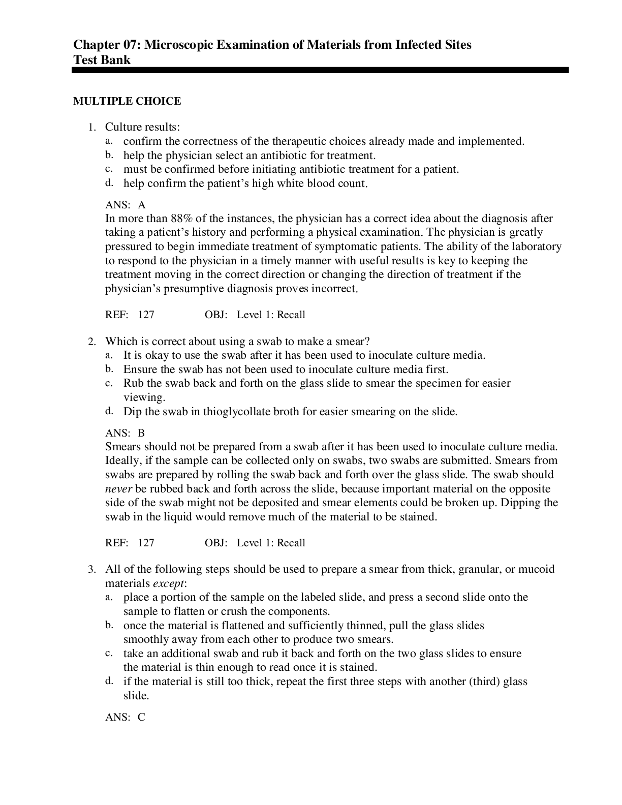
MULTIPLE CHOICE
1. Culture results:
a. confirm the correctness of the therapeutic choices already made and implemented.
b. help the physician select an antibiotic for treatment.
c. must be confirmed before initiating antibiotic treatment for a patient.
d. help confirm the patient’s high white blood count.
A
In more than 88% of the instances, the physician has a correct idea about the diagnosis after taking a patient’s history and performing a physical examination. The physician is greatly pressured to begin immediate treatment of symptomatic patients. The ability of the laboratory to respond to the physician in a timely manner with useful results is key to keeping the treatment moving in the correct direction or changing the direction of treatment if the physician’s presumptive diagnosis proves incorrect.
REF: 127 OBJ: Level 1: Recall
2. Which is correct about using a swab to make a smear?
a. It is okay to use the swab after it has been used to inoculate culture media.
b. Ensure the swab has not been used to inoculate culture media first.
c. Rub the swab back and forth on the glass slide to smear the specimen for easier viewing.
d. Dip the swab in thioglycollate broth for easier smearing on the slide.
B
Smears should not be prepared from a swab after it has been used to inoculate culture media. Ideally, if the sample can be collected only on swabs, two swabs are submitted. Smears from swabs are prepared by rolling the swab back and forth over the glass slide. The swab should never be rubbed back and forth across the slide, because important material on the opposite side of the swab might not be deposited and smear elements could be broken up. Dipping the swab in the liquid would remove much of the material to be stained.
REF: 127 OBJ: Level 1: Recall
3. All of the following steps should be used to prepare a smear from thick, granular, or mucoid materials except:
a. place a portion of the sample on the labeled slide, and press a second slide onto the sample to flatten or crush the components.
b. once the material is flattened and sufficiently thinned, pull the glass slides smoothly away from each other to produce two smears.
c. take an additional swab and rub it back and forth on the two glass slides to ensure the material is thin enough to read once it is stained.
d. if the material is still too thick, repeat the first three steps with another (third) glass slide.
C
Once the material is flattened between the two slides and the glass slide is pulled away to make two smears, you never put a swab on the specimen to spread it out. If it is still too thick, then an additional slide is used to flatten the material out and another pull slide is made. This slide should be adequate to stain and read.
REF: 128 OBJ: Level 1: Recall
4. Which is correct about making slides of thin materials such as urine and cerebrospinal fluid (CSF)?
a. Spread the material once, let the specimen dry, then spread another layer on top to ensure enough specimen to stain.
b. Use the two-slide pull method to ensure evenly distributed material over the slides.
c. Dip a swab into the specimen, then roll the swab over the glass slide to ensure adequate coverage for staining.
d. Mark the area of the sample drop on the reverse side with a wax pencil, then place a drop of the specimen into the marked area.
D
Thin materials such as urine and CSF are difficult to view on a glass slide. Marking the underneath area with a wax pencil will allow the technician to know exactly where the sample was placed. This may be helpful after staining the specimen. Never spread two layers of a specimen on a slide. A drop on a slide contains enough material to be stained and visualized. The two-slide pull method is for thick specimens, not thin. Never use a swab to apply a specimen when there is enough fluid to be dropped onto a slide.
REF: 128 OBJ: Level 1: Recall
5. A cytocentrifuge is an excellent method for preparing nonviscous fluids, because:
a. it deposits cellular elements and microorganisms from the specimen onto the surface of a glass slide as a monolayer.
b. it concentrates the protein evenly throughout the slide surface.
c. the sediment is mixed with 70% albumin before spreading it on the surface of a slide.
d. the cytocentrifuge not only spreads the specimen over the surface of a slide but also stains the specimen.
A
The cytocentrifuge is a centrifuge that deposits a uniform layer of the specimen onto a glass slide. It does not concentrate protein, nor does it mix the specimen with 70% albumin. The sediment is fixed and decontaminated in 70% alcohol for 5 minutes. The cytocentrifuge is only a centrifuge—it is not a stainer and does not stain the slide.
REF: 129 OBJ: Level 1: Recall
6. The purpose of a simple stain is to:
a. color specific components of specific elements.
b. color the forms and shapes present.
c. specifically identify an organism.
d. identify the types of DNA and RNA present in the nucleus of a cell.
B
Simple stains identify colors and forms. Differential stains color specific components of those elements present. DNA probe-mediated stains are directed specifically at an organism’s identification.
REF: 129 OBJ: Level 1: Recall
7. In the microbiology laboratory, which instrument is routinely used to examine smears for structures that are too small to be seen with the unaided eye?
a. Cytocentrifuge
b. Thermal cycler
c. Compound microscope
d. Electron microscope
C
A compound light microscope is routinely used in the microbiology laboratory for viewing specimens and bacterial colonies. A cytocentrifuge makes monolayer smears from specimens. A thermal cycler carries out DNA amplification. An electron microscope is a specialized piece of equipment available in some laboratories for viewing special specimens.
REF: 129 OBJ: Level 1: Recall
8. A technician is working the night shift and receives a CSF specimen on an infant. The technician makes a Gram stain of the spinal fluid, then reads the smear under the microscope. The report the technician sends to the physician reads as follows: “gram-negative bacillus, small and pleomorphic.” What bacteria are being implied as the infecting agent?
a. Escherichia coli
b. Legionella pneumophila
c. Staphylococcus aureus
d. Haemophilus influenzae
D
Microorganisms can be described in such a way that, based on prevalence, the description implies the identification of the organism. A gram-negative bacillus, small and pleomorphic, brings to mind a miscellaneous gram-negative bacillus. Because it is in the spinal fluid of an infant where H. influenzae is known to cause meningitis, the physician would base his or her treatment upon this fact.
REF: 133 OBJ: Level 3: Synthesis
9. A patient comes to the emergency department with a productive cough, shortness of breath, and a fever of 102° F. The patient is diagnosed with pneumonia. What organism commonly encountered in the microbiology laboratory can cause this disease?
a. Streptococcus pneumoniae
b. Staphylococcus aureus
c. Escherichia coli
d. Haemophilus influenzae
A
Several common organisms are encountered in the microbiology laboratory. Infections caused by common single species, or by classic infectious agents, include S. pneumoniae pneumonia, S. aureus abscesses or pyodermas, H. influenzae tracheobronchitis or meningitis in infants, Clostridium perfringens gas gangrene, Nocardia spp. lung abscesses, and gonococcal urethritis.
REF: 133 OBJ: Level 3: Synthesis
10. What is the most common causative agent of urethritis?
a. Staphylococcus aureus
b. Neisseria gonorrhoeae
c. Clostridium perfringens
d. Haemophilus influenzae
B
Several common organisms are encountered in the microbiology laboratory. Infections caused by common single species, or by classic infectious agents, include Streptococcus pneumoniae pneumonia, Staphylococcus aureus abscesses or pyodermas, H. influenzae tracheobronchitis or meningitis in infants, C. perfringens gas gangrene, Nocardia spp. lung abscesses, and gonococcal urethritis.
REF: 133 OBJ: Level 2: Interpretation
11. What is the most common causative agent of gas gangrene?
a. Staphylococcus aureus
b. Neisseria gonorrhoeae
c. Clostridium perfringens
d. Haemophilus influenzae
C
Several common organisms are encountered in the microbiology laboratory. Infections caused by common single species, or by classic infectious agents, include C. perfringens gas gangrene, Streptococcus pneumoniae pneumonia, Staphylococcus aureus abscesses or pyodermas, H. influenzae tracheobronchitis or meningitis in infants, Nocardia spp. lung abscesses, and gonococcal urethritis.
REF: 133 OBJ: Level 2: Interpretation
12. What is the most common causative agent of pyoderma?
a. Staphylococcus aureus
b. Neisseria gonorrhoeae
c. Clostridium perfringens
d. Haemophilus influenzae
A
Several common organisms are encountered in the microbiology laboratory. Infections caused by common single species, or by classic infectious agents, include Staphylococcus aureus abscesses or pyodermas, Streptococcus pneumoniae pneumonia, H. influenzae tracheobronchitis or meningitis in infants, C. perfringens gas gangrene, Nocardia spp. lung abscesses, and gonococcal urethritis.
REF: 133 OBJ: Level 2: Interpretation
13. Polymicrobial presentations in smears require more interpretation and must take into account all the following except:
a. smear background.
b. morphology of the organisms.
c. anatomic location of the suspected infection.
d. method used to make the smear.
D
Polymicrobial presentations in smears require more interpretation and must take into account smear background, the morphology of the organisms, and the anatomic location of the suspected infection, as well as accompanying clinical symptoms. Polymicrobial infections usually arise from displaced normal or altered flora, and a culture will yield the same species that can be isolated in a culture from uninfected but contaminated specimens. The method used to make the smear has no bearing on smear interpretation.
REF: 133 OBJ: Level 2: Interpretation
14. Commonly encountered polymicrobial infections are:
a. perirectal abscesses.
b. indwelling catheter infections.
c. infant meningitis.
d. food poisoning.
A
The rectal area contains a lot of different types of normal bacteria called fecal flora. These bacteria can invade infected tissue once the body’s defenses are lowered. Indwelling catheter infections are a colonization, not usually caused by skin flora. Infant meningitis and food poisoning are usually single-organism infections that are not caused by normal flora.
REF: 133 OBJ: Level 1: Recall
15. When a laboratory professional examines a specimen smear, he or she should be looking for all of the following except:
a. squamous epithelial cells.
b. mucus.
c. bacteria without the cells of inflammation.
d. debris.
B
Mucus is a common lubricant of the mucous membranes and may be found in acceptable (not contaminated) and unacceptable (contaminated) specimens. Squamous epithelial cells, bacteria without the cells of inflammation, food, or other debris on a smear may indicate contamination by normal (resident) microbial flora.
REF: 133 OBJ: Level 1: Recall
16. A specimen that is spread on a slide and appears to have a homogenous constitution is said to:
a. be clumped at different areas on the slide.
b. contain pathogens only in one visual field.
c. have material spread evenly throughout the slide.
d. contain normal flora.
C
Specimens can be homogenous or heterogenous and may contain pathogens evenly distributed throughout the specimen or limited to one visual field. Homogenous constitution is said to have an even consistency throughout.
REF: 133 OBJ: Level 1: Recall
17. Why should laboratory professionals look for contamination of a specimen by normal microbial flora?
a. Contaminated specimens will have much amorphous debris that also goes with them.
b. Normal flora always preclude the possibility of a pathogen infecting the area where they reside.
c. The normal flora will be found in clumps, and any pathogenic bacteria will be found with the normal flora.
d. Contamination of specimens with normal flora that are not collected from sterile sites diminishes the value of the culture studies.
D
Normal flora can indicate contamination of a culture. If there is contamination of a culture, the pathogenic organism may be close to impossible to identify in that culture. The normal flora may also overgrow the culture and kill the pathogen.
REF: 133 OBJ: Level 2: Interpretation
18. Why should the laboratory professional look for unexpected structures in smears?
a. Large granules, grains, or fungal forms, such as spherules or fungal mats, can best be recognized at low power and give the technician and the physician an insight to the infectious process occurring at that site.
b. Small numbers of organisms in samples from sterile sites must be seriously considered.
c. The inflammatory cells that do migrate into the area of the infection can be lysed, and patients with leukopenia may also have fewer inflammatory cells within their inflammatory debris.
d. Debris is characteristic of particular organisms.
A
Curschmann’s spiral is most often confused with parasitic larvae, which can also be found in sputum through use of low-power magnification. Some distinguishing structures, such as granules or fungal mats, give the technician a hint into what types of pathogens are present in the specimen.
REF: 135 OBJ: Level 2: Interpretation
19. When examining specimen smears for pathogenic bacteria, what is important to note?
a. Wright’s stain reaction
b. Intracellular and extracellular forms
c. Details of the microbe’s nuclear structure
d. The amount of fecal material on the slide
B
Intracellular and extracellular forms are important to note because they point to definite processes in the infection. If the white blood cells are alive and phagocytic, they would engulf the bacteria causing the infection. This can be easily observed under oil immersion on a slide. Most of the time, bacteriologic smears are not stained with Wright’s stain, unless a bloodborne parasite or pathogen is being sought. Unfortunately, details of the microbe’s nuclear structure are not visible under bright light microscopy, but only electron microscopy. Fecal material will only be present in fecal smears.
REF: 135 OBJ: Level 1: Recall
20. Because cell wall–damaged bacteria, antibiotic-treated bacteria, or dead bacteria may appear falsely gram-negative, what are “critical cocharacteristics”?
a. Color and cell wall composition
b. Growth characteristics
c. Shape and size
d. Size and color
C
When bacteria die, their Gram-staining characteristics are different than a living organism. For example, when Streptococcus pneumoniae dies, it can appear to be gram-negative, lancet-shaped diplococci. Normally S. pneumoniae is a gram-positive, lancet-shaped diplococci. One cannot always rely on color, but the size and shape of bacteria will always be the same.
REF: 135 OBJ: Level 2: Interpretation
21. If a symptomatic patient presents to a physician with an infection, the physician will treat the patient with antibiotics. What role does the laboratory play in antibiotic selection if the physician has already treated the patient before receiving the culture results?
a. The physician has only given the patient antibiotic samples, and they will only last for a couple days—until the culture results are ready and a proper prescription can be written.
b. Physicians will only write antibiotic prescriptions for patients who are infected with a large number of bacteria, so the laboratory needs to tell the physician which antibiotic to use to treat the patient infected with a small number of bacteria.
c. The physician can treat the patient for bacterial infections, but if the patient has a fungal infection, the physician relies on the laboratory to tell her or him if the patient has a fungal infection.
d. The physician expects the laboratory to affirm or reject the antibiotic choice made after the presumptive diagnosis.
D
The physician will treat all symptomatic patients with antibiotics before the culture results are available. Once the culture results are available, physicians need to know if they prescribed the correct antibiotic to kill the bacteria causing the infection.
REF: 135 OBJ: Level 2: Interpretation
22. All of the following are signs of acute inflammation on a bacterial smear except:
a. mucus.
b. red blood cells.
c. neutrophils.
d. protein background.
A
Red blood cells, neutrophils, protein background, and necrosis are all signs of acute inflammation on a bacterial smear. Mucus can be found on many bacterial smears, especially those taken from mucus membranes. Mucus is a normal substance that flows over mucous membranes, making sure they are clean and moist.
REF: 135 OBJ: Level 1: Recall
23. All the following cells reflect chronic inflammation except:
a. lymphocytes.
b. neutrophils.
c. monocytes.
d. macrophages.
B
Although red blood cells, neutrophils, protein background, and necrosis are the hallmarks of acute inflammation, mononuclear cells, including lymphocytes, monocytes, and macrophages, are the hallmarks of chronic inflammation.
REF: 135 OBJ: Level 1: Recall
24. By looking at a bacterial smear, how can you tell if the infection is polymicrobial?
a. Mucus, red blood cells, neutrophils, and necrosis are on the smear.
b. Lymphocytes, monocytes, and macrophages are on the smear.
c. More than one morphologic type of bacteria is present on the smear.
d. Both gram-negative rods and gram-positive cocci are present on the smear.
C
To have a polymicrobial infection, there must be more than one morphologic type of organism on the smear. This means gram-positive rods, gram-positive cocci, gram-negative rods, gram-negative cocci, yeast, and even fungus can be present on the smear. Polymicrobial means more than one type of microbe, not only bacteria.
REF: 135 OBJ: Level 2: Interpretation
25. The direct microscopic examination of infected materials, along with specimen site and historical information, may suggest modifications in routine culture techniques to allow the isolation of a suspected pathogen. Common modifications include all of the following except:
a. ordering other culture tests.
b. adding special media.
c. changing incubation temperature or atmosphere.
d. changing the amount of humidity in the incubator.
D
Changing the amount of humidity in the incubator will not assist the laboratory professional in increasing the chances to isolate a particular pathogen. The options for increasing isolation chances include ordering other culture tests, adding special media, changing incubation temperature or atmosphere, or changing incubation time.
REF: 136 OBJ: Level 1: Recall
26. Some pathogens will not grow in culture and are usually unsuspected. This pathogen can be seen in the sputum and bronchoalveolar fluid of patients with a hyperinfestation syndrome. The name of this pathogen is:
a. Strongyloides stercoralis.
b. Giardia lamblia.
c. Staphylococcus aureus.
d. Klebsiella pneumoniae.
A
S. stercoralis is an intestinal nematode that goes through a stage of infection called larva migrans when the rhabditiform larvae travel through the walls of the intestines into the bloodstream and can be found in the lungs. Usually to diagnosis this disease, an ova and parasite examination on the patient’s stool is necessary.
REF: 136 OBJ: Level 1: Recall
27. Pathogens that will grow in culture, but not in routine bacterial culture, can be recognized on the smear and include all the following except:
a. Legionella spp.
b. Mycobacterium spp.
c. Bartonella spp.
d. Klebsiella spp.
D
Klebsiella will grow on routine bacterial media. Legionella, Mycobacterium, and Bartonella all require special media to be grown in culture.
REF: 136 OBJ: Level 1: Recall
28. Bartlett’s method for scoring sputum and the Murray-Washington method for contamination assessment associate what with unacceptable sputum specimens?
a. 10 to 25 neutrophils/10 field
b. Greater than 25 neutrophils/10 field
c. 10 to 20 squamous epithelial cells/10 field
d. Less than 10 squamous epithelial cells/10 field
C
If there are 10 to 20 squamous epithelial cells/10 field, the specimen is contaminated with cells and bacteria from the mouth and is not suitable for processing. If there are 10 to 25 neutrophils per 10 field, this specimen is not contaminated and shows significant infection, so it is suitable for processing.
REF: 136 OBJ: Level 1: Recall
29. Contaminating materials include all the following except:
a. crystallized Gram stain.
b. materials not coming from the collection site.
c. materials not contributed by the inflammatory response from the tissues.
d. materials not likely to contain the infecting organism.
A
Crystallized Gram stain is an artifact that occurs on a slide, not a contaminating material. Contaminating materials are biologic, but they do not come from the site, are not contributions from the inflammatory response, and have little possibility of containing the infecting organism.
REF: 136 OBJ: Level 1: Recall
30. The most bothersome contaminating materials for the laboratory professional are those that:
a. make it hard to see the pathogenic bacteria through all the mucus.
b. contain microorganisms that will grow in culture and confuse the culture evaluation.
c. make processing the specimen through digestion and straining extremely time consuming.
d. appear to resemble fungus on the direct smear.
B
Any contaminating microorganisms (such as normal flora) that will grow in culture and confuse the technician reading the culture are the most bothersome. Much experience is needed to differentiate pathogenic organisms through the many bacterial species present in normal flora.
REF: 136 OBJ: Level 1: Recall
31. Local materials that may be added in the criteria for accepting or rejecting a respiratory specimen include all the following except:
a. mucus.
b. macrophages.
c. bacterial ropes.
d. ciliated columnar cells.
C
Local constituents of the lower respiratory tract include mucus, macrophages, ciliated columnar cells, goblet cells, and occasionally metaplastic epithelial cells. Bacterial ropes are usually introduced into the specimen from the mouth area.
REF: 137 OBJ: Level 1: Recall
32. To assist the physician and the laboratory professional in interpreting sputum smears, all of the following local materials are quantitated except:
a. microbial flora.
b. neutrophils.
c. epithelial cells.
d. mucus.
D
Microbial flora, neutrophils, and epithelial cells all influence the quality of the culture results reported by the laboratory. Mucus does not influence the culture results one way or another, so it is not quantitated in bacterial smears.
REF: 137 OBJ: Level 1: Recall
33. What elements should be included in the direct examination of a microbial smear?
a. Only those elements useful in characterizing the specimen
b. Only elements pertaining to the necrosis present in the specimen
c. Only those elements pertaining to the amount of gram-positive cocci present in the specimen
d. Only those elements of contaminating material present in the specimen
A
The direct examination of a microbial smear is used to evaluate the adequacy of the specimen and the presence of contaminating material in the specimen.
REF: 137 OBJ: Level 1: Recall
34. You are working in a microbiology laboratory and receive a sputum for culture. You perform a Gram stain on the sputum and get the following results: 3+ gram-positive cocci, greater than 25 epithelial cells/10 field, less than 10 neutrophils/10 field, and heavy mucus. What do these results indicate?
a. An acceptable specimen that should be worked up with definitive identification for all bacteria present
b. An unacceptable specimen and workup that should be limited to basic identification of bacteria
c. An unacceptable specimen that should be worked up with definitive identification for all bacteria present
d. An acceptable specimen and workup that should be limited to the basic identification of bacteria
B
The direct examination shows that 3+ contaminating bacteria are present because there are greater than 25 epithelial cells and less than 10 neutrophils/10 fields. The bacteria are probably obtained from the normal flora in the mouth because there are not enough neutrophils present to constitute pus or acute inflammation. Therefore, the bacteria should be minimally identified and no antimicrobial susceptibility performed.
REF: 136 OBJ: Level 3: Synthesis
[Show More]

 Questions with Complete Answers.png)
.png)



.png)
 Med Surg test questions Chapter 07 Overview of Transcultural Nursing.png)
 Med Surg Test Questions and Answers with Explanations Chapter 07 Overview of Transcultural Nursing.png)