NR602 MIDTERM STUDYGUIDE 2022[APPROVED]
Document Content and Description Below
Signs of pregnancy
presumptive (subjective signs) Amenorrhea, nausea, vomiting, increased urinary frequency, excessive fatigue, breast tenderness, quickening at 18–20 weeks
probable (objective signs) Goodell sign (soft
...
[Show More]
Last updated: 3 years ago
Preview 1 out of 77 pages
Instant download
![Preview of NR602 MIDTERM STUDYGUIDE 2022[APPROVED]](https://scholarfriends.com/storage/NR602 MIDTERM STUDYGUIDE 2022[APPROVED].png)
Buy this Document to get the Full Access Instantly
Provided by Students Who Aced it
We Verify Document Content to Gurantee Accuracy
Reviews( 0 )
Document information
Connected school, study & course
About the document
Uploaded On
Nov 04, 2022
Number of pages
77
Written in
All

Additional information
This document has been written for:
Uploaded
Nov 04, 2022
Downloads
0
Views
68
Document Keyword Tags
Recommended For You
Get more on EXAM »


$8
4 Pages
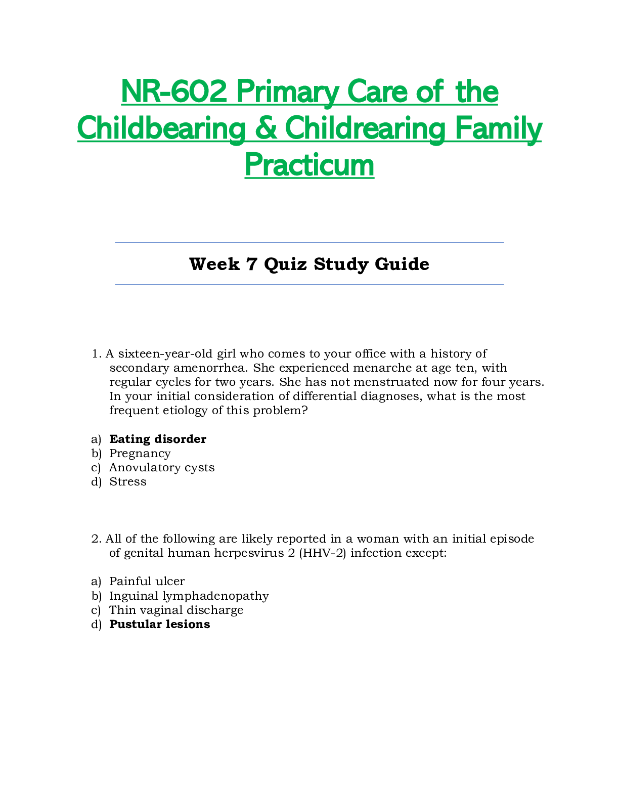
NR602 / NR-602 Week 7 Quiz Study Guide (Latest): Primary Care...
$8
5 Pages
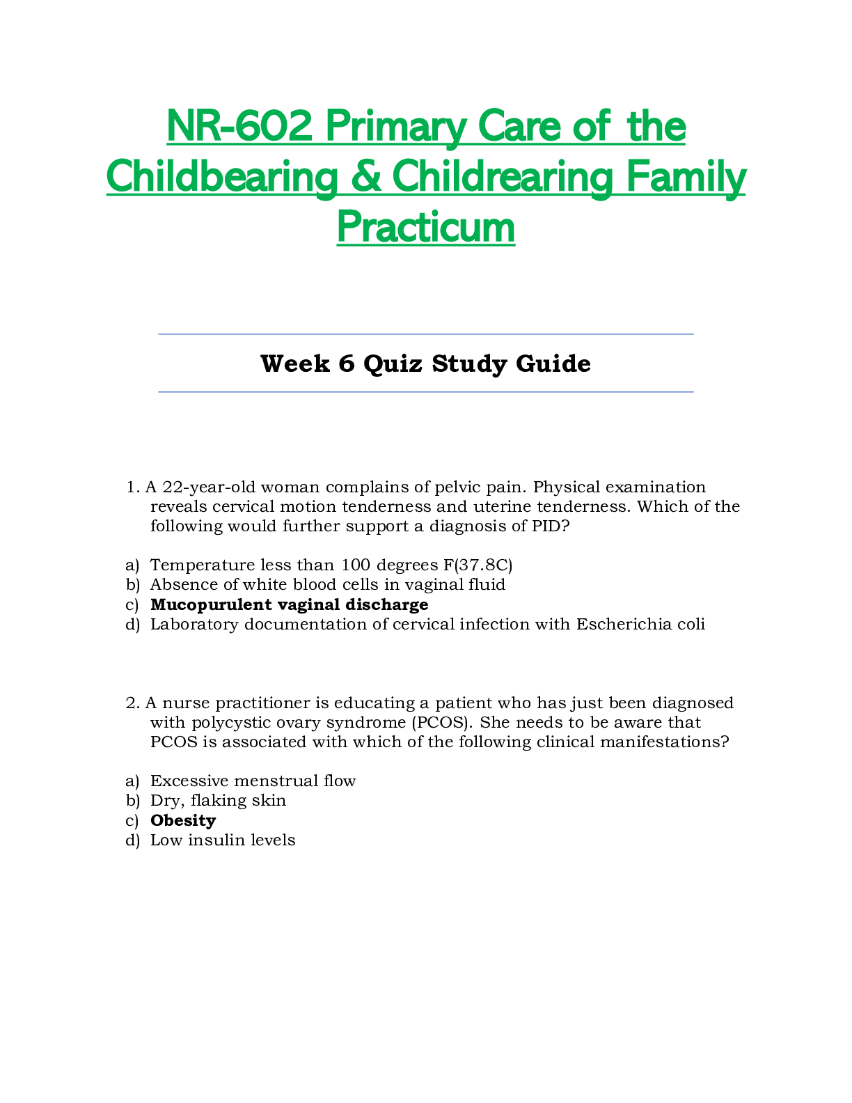
NR602 / NR-602 Week 6 Quiz Study Guide (Latest): Primary Care...

$8
7 Pages
NR602 / NR-602 Week 3 Quiz Study Guide (Latest): Primary Care...

$13
7 Pages
NR 602 Midterm Exam-Questions and Answers, GRADED A,2020.

$12
6 Pages
NR 602 Midterm Exam-Questions and Answers, GRADED A,2020/2021
$12.5
11 Pages
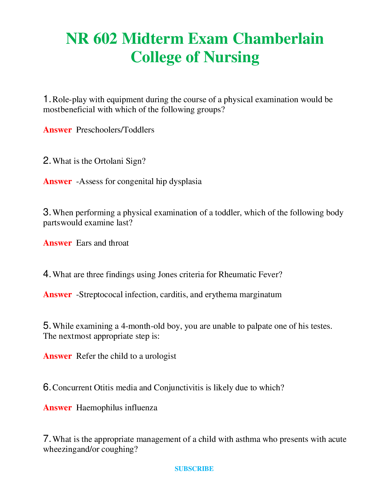
NR 602 Midterm Exam Chamberlain College of Nursing Question...
$12.5
22 Pages
NR 602 Final Exam - Chamberlain College of Nursing Question...

![Preview of WEEK **5 [Chamberlain university]NR 602 IHUMAN Case Week #5 Deborah Arnaudin 54 Year Old](https://browseimages.nyc3.digitaloceanspaces.com/paper-images/2026/03/31/zbQKePwt2026-03-31-11-4669cc328fcad96.png)
More related documents below


 Complete solutions.png)
 NURS 3247Pharmacology - Proctored Assessment,.png)