Thymoma
most common neoplasm of the anterior mediastinum, accounting for 20-25% of all mediastinal tumors and 50% of anterior mediastinal masses. Its peak incidence occurs in the fourth and fifth decades of life; the me
...
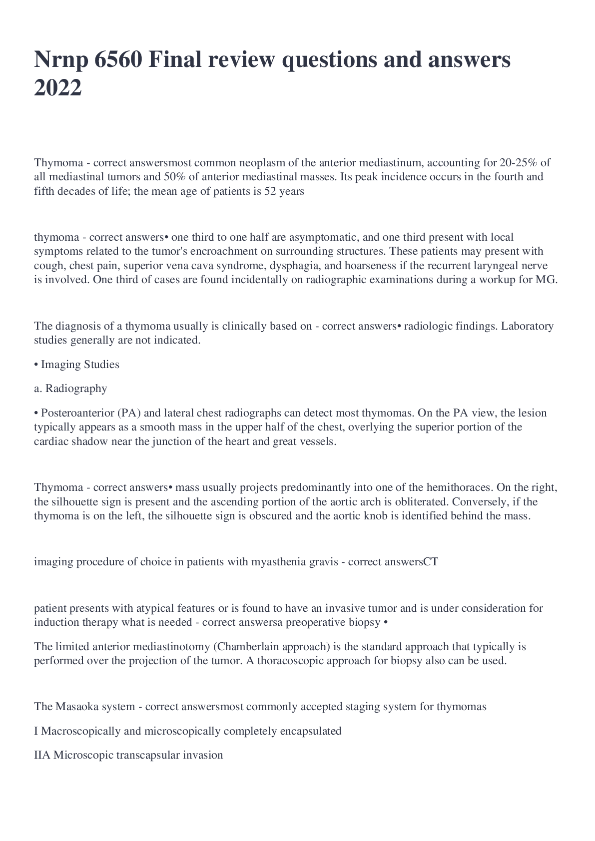
Thymoma
most common neoplasm of the anterior mediastinum, accounting for 20-25% of all mediastinal tumors and 50% of anterior mediastinal masses. Its peak incidence occurs in the fourth and fifth decades of life; the mean age of patients is 52 years
thymoma
• one third to one half are asymptomatic, and one third present with local symptoms related to the tumor's encroachment on surrounding structures. These patients may present with cough, chest pain, superior vena cava syndrome, dysphagia, and hoarseness if the recurrent laryngeal nerve is involved. One third of cases are found incidentally on radiographic examinations during a workup for MG.
The diagnosis of a thymoma usually is clinically based on
• radiologic findings. Laboratory studies generally are not indicated.
• Imaging Studies
a. Radiography
• Posteroanterior (PA) and lateral chest radiographs can detect most thymomas. On the PA view, the lesion typically appears as a smooth mass in the upper half of the chest, overlying the superior portion of the cardiac shadow near the junction of the heart and great vessels.
Thymoma
• mass usually projects predominantly into one of the hemithoraces. On the right, the silhouette sign is present and the ascending portion of the aortic arch is obliterated. Conversely, if the thymoma is on the left, the silhouette sign is obscured and the aortic knob is identified behind the mass.
imaging procedure of choice in patients with myasthenia gravis
CT
patient presents with atypical features or is found to have an invasive tumor and is under consideration for induction therapy what is needed
a preoperative biopsy •
The limited anterior mediastinotomy (Chamberlain approach) is the standard approach that typically is performed over the projection of the tumor. A thoracoscopic approach for biopsy also can be used.
The Masaoka system
most commonly accepted staging system for thymomas
I Macroscopically and microscopically completely encapsulated
IIA Microscopic transcapsular invasion
IIB Macroscopic invasion into surrounding fatty tissue or grossly adherent to but not through mediastinal pleura or pericardium
III Macroscopic invasion into neighboring organs (ie, pericardium, great vessels, or lung)
IVA Pleural or pericardial dissemination
IVB Lymphogenous or hematogenous metastasis
WHO histologic classification Type and Histologic description
Stage Definition
A Tumor in which foci having features of type A thymoma are admixed with foci rich in lymphocytes
B1 Tumor resembles normal functional thymus; combines large expanses having an appearance practically indistinguishable from that of normal thymic cortex with areas resembling thymic medulla
B2 Tumor in which neoplastic epithelial component appears as scattered plump cells with vesicular nuclei and distinct nucleoli among a heavy population of lymphocytes; perivascular spaces are common and sometimes very prominent; a perivascular arrangement of tumor cells resulting in a palisading effect may be seen.
B3 Thymoma predominantly composed of epithelial cells having a round or polygonal shape and exhibiting no or mild atypia; they are admixed with a mild component of lymphocytes, resulting in a sheetlike growth of the neoplastic epithelial cells
C Thymic tumor exhibiting clear-cut cytologic atypia and a set of cytoarchitectural features no longer specific to the thymus, but rather analogous to those seen in carcinomas of the other organs; type C thymomas lack immature lymphocytes; whatever lymphocytes may be present are mature and usually admixed with plasma cells
Treatment and management of thymomas
A. Chemotherapy
B. Corticosteroids
Tuberculosis medication regimen
1. Isoniazid 300mg, rifampin 600mg, pyrazinamide 1.5-2.0 gm and ethambutol 15mg/kg or streptomycin 15mg/kg daily initially.
2. If the isolate proves to be fully susceptible to INH and RIF, then the fourth drug may be dropped.
3. Continue the first three drugs daily for 2 months, then 4 more months of INH and RIF daily.
• Persons with HIV should be treated for nine months.
• A variety of DOT options are also available at twice/thee times weekly dosing.
Those with positive TB skin test should receive ___ months of INH.
6 mos
PPD positive if:
1. A positive test is 5mm for HIV infected persons, contacts of a known case, or persons with a chest film typical for TB.
2. A positive test is 10mm for immigrants from high prevalence areas, or those in high risk groups, including health care workers.
3. A positive test is 15mm for all others not in high prevalence groups.
hypovolemic shock
results from low cardiac output due to low blood volume, such as in a massive hemorrhage or fluid loss from severe burns
cardiogenic shock
results from low cardiac output due to myocardial pump failure
Treatment for neurogenic shock
Fluid replacement and alpha agonists
Central Venous Pressure
A measure of the pressure exerted by fluid in the right atrium; indicative of right sided heart function
Normal: 0-6
CVP Elevation
Ex. Fluid overload, cardiogenic shock
Conditions that cause an increase in the amount of fluid in the R atrium
CVP Decreased
Ex. Dehydration, Distributive shock
Conditions that cause a decrease in the amount of fluid in the R atrium
Pulmonary Artery Pressure
a measure of the systolic and diastolic pressures in the pulmonary artery
Normal: 15-25/5-15
PAP Elevation
Ex. Hypervolemia, pulmonary HTN
Conditions that cause an increase in the amount of fluid in the pulmonary artery OR conditions that decrease the elasticity of the pulmonary artery
PAP Decreased
Ex. Hypovolemia
Conditions that cause a decrease in the amount of fluid in the pulmonary artery
Pulmonary Capillary Wedge Pressure aka Pulmonary Artery Occlusion Pressure
Measure of the pressure in the L ventricle at end-diastole (maximal stretch); indicative of left-sided heart function
-Reflection of the tendency to develop pulmonary edema. To optimize cardiac performance and minimize tendency for pulmonary edema, the PCWP should be kept at the lowest point at which cardiac performance is acceptable.
Normal 6-12
PCWP Elevation
Ex. Hypovolemia
Conditions that decrease the pressure in the L ventricle at end diastole
Cardiac Output
The amount of fluid in liters per minute that the heart pumps into systemic circulation. Product of HR x SV
Normal 4-8
CO Elevation
Ex. Inotropic agents, excess fluid
Factors that increase the HR or increase the amount of blood that the heart puts out with each beat
CO Decreased
Ex. Drugs that decrease contractility, hypovolemia
Factors that decrease HR or decrease the amount of blood that the heart puts out with each beat
Cardiac Index
The cardiac output/body surface area; more accurate measure than CO because this value takes body surface area into account
Normal 2.5-4 L/min
Systemic Vascular Resistance
The resistance provided by the systemic circulation against which the L ventricle must pump blood; calculated by the following formula: MAP- mean CVP x 80)/CO
Normal 800-1200 dynes/sec/cm-5
Labs and Diagnostics of Hypovolemic Shock
Decreased CO/CI
Decreased CVP
Decreased PWCP
*Increased SVR
Decreased SvO2
Management of Hypovolemic Shock
Fluid resuscitation is mainstay of treatment
PRBCs when indicated by Hgb/Hct
Cardiogenic Shock
A loss of effective contractile function results in impaired cardiac output, impaired O2 delivery, and reduced tissue perfusion
Causes: "acute pump failure"
Acute MI
Ventricular aneurysm
Dysrhythmia
Pericardial tamponade
Hypoxemia
Pulmonary edema
Acute valvular regurgitation
Acute VSD
Labs and diagnostics of Cardiogenic Shock
Decreased CO/CI
Increased CVP
Increased PWCP
Increased SVR
Decreased SvO2
[Show More]