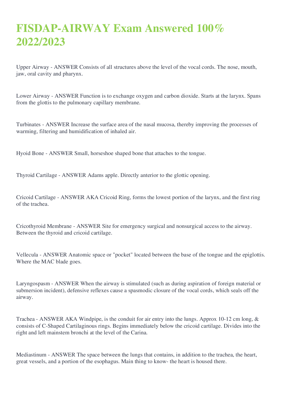
Upper Airway - ANSWER Consists of all structures above the level of the vocal cords. The nose, mouth, jaw, oral cavity and pharynx.
Lower Airway - ANSWER Function is to exchange oxygen and carbon dioxide. Starts at th
...
Upper Airway - ANSWER Consists of all structures above the level of the vocal cords. The nose, mouth, jaw, oral cavity and pharynx.
Lower Airway - ANSWER Function is to exchange oxygen and carbon dioxide. Starts at the larynx. Spans from the glottis to the pulmonary capillary membrane.
Turbinates - ANSWER Increase the surface area of the nasal mucosa, thereby improving the processes of warming, filtering and humidification of inhaled air.
Hyoid Bone - ANSWER Small, horseshoe shaped bone that attaches to the tongue.
Thyroid Cartilage - ANSWER Adams apple. Directly anterior to the glottic opening.
Cricoid Cartilage - ANSWER AKA Cricoid Ring, forms the lowest portion of the larynx, and the first ring of the trachea.
Cricothyroid Membrane - ANSWER Site for emergency surgical and nonsurgical access to the airway. Between the thyroid and cricoid cartilage.
Vellecula - ANSWER Anatomic space or "pocket" located between the base of the tongue and the epiglottis. Where the MAC blade goes.
Laryngospasm - ANSWER When the airway is stimulated (such as during aspiration of foreign material or submersion incident), defensive reflexes cause a spasmodic closure of the vocal cords, which seals off the airway.
Trachea - ANSWER AKA Windpipe, is the conduit for air entry into the lungs. Approx 10-12 cm long, & consists of C-Shaped Cartilaginous rings. Begins immediately below the cricoid cartilage. Divides into the right and left mainstem bronchi at the level of the Carina.
Mediastinum - ANSWER The space between the lungs that contains, in addition to the trachea, the heart, great vessels, and a portion of the esophagus. Main thing to know- the heart is housed there.
Carina - ANSWER Where the right and left mainstem bronchi branch off.
Goblet Cells - ANSWER Mucous producing cells, that are lined in the trachea and bronchi. They trap small particles and other potential contaminants.
Beta-2 Adrenergic Receptors - ANSWER Stimulate bronchodilation.
# of Lobes in each Lung - ANSWER Right lung- 3, Left lung- 2.
Visceral pleura - ANSWER Thin, slippery, outer membrane covering the lungs.
Parietal Pluera - ANSWER Lines the inside of the thoracic cavity.
Bronchioles - ANSWER Made of smooth muscle & lined with beta-2 receptors, which can dilate and constrict based on stimuli.
Alveoli - ANSWER Balloon-like clusters of single-layer air sacs, and serve as the functional site for the exchange of oxygen and CO2. This exchange occurs by simple diffusion over the pulmonary capillaries.
Ventilation - ANSWER Process of moving air in and out of the lungs. Consist of two phases- inhalation and exhalation.
Oxygenation - ANSWER Process of loading O2 molecules onto hemoglobin molecules in the bloodstream.
Respiration - ANSWER Actual exchange of O2 and CO2 in the alveoli and the tissues of the body.
Inhalation - ANSWER Air enters the body, the diaphragm and intercostal muscles contract. When the diaphragm contracts, it descends and enlarges the thoracic cage from top to bottom. When the intercostal muscles contract, they lift the ribs up and out.
Diaphragm - ANSWER Stimulated by the Phrenic Nerve, it is a voluntary and involuntary muscle.
Accessory Muscles - ANSWER Secondary muscles of breathing, and include the sternocleidomastoid and trapezius muscles of the neck.
Negative Pressure Ventilation - ANSWER The air outside the body, is normally higher in pressure than the air within the thorax. During inhalation, the thoracic cage expands and the air within the thorax decreases, creating a slight vacuum. The vacuum pulls the air in through the trachea, causing the lungs to fill.
Positive Pressure Ventilation - ANSWER With ineffective chest movement, or no chest movement, negative intrathoracic pressure cannot be created. When this occurs, the only way to move air into the lungs is by PPV, the forcing of air into the lungs.
Tidal Vol/Dead Space/Residual Vol/Total Lung Capacity - ANSWER Tidal- 500ml
Total Lung Capacity- 6,000ml/5-6L
Dead Space- 150ml
Residual Vol- 1,200ml
Hering- Breuer Reflex - ANSWER Terminates inhalation to prevent over-expansion of the lungs.
Medulla - ANSWER Primary involuntary (autonomic) respiratory center. Connected to the respiratory muscles by the vagus nerve. The medullary respiratory center controls the rate, depth, and rhythm of breathing.
Chemoreceptors - ANSWER Receptors that monitor the chemical composition (pH, CO2,) of body fluids that are located throughout the body. They measure the amount of CO2 in arterial blood and pH in CSF, and if sensed any changes will send signals to the respiratory center.
Dorsal Respiratory Group - ANSWER Responsible for initiating inspiration based on the information received in the chemoreceptors.
Ventral Respiratory Group - ANSWER Responsible for motor control of the inspiratory and expiratory muscles.
Hypoxic Drive - ANSWER Pt's with COPD have a hard time eliminating CO2, therefore always have higher levels of it. The respiratory centers gradually accommodate elevated CO2 levels. The body uses a backup system, that stimulates breathing when the arterial O2 level falls, but then nerves and receptors are easily satisfied with minimal O2.
Aerobic Metabolism - ANSWER In the presence of oxygen, the cells convert glucose into energy through this process.
Anaerobic Metabolism - ANSWER Without adequate O2, the cells do not completely convert glucose into energy and lactic acid and other toxins accumulate into the cell. If this is not corrected (with adequate perfusion and ventilation) the cells will die.
Hypoxia - ANSWER Dangerous condition in which the cells in the tissues do not receive enough O2.
V/Q Mismatch - ANSWER Ventilation and perfusion must be matched. A failure to match ventilation and perfusion, lies behind most abnormalities in oxygen and carbon dioxide exchange.
Hypoventilation - ANSWER Minute vol- decreases. CO2 elimination- decreases, leading to hypercarbia. The level of CO2 in the pt's blood will exceed normal limits.
Hyperventilation - ANSWER Minute vol- increases. CO2 elimiation- increases, leading to hypocarbia. The level of CO2 in pt's blood will fall below normal, leading to dizziness, and numbness in the face and extremities.
Intrapulmonary Shunting - ANSWER Blood entering the lungs from the right side of the heart, in an unoxygenated state.
Resp Acidosis - ANSWER Anytime a pt is in resp distress or is unable to breathe, acidosis quickly develops, because of a buildup in CO2.
Resp Alkalosis - ANSWER Alkalosis can develop if respiration's are too high, and there is not enough CO2 in the body, the body will be high in base.
Orthopnea - ANSWER Severe dyspnea experienced when recumbent and relieved by sitting or standing up.
Apnuestic Breathing - ANSWER Prologned, gasping inhalation followed by extremely short, ineffective exhalation; associated with brainstem insult.
Adventitious Respirations - ANSWER Abnormal airway sounds.
Retractions - ANSWER Skin pulling between and around the ribs during inhalation.
Paradoxical Motion - ANSWER The inward movement of a segment of the chest during inhalation and outward movement of the chest during exhalation, opposite normal chest movement and an indication of a flail chest.
Pulsus Paradoxus - ANSWER A clinical finding in which the systolic blood pressure drops more than 10mmHg, during inhalation. A change in pulse quality, or a disappearance of a pulse during inhalation, may also be detected. COPD, pericaridal tamponade, tension pnuemothorax, & severe asthma attack.
End- Tidal CO2 - ANSWER Detects the presence of CO2 in exhaled air and are important adjuncts for determining ventilation adequacy. Normally range between 35-45.
Capnographer - ANSWER Performs the same function and attaches in the same way as the capnometer, but it provides a graphic representation of exhaled carbon dioxide.
Capnometer - ANSWER Capnometer provides a numeric reading of exhaled carbon dioxide.
Phase A of Waveform - ANSWER First phase, is the respiratory baseline. The initial stage of exhalation; the gas sample is dead space gas, free of CO2.
Phase B of Waveform - ANSWER Second phase, abrupt rise in CO2 due to a mixture of alveolar gas with dead space gas, called the upslope. Also phase B-C.
Phase C of Waveform - ANSWER Expiratory or alveolar plateau is represented by phase C-D, and the gas sampled is essentially alveolar.
Phase D of Waveform - ANSWER Point D is maximal ETCO2 level, the best reflection of teh alveolar CO2 level.
Phase E of Waveform - ANSWER Fresh gas is introduced during the inspiratory downstroke (phase D-E), and the waveform returns to the baseline level of CO2- approx 0.
Bag Valve Mask - ANSWER Whenever possible, you and your partner should work together to provide ventilation's with the BVM, to ensure the artificial ventilations are adequate, you must look for CHEST RISE AND FALL, make sure the RATE IS NOT TOO FAST or SLOW, and make sure the PULSE RATE IMPROVES.
CPAP - ANSWER CHF pt's, pulm edema, COPD, and acute bronchospasm (acute asthma), submersion accidents, pulse ox less than 90, and rapid breathing, are indications for CPAP. Do not use if pt has abnormal LOC, hypoventilation, pneumothorax, tracheostomy, and active GI bleed.
PEEP - ANSWER Positive end expiratory pressure; during the epxiratory phase the pt exhales against this resistance. A PEEP of 5-10 is generally what is used.
Suctioning a Trache - ANSWER Preoxygenate, insert 3ml of saline into stoma, instruct the pt to exhale while inserting the catheter, then begin suctioning on the way out.
Ventilating a Trache Stoma - ANSWER Neither head tilt or chin lift is required for pt w/ a stoma. If using a BVM, use a pediatric mask over the stoma to get a good seal, then seal the pt's nose and mouth to prevent leakage.
Tracheostomy Tube - ANSWER A plastic tube placed within the trachea site (stoma), and is compatible with BVM 15/22mm, so your BVM will connect right too it. May need to suction the trache tube before ventilating.
Stenosis - ANSWER When the trache tube becomes dislodged, and is potentially life threatening because soft tissue damage may occur. You may have to insert an ET tube into the stoma before it becomes totally occluded.
How to fix Stenosis - ANSWER Lubricate the same size trache tube or ET tube, instruct the pt to exhale and gently insert the tube approx 1 to 2 cm beyond the balloon cuff. Inflate the balloon cuff. Confirm patency and proper placement of the tube, auscultate lung sounds.
Predicting the Difficult Airway - ANSWER LEMON- Look externally, Evaluate 3-3-2, Mallampati, Obstruction, Neck mobility.
3-3-2 RULE - ANSWER The first 3 refers to mouth opening, ideally a pt's mouth should open at least 3 fingerwidths (5cm). The second 3 refers to the length of the mandible, at least 3 fingerwidths is optimal. The 2 part refers to the distance from the hyoid bone to the thyroid notch; it should be at least 2 fingers wide.
Complications of an ET tube - ANSWER Bleeding, hypoxia, laryngeal swelling, laryngospasm, vocal cord damage, mucosal necrosis and barotrauma.
*NEVER take your hand off the ET tube before it has been secured in place.
Straight Blade (Miller) - ANSWER Designed to lift up the epiglottis, and goes beneath it. Beneficial for children who have large epiglottis, but requires great care and if used improperly it is more likely to damage teeth.
Curved Blade (MAC) - ANSWER Goes into the vallecular space, between the tongue and the glottis, and indirectly lifts the epiglottis. Less likely to be levered against the teeth.
Confirmation of ET tube placement - ANSWER Visualizing the passes of the tube through the vocal cords is your first confirmation and most reliable, listening to lung sounds and gastric sounds, resistance while BVM'ing, capnography readings, and the esophageal detector device.
Steps for an ET tube - ANSWER Preparation (gathering all your supplies), preoxygenating (at least 2 minutes with an airway adjunct), positioning the patient (sniffing position, or ear to sternum), insert the blade, insert the tube *you must see the tube pass through the vocal cords, if you don't see the vocal cords do not pass the tube, after you pass it through, remove your blade*, hold the tube securely, and remove the stylet, inflate the cuff 5-10cc, and begin ventilations.
BURP Maneuver - ANSWER Backward, upward, rightward position. Done by partner to help you visualize the vocal cords.
Tracheobronchial Suctioning - ANSWER *Sterile procedure, do not do if you do not have too, unless the secretions are so severe they are interfering with ventilation. Suctioning the trachea can cause dysrhythmias, so make sure you have the pt on a 12 lead. Preoxygenation is very important. Lubricate the suctioning tube. May be necessary to insert 3-5ml of saline to loosen the secretions. Gently insert until resistance is felt (when you hit the carina), and suction on the way out for no more than 10 seconds.
Extubation - ANSWER Process of removing the ET tube. Consider sedating the pt before remove the tube. Complications of removing the tube are vomiting, a laryngospasm, and airway swelling. Do not remove the tube if there is any risk of recurrent resp failure or uncertainty about a pt's ability to maintain their own airway.
20 Seconds or Less - ANSWER *A single pediatric intubation should not exceed more than 20 seconds. If intubation cannot be performed at this time, abort the attempt and resume bag-mask ventilations.
Pediatric Laryngoscope Blades - ANSWER Premature newborn- 0 straight
Full-term newborn- size 1 straight
2 yrs to adolescent- size 2 straight
[Show More]