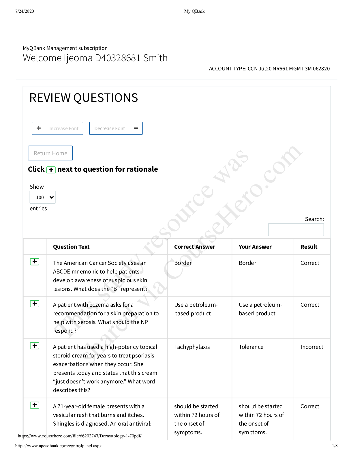
1. List the functions of the heart.
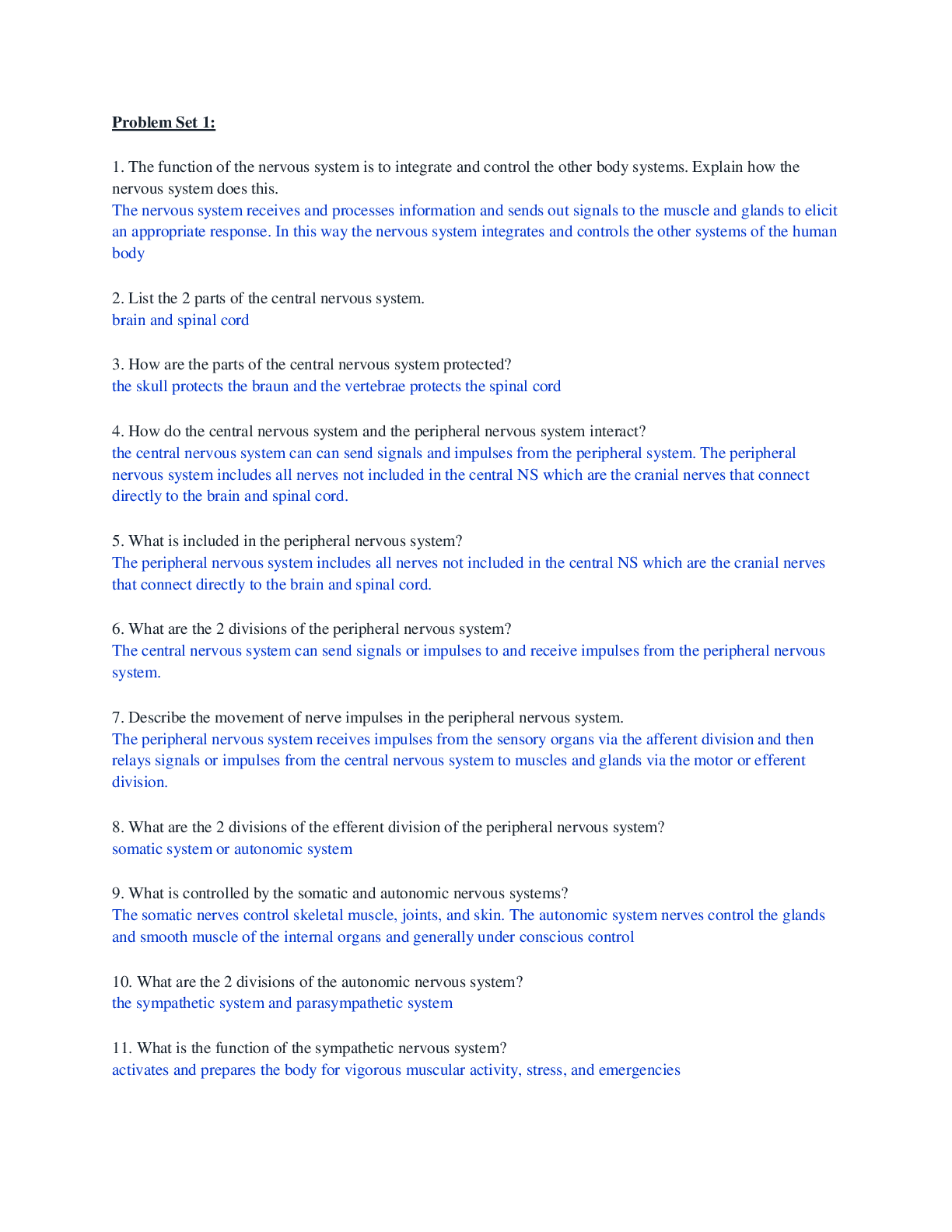
A: The functions of the heart are: generating blood pressure, routing blood, and regulating blood supply.
2. Define the pulmonary and systemic circulations and describe the gas exch
...
1. List the functions of the heart.
A: The functions of the heart are: generating blood pressure, routing blood, and regulating blood supply.
2. Define the pulmonary and systemic circulations and describe the gas exchange taking place in each of them.
A: The pulmonary circulation is the flow of blood from the heart through the lungs back to the heart. This blood picks up oxygen and releases carbon dioxide in the lungs. The systemic circulation is the flow of blood from the heart through the body back to the heart. This blood delivers oxygen and picks up carbon dioxide in the body’s tissues.
3. Describe the location, shape, and approximate size of the heart.
A: The heart is located in the thoracic cavity between the lungs as part of the midline partition known as the mediastinum. The heart is shaped like a blunt cone with an apex rounded point and a flat part on the opposite side as the base. It is approximately the size of a closed fist, but it can be larger in more active adults.
4. What is the pericardium? Describe its parts and their function.
A: The pericardium, also known as the pericardial sac, holds the pericardial fluid which helps reduce friction as the heart moves within the sac. It consists of two layers. An outer fibrous pericardium, that is a tough and fibrous connective tissue, anchors the heart to the mediastinum. The inner serous pericardium that surrounds the heart is simple squamous epithelium overlying a layer of loose connective tissue and fat. The part in contact with the fibrous pericardium is the parietal pericardium while the part covering the heart is the visceral pericardium. In between the visceral and parietal pericardia, there is the pericardial cavity filled with a thin pericardial fluid produced by the serous pericardium that helps reduce friction.
5. Describe the three layers of the heart, and state their functions.
A: The three layers of the heart are epicardium, myocardium, and endocardium. The epicardium (an interchangeable term for the structure that is also called the visceral pericardium) is a thin serous membrane that forms the smooth outer surface of the heart. It consists of simple squamous epithelium overlying a layer of loose connective tissue and fat. It is part of the inner serous pericardium surrounding the heart and is important in forming the cavity that holds the pericardial fluid which reduces friction during movement. The myocardium is the thick middle layer of the heart. It is composed of cardiac muscle cells and is responsible for contractions of the heart chambers. The endocardium is the inner surface of the heart chambers. It consists of simple squamous epithelium over a layer of connective tissue. Its smoothness allows blood to move easily over its surface through the heart.
6. Name the muscular ridges found on the interior walls of the ventricles and atrium.
A: Trabeculae carneae are the ridges and columns of cardiac muscle that make up the surfaces of the interior walls of the ventricles. The pectinate muscles are smaller, muscular ridges that are found in portions of the atria.
7. Name the chambers of the heart and describe their locations as seen from the outside of the heart.
A: The four chambers of the heart are two atria and two ventricles. The right and left atria are located at the base of the heart, and the right and left ventricles extend from the base of the heart toward the apex.
8. Name the major blood vessels that enter and leave the heart. Which chambers of the heart do they enter or exit.
A: There are six large veins that carry blood to the heart: the superior vena cava and the inferior vena cava (carry blood from the body to the right atrium) and four pulmonary veins (carry blood from the lungs to the left atrium). Two arteries, the pulmonary trunk and the aorta, carry blood away from the heart. The pulmonary trunk arises from the right ventricle and splits into the right and left pulmonary arteries known for carrying blood to the lungs. The aorta arises from the left ventricle and carries blood to the rest of the body.
9. Describe the openings through which blood enters the atria. What structure separates the atria from each other?
A: The atria receive blood from veins. The right atrium has three major openings through which blood enters: the openings from the superior vena cava and the inferior vena cava both receive blood from the body and the opening of the coronary sinus receives blood from the heart itself. The left atrium has four openings that receive blood from the four pulmonary veins from the lungs. The two atria are separated by the interatrial septum, although in the embryo and fetus there was an opening between the atria to allow blood flow and the bypass of the pulmonary circulation.
10. What are the fossa ovalis and the foramen ovale?
A: The fossa ovalis is a slight, oval depression on the right side of the interatrial septum that marks the former location of the foramen ovale, an opening between the right and left atria in the embryo and fetus that allowed blood to flow from the right to the left atrium in the fetus to bypass the pulmonary circulation
[Show More]


.png)