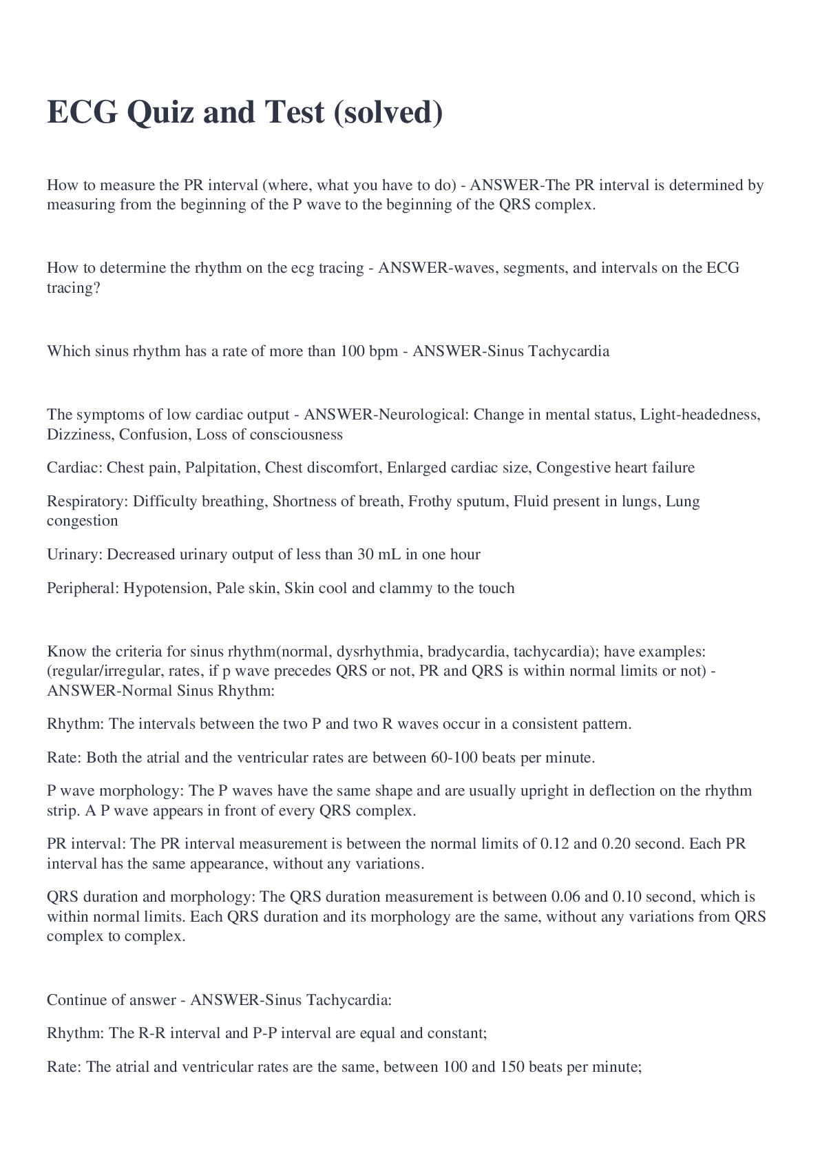
How to measure the PR interval (where, what you have to do) - ANSWER-The PR interval is determined by measuring from the beginning of the P wave to the beginning of the QRS complex.
How to determine the rhythm on the
...
How to measure the PR interval (where, what you have to do) - ANSWER-The PR interval is determined by measuring from the beginning of the P wave to the beginning of the QRS complex.
How to determine the rhythm on the ecg tracing - ANSWER-waves, segments, and intervals on the ECG tracing?
Which sinus rhythm has a rate of more than 100 bpm - ANSWER-Sinus Tachycardia
The symptoms of low cardiac output - ANSWER-Neurological: Change in mental status, Light-headedness, Dizziness, Confusion, Loss of consciousness
Cardiac: Chest pain, Palpitation, Chest discomfort, Enlarged cardiac size, Congestive heart failure
Respiratory: Difficulty breathing, Shortness of breath, Frothy sputum, Fluid present in lungs, Lung congestion
Urinary: Decreased urinary output of less than 30 mL in one hour
Peripheral: Hypotension, Pale skin, Skin cool and clammy to the touch
Know the criteria for sinus rhythm(normal, dysrhythmia, bradycardia, tachycardia); have examples: (regular/irregular, rates, if p wave precedes QRS or not, PR and QRS is within normal limits or not) - ANSWER-Normal Sinus Rhythm:
Rhythm: The intervals between the two P and two R waves occur in a consistent pattern.
Rate: Both the atrial and the ventricular rates are between 60-100 beats per minute.
P wave morphology: The P waves have the same shape and are usually upright in deflection on the rhythm strip. A P wave appears in front of every QRS complex.
PR interval: The PR interval measurement is between the normal limits of 0.12 and 0.20 second. Each PR interval has the same appearance, without any variations.
QRS duration and morphology: The QRS duration measurement is between 0.06 and 0.10 second, which is within normal limits. Each QRS duration and its morphology are the same, without any variations from QRS complex to complex.
Continue of answer - ANSWER-Sinus Tachycardia:
Rhythm: The R-R interval and P-P interval are equal and constant;
Rate: The atrial and ventricular rates are the same, between 100 and 150 beats per minute;
P wave morphology: The P waves have the same shape and usually are upright in deflection on the rhythm strip. There is a P wave in front of every QRS complex;
PR interval: The PR interval measurement is between the normal limits of 0.12 and 0.20 second. Each PR interval is the same, without any variations;
QRS duration and morphology: The QRS duration measurement is between the normal limits of 0.06 and 0.10 second. Each QRS duration and its morphology are the same, without any variations from QRS complex to complex.
Continue of Answer - ANSWER-Sinus Bradycardia:
Rhythm: The R-R interval and P-P interval occur on a regular and constant basis;
Rate: The atrial and ventricular rates are equal and less than 60 beats per minute;
P wave morphology: The shape of each P wave is upright and uniform. There is a P wave in front of each QRS complex. No additional P waves or QRS complexes are noted;
PR interval: The PR interval measurement is between the normal limits of 0.12 and 0.20 second. All PR intervals are the same;
QRS duration and morphology: The QRS duration measurement is between the normal limits of 0.06 and 0.10 second. Each QRS duration and its morphology are the same, without any variations from QRS complex to complex.
Continue - ANSWER-Sinus Dysrhythmia:
Rhythm: The interval between the P-P and R-R waves occur at irregular periods.
Rate: The atrial and ventricular rates are the same, between 60 and 100 beats per minute.
P wave morphology: The P waves have the same shape and usually are upright in deflection on the rhythm strip. There is a P wave in front of every QRS complex.
PR interval: The PR interval measurement is between the normal limits of 0.12 and 0.20 second. Each PR interval is the same, without any variations.
QRS duration and morphology: The QRS duration measurement is between 0.06 and 0.10 second. Each QRS duration and its morphology are the same, without any variations from QRS complex to complex.
Is an ECG considered an legal document; where do you put it; four items that need to be listed on the ecg - ANSWER-Yes; in patient medical record; name, date, time, and the initials of the person performing the ECG
The normal range/length for the QRS complex duration - ANSWER-Narrow: 0.06 to 0.10 second
Wide: 0.12 and greater
What is the normal PR interval measurement is - ANSWER-0.12-0.20 sec
What components the ECG technician evaluates on the ECG rhythm strip - ANSWER-Rhythm, rate, P wave morphology, PR interval, QRS duration and morphology
What is low cardiac output; what type of patients are likely to have this - ANSWER-heart is not pumping adequately; if the patient's rhythm returns to sinus rhythm from another dysrhythmia
Why the shape of the P wave in analyzed; when you determine the shape what is it telling you - ANSWER-the atrial current is moving in the same pathway
The signs and symptoms of adequate cardiac output - ANSWER-an alert and oriented patient with no difficulty breathing, no chest pain or pressure, and a stable blood pressure
If you are looking at sinus brady or tachy they almost have the same criteria as normal rhythm, what are their differences that makes them them (Know the rate/characteristics of sinus brady, tachy, and normal) - ANSWER-Their heart rates: (brady<60, tachy100)
What is evaluated and classified when determining dysrhythmias - ANSWER-The EC waveform has various components such as waves, segments, and intervals that are evaluated and classified based on their size, length of time, and location on the tracing. All of the different components determine the type of cardiac rhythm. In order to evaluate a rhythm you must first understand each component and its normal appearance. When these components differ from the expected norm, a dysrhythmia (or arrhythmia) is indicated. The tracing must be evaluated for artifact prior to the evaluation of the heart rhythm.
What does the QRS duration measures - ANSWER-Ventricular depolarization and ventricular contraction measured from the beginning of the Q wave to the end of the S wave Duration
The QRS complex represents the time it takes for what certain action to happen - ANSWER-For the ventricles to depolarize or contract
What question is necessary to analyze a P wave on and ECG tracing - ANSWER-Are the shapes and waveforms all the same?
Does each P wave have a QRS complex following it?
If you can't identify an R wave on ECG tracing, which wave or waves would you use to determine the regularity of the ventricles - ANSWER-point of the Q and S wave junction as an easy point of evaluation
If you are monitoring a patient that is very pale, breathing very fast, and their monitor indicates that they are in normal sinus rhythm, what do you do - ANSWER-Report his condition to a licensed practitioner immediately.
Know the difference between normal ventricular conduction and delayed ventricular conduction - ANSWER-Normal ventricular conduction: when QRS complex is narrow
Delayed ventricular conduction: when QRS complex is wide
Test - ANSWER-
What kind of conditions you might see tachycardia in (may be regular or irregular) - ANSWER-Regular
Tachycardia can b
[Show More]












![Preview of THE TEFL ACADEMY LEVEL 5 - ASSIGNMENT C - AUTHENTIC MATERIAL [2026]](https://browseimages.nyc3.digitaloceanspaces.com/paper-images/2026/04/10/HpVkRcWV2026-04-10-11-5769d96426bd64f.png)