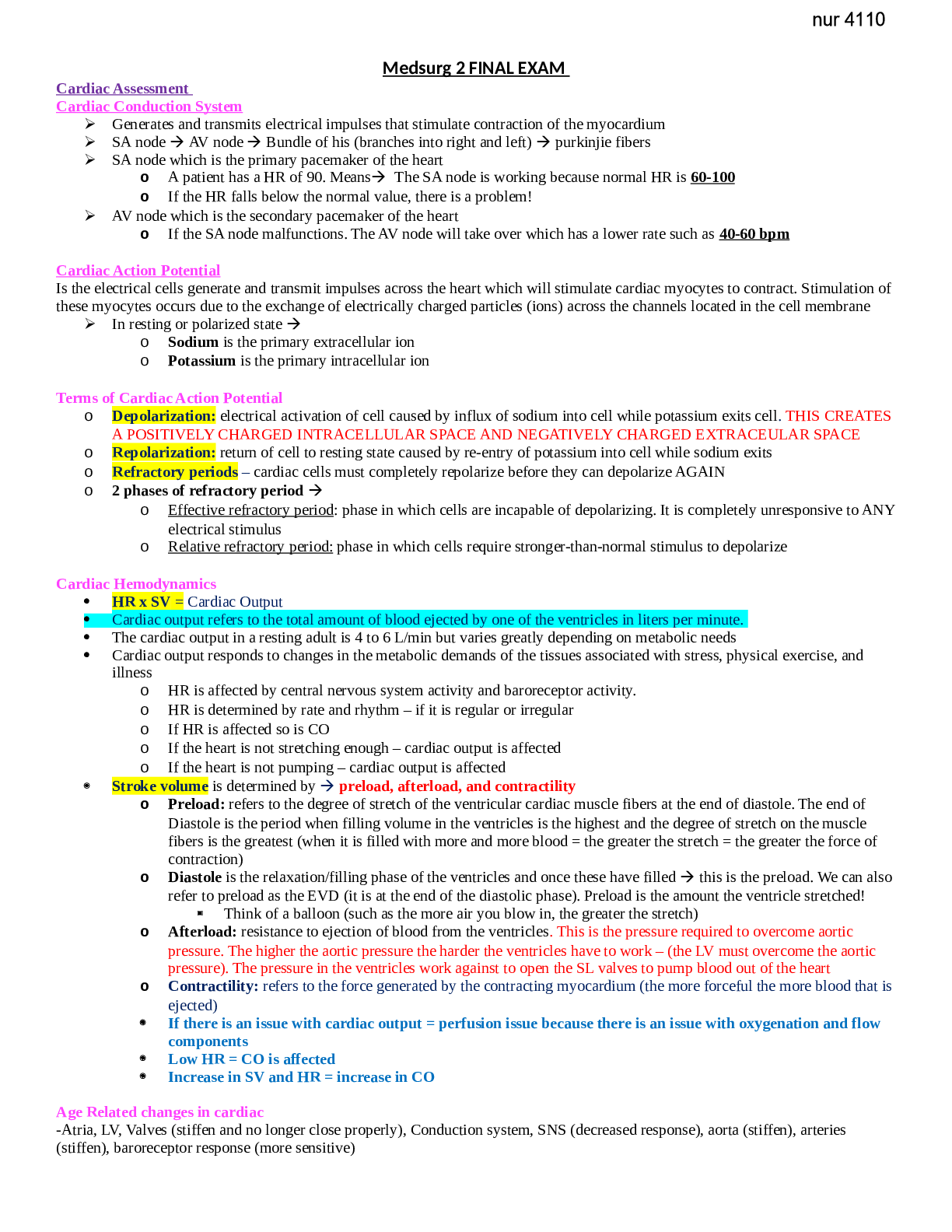
Medsurg 2 FINAL EXAM
Cardiac Assessment
Cardiac Conduction System
Generates and transmits electrical impulses that stimulate contraction of the myocardium
SA node AV node Bundle of his (branches into right an
...
Medsurg 2 FINAL EXAM
Cardiac Assessment
Cardiac Conduction System
Generates and transmits electrical impulses that stimulate contraction of the myocardium
SA node AV node Bundle of his (branches into right and left) purkinjie fibers
SA node which is the primary pacemaker of the heart
o A patient has a HR of 90. Means The SA node is working because normal HR is 60-100
o If the HR falls below the normal value, there is a problem!
AV node which is the secondary pacemaker of the heart
o If the SA node malfunctions. The AV node will take over which has a lower rate such as 40-60 bpm
Cardiac Action Potential
Is the electrical cells generate and transmit impulses across the heart which will stimulate cardiac myocytes to contract. Stimulation of
these myocytes occurs due to the exchange of electrically charged particles (ions) across the channels located in the cell membrane
In resting or polarized state
o Sodium is the primary extracellular ion
o Potassium is the primary intracellular ion
Terms of Cardiac Action Potential
o Depolarization: electrical activation of cell caused by influx of sodium into cell while potassium exits cell. THIS CREATES
A POSITIVELY CHARGED INTRACELLULAR SPACE AND NEGATIVELY CHARGED EXTRACEULAR SPACE
o Repolarization: return of cell to resting state caused by re-entry of potassium into cell while sodium exits
o Refractory periods – cardiac cells must completely repolarize before they can depolarize AGAIN
o 2 phases of refractory period
o Effective refractory period: phase in which cells are incapable of depolarizing. It is completely unresponsive to
ANY electrical stimulus
o Relative refractory period: phase in which cells require stronger-than-normal stimulus to depolarize
Cardiac Hemodynamics
HR x SV = Cardiac Output
Cardiac output refers to the total amount of blood ejected by one of the ventricles in liters per minute.
The cardiac output in a resting adult is 4 to 6 L/min but varies greatly depending on metabolic needs
Cardiac output responds to changes in the metabolic demands of the tissues associated with stress, physical exercise, and
illness
o HR is affected by central nervous system activity and baroreceptor activity.
o HR is determined by rate and rhythm – if it is regular or irregular
o If HR is affected so is CO
o If the heart is not stretching enough – cardiac output is affected
o If the heart is not pumping – cardiac output is affected
Stroke volume is determined by preload, afterload, and contractility
o Preload: refers to the degree of stretch of the ventricular cardiac muscle fibers at the end of diastole. The end of
Diastole is the period when filling volume in the ventricles is the highest and the degree of stretch on the muscle
fibers is the greatest (when it is filled with more and more blood = the greater the stretch = the greater the force of
contraction)
o Diastole is the relaxation/filling phase of the ventricles and once these have filled this is the preload. We can also
refer to preload as the EVD (it is at the end of the diastolic phase). Preload is the amount the ventricle stretched!
Think of a balloon (such as the more air you blow in, the greater the stretch)
o Afterload: resistance to ejection of blood from the ventricles. This is the pressure required to overcome aortic
pressure. The higher the aortic pressure the harder the ventricles have to work – (the LV must overcome the aortic
pressure). The pressure in the ventricles work against to open the SL valves to pump blood out of the heart
o Contractility: refers to the force generated by the contracting myocardium (the more forceful the more blood that is
ejected)
If there is an issue with cardiac output = perfusion issue because there is an issue with oxygenation and flow
components
Low HR = CO is affected
Increase in SV and HR = increase in CO
Age Related changes in cardiac
-Atria, LV, Valves (stiffen and no longer close properly), Conduction system, SNS (decreased response), aorta (stiffen), arteries
(stiffen), baroreceptor response (more sensitive)
History and Physical
Health History which refers to the patient’s ability to recognize cardiac symptoms to know what to do when they occur it is essential
for effective self-care management
Want CLEAR information when talking to the patient Chief complaint, Hx present illness & past medical history/social
history, Home meds, Nutrition, Allergies
Physical Assessment (Cardiac specific)
General appearance, Skin and extremities
Blood pressure pulse, and postural BP Below 30 is reduction in CO
Arterial pulses rate, rhythm, amplitude
Jugular venous pulsations fluid accumulation/overload
Heart inspection and auscultation
Any deviations from normal? Meaning changes in how the patient responds to how they are feeling. Such as a patient with
HF or circulatory issues will change their shoes due to their feet being swollen
o Heart as a pump
o Atrial/ventricular filling volumes
o Cardiac output
o Compensatory mechanisms
What position does the patient lay in when they sleep?
Tripod position patient is leaning forward. This is an indication of respiratory distress
MOST common clinical manifestation
Ask why the pt would feel like this?
Chest pain
SOB, dyspnea fluid overload/pulmonary issue can lead to dyspnea
Peripheral edema
Weight gain
Abd distention
Fatigue
Dizziness, Syncope, Changes LOC due to perfusion and oxygenation issues (brain likes oxygen)
We know diuretic is working if the pt is peeing a lot and they feel less fatigue/dyspnea
Chest Pain
Identify Quantity, location, quality, radiation, duration of pain , Associated S/S
Assess for other cardiac conditions and for other significant conditions
Angina Pectoris: uncomfortable pressure, squeezing, or fullness in substernal chest area. It can radiate across chest to the
medial aspect of one of both arms and hands, jaw, shoulders, upper back, or epigastrium. (BOOK)
ACS: is the same as angina pectoris. Pain and discomfort ranges from mild to severe associated with SOB, diaphoresis,
palpitations, unusual fatigue, and n/v (BOOK)
Pericarditis: sharp, severe substernal or epigastric pain which can radiate to necks, arms, and back.
Pneumonia, pulmonary embolism sharp, severe substernal or epigastric pain arising from interior portion of pleura
Hiatal hernia, GERD substernal pain described as sharp, burning, or heavy. Often mimics angina and can radiate to
neck, arm, or shoulders.
Costochondritis musculoskeletal disorders. This is a sharp or stabbing pain localized in anterior chest. Most often is
unilateral and can radiate across chest to epigastrium or back
Vascular
[Show More]















 (1).png)