*NURSING > SHADOW HEALTH > Shadow health Physical Assessment Head Eyes Ears Nose Mouth and Neck (All)
Shadow health Physical Assessment Head Eyes Ears Nose Mouth and Neck
Document Content and Description Below
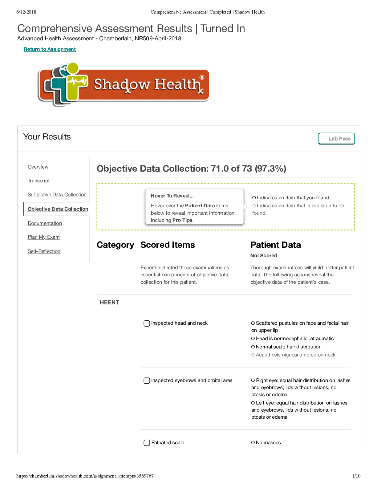
Normal range of findings Abnormal findings HEAD Wash hands To minimize transmission of infection INSPECT the head for size, shape, skin characteristics. Skull is symmetric and appropriately propor... tioned for the size of the body- normocephalic Microcephaly (abnormally small head) and macrocephaly (abnormally large head- found in hydrocephalus) INSPECT the facial structures for size, symmetry, movement, skin characteristics, and facial expression. Look for the symmetry in facial bones, and other structures such as eyebrows, nasolabial folds, especially with calm facial expression. Asymmetry of facial features may be noted in facial palsy. PALPATE the structures of the skull for contour, tenderness, and intactness, if there is any history of injury or pain. Depressions or unevenness of the skull may occur due to skull injury. PALPATE the bony structures of the face and jaw, noting tenderness and jaw movement (done only if patient reports pain/ injury/ irregularity) To palpate jaw movement, place two fingers in front of each ear and ask the patient to slowly open and close the mouth and move the lower jaw from side to side. The jaw should move smoothly and without pain. Pain associated with palpation, limited movement, pain with movement, and a jaw that clicks or catches with movement may indicate temporomandibular joint disease. PALPATE the temporal arteries for pulsation, texture, and tenderness. Palpate over the temporal bone on each side of the head lateral to each eyebrow for the temporal artery. The artery should be smooth and non-tender, with pulsation noted. Auscultate the temporal arteries using the bell of stethoscope. Normally no sound is heard. Tender, edematous, or hardened temporal arteries with redness over the temporal region suggest temporal arteritis. A bruit (a low-pitched blowing sound) heard during auscultation indicates a vascular abnormality. EYES TEST visual acuity (distance vision) (tests cranial nerve II). Place Snellen chart on the wall in a well-lighted room. The patient may sit or stand 20 feet (6 m) from the chart. If the patient wears contact lenses or glasses, he or she should leave them in place. Have the patient cover one eye with an opaque card Note any hesitancy, squinting, leaning forward, blinking, or facial expressions indicating that the patient is struggling to see. The larger the denominator, the poorer the vision. If vision is poorer than 20/30 [Show More]
Last updated: 2 years ago
Preview 1 out of 11 pages

Buy this document to get the full access instantly
Instant Download Access after purchase
Buy NowInstant download
We Accept:

Reviews( 0 )
$11.50
Can't find what you want? Try our AI powered Search
Document information
Connected school, study & course
About the document
Uploaded On
Mar 05, 2023
Number of pages
11
Written in

Additional information
This document has been written for:
Uploaded
Mar 05, 2023
Downloads
0
Views
70