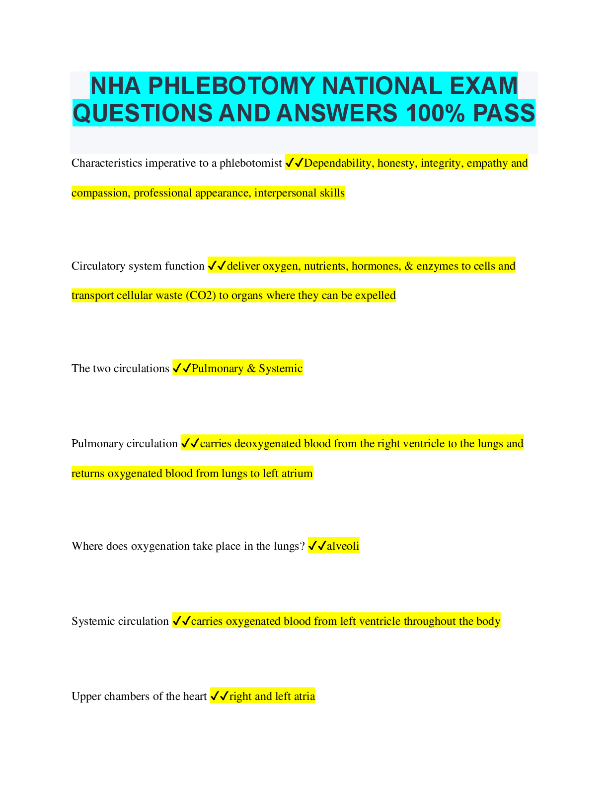
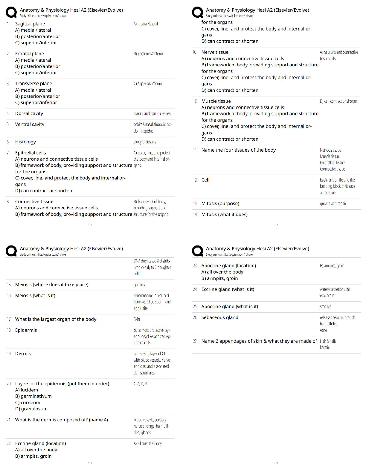
Abdomen ARDMS _ MOCK exam Already
Passed
Progression of which of the following abnormalities flattens the portal veins? ✔✔Biliary
obstruction
The wall thickness in a normal fasting gallbladder should not exceed ✔✔3 m
...
Abdomen ARDMS _ MOCK exam Already
Passed
Progression of which of the following abnormalities flattens the portal veins? ✔✔Biliary
obstruction
The wall thickness in a normal fasting gallbladder should not exceed ✔✔3 mm
This color Doppler sonogram is most likely demonstrating which of the following abnormalities?
✔✔Pseudoaneurysm
This color Doppler image demonstrates turbulent swirling blood flow within a fluid collection,
classic sonographic findings of a common femoral artery pseudoaneurysm.
A patient presents with sudden onset of upper abdominal pain. Ultrasound demonstrates
prominence in the stomach rugae. These findings are most suspicious for which of the following
conditions? ✔✔Gastritis
Prominence of the stomach rugae in a patient with upper abdominal pain is most suspicious for
gastritis. Hypervascular, thick gastric walls are sonographic findings associated with gastric
ulcers.
Which of the following is a complication of acute pancreatitis? ✔✔Duodenal obstruction
Complications of acute pancreatitis may include abscess formation, duodenal obstruction,
hemorrhage, phlegmon, and pseudocyst formation. Cholecystitis is a possible etiology of acute
pancreatitis.
A patient presents with a history of hematuria. The findings in this duplex image are most
suspicious for which of the following pathologies? ✔✔Carcinoma
A vascular echogenic mass is identified protruding from the posterior wall of the urinary bladder.
Bladder carcinoma commonly presents with a history of painless hematuria. Based on the
clinical history, the sonographic findings are suspicious for a malignant mass.
A sagittal sonogram medial to the porta hepatis is demonstrating which of the following
abnormalities? ✔✔Dilated common bile duct
A hypoechoic mass identified by the calipers is obstructing the common bile duct resulting in
dilatation. The mass is most likely a malignancy in the head of the pancreas.
The pathology in this sonogram is most likely a/an ✔✔Pseudocyst
A complex fluid collection is identified posterior to the tail of the pancreas. This is most likely a
pancreatic pseudocyst. Phlegmons and islet cell tumors appear as hypoechoic masses on
ultrasound. A pancreatic hemorrhage is a differential consideration but not the most likely
pathology.
Which of the following abnormalities is demonstrated in this transverse sonogram? ✔✔Stones in
the duct of Wirsung
Multiple stones are located in main pancreatic duct (duct of Wirsung).
Which of the following is a clinical symptom of hypothyroidism? ✔✔Muscle cramps
Muscles cramping is a symptom of hypothyroidism. Other symptoms may include weight gain,
mental and physical lethargy, arthritis, skin dryness, feeling cold, slow metabolic rate, and
decreased heart rate. Symptoms commonly associated with hyperthyroidism include weight loss,
palpitations, nervousness, exophthalmos, constant hunger, tremors, increased heart rate, and
intolerance to heat.
A 20-year-old patient presents with a palpable left scrotal mass. The sonographic findings are
most suspicious for which of the following pathologies? ✔✔Malignant neoplasm
A malignant neoplasm is the most likely diagnosis in a young adult demonstrating a hypoechoic
intratesticular mass. The patient is afebrile excluding a testicular abscess from the differential
considerations.
A patient presents with a history of a palpable neck mass. Which of the following terms best
describes the sonographic findings? ✔✔Heterogeneous thyroid gland
The sonographer's technical report should describe the right thyroid lobe as demonstrating an
irregular and heterogeneous echo texture.
A patient presents with a history of cirrhosis. The arrows are identifying the ✔✔coronary
ligament
A hyperechoic linear structure is identified dividing the right subphrenic space from the
subhepatic space. This is consistent with the right coronary ligament. The right coronary
ligament serves as a barrier between these two peritoneal spaces.
[Show More]
.png)

.png)