Relias Dysrhythmia Basic Test Answers normal sinus rhythm heart rhythm originating in the sinoatrial node with a rate in patients at rest of 60 to 100 beats per minute Sinus Arrhythmia Appearance is ALMOST NORMAL: Respir
...
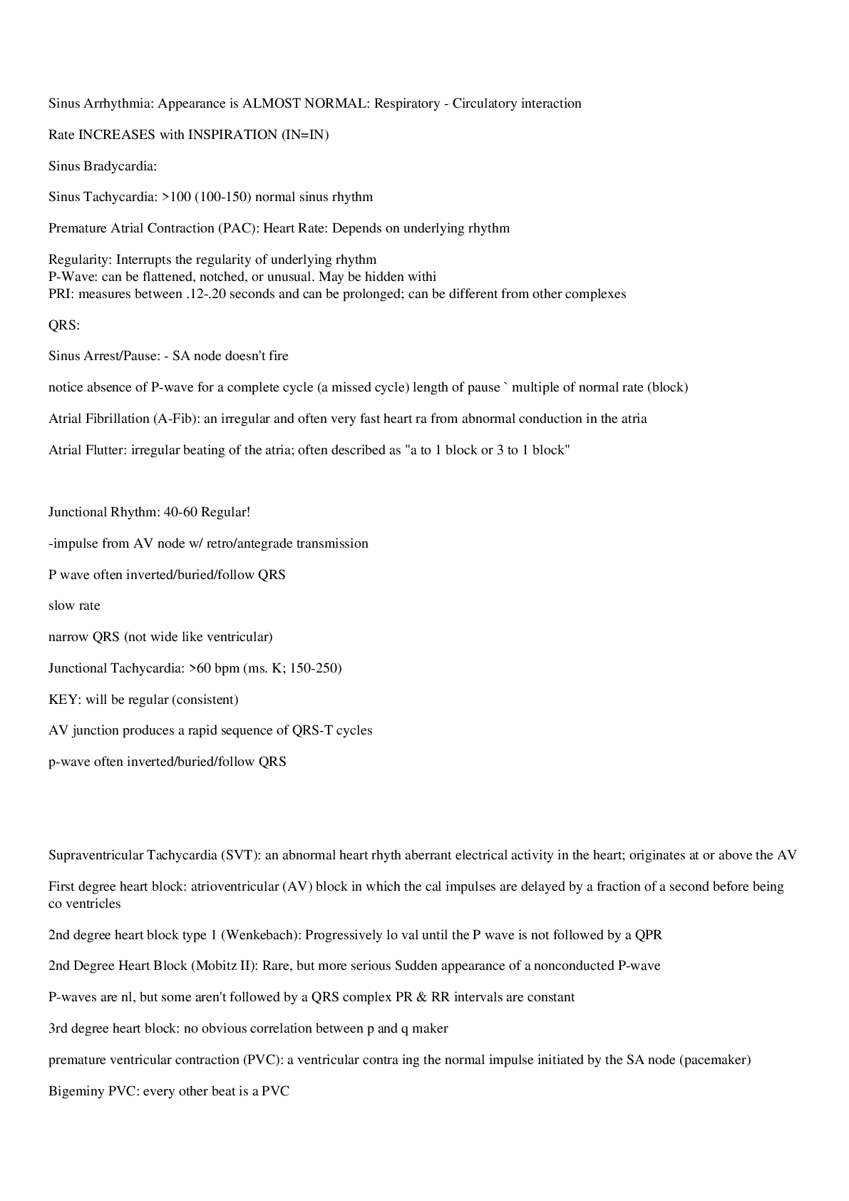
Relias Dysrhythmia Basic Test Answers normal sinus rhythm heart rhythm originating in the sinoatrial node with a rate in patients at rest of 60 to 100 beats per minute Sinus Arrhythmia Appearance is ALMOST NORMAL: Respiratory – Circulatory interaction Rate INCREASES with INSPIRATION (IN=IN) Sinus Bradycardia <60 normal sinus rhythm Sinus Tachycardia >100 (100-150) normal sinus rhythm Premature Atrial Contraction (PAC) Heart Rate: Depends on underlying rhythm Regularity: Interrupts the regularity of underlying rhythm P-Wave: can be flattened, notched, or unusual. May be hidden within the T wave PRI: measures between .12-.20 seconds and can be prolonged; can be different from other complexes QRS: <.12 seconds Sinus Arrest/Pause – SA node doesn’t fire – notice absence of P-wave for a complete cycle (a missed cycle) length of pause ≠ multiple of normal rate (block) Atrial Fibrillation (A-Fib) an irregular and often very fast heart rate originating from abnormal conduction in the atria Atrial Flutter irregular beating of the atria; often described as “a-flutter with 2 to 1 block or 3 to 1 block” Junctional Rhythm 40-60 Regular! -impulse from AV node w/ retro/antegrade transmission – P wave often inverted/buried/follow QRS – slow rate – narrow QRS (not wide like ventricular) Junctional Tachycardia >60 bpm (ms. K; 150-250) – KEY: will be regular (consistent) – AV junction produces a rapid sequence of QRS-T cycles – p-wave often inverted/buried/follow QRS Premature Junctional Contraction Inverted p wave or hidden p wave PRI<0.12 or none Normal QRS Supraventricular Tachycardia (SVT) an abnormal heart rhythm arising from aberrant electrical activity in the heart; originates at or above the AV node First degree heart block atrioventricular (AV) block in which the atrial electrical impulses are delayed by a fraction of a second before being conducted to the ventricles 2nd degree heart block type 1 (Wenkebach) Progressively longer PR interval until the P wave is not followed by a QPR 2nd Degree Heart Block (Mobitz II) Rare, but more serious Sudden appearance of a nonconducted P-wave P-waves are nl, but some aren’t followed by a QRS complex PR & RR intervals are constant 3rd degree heart block no obvious correlation between p and qrs, need pace maker premature ventricular contraction (PVC) a ventricular contraction preceding the normal impulse initiated by the SA node (pacemaker) Bigeminy PVC every other beat is a PVC PVC couplets PVC occurring in pairs, no adequate C.O. when this occurs monomorphic ventricular tachycardia presents with wide QRS complexes of a common shape. Torsades de pointes Rate: 120 – 200 usually P wave: Obscured by ventricular waves QRS: Wide QRS – “Twisting of the Points” Conduction: Ventricular only Rhythm: Slightly irregular Ventricular fibrillation (V-fib) abnormal heart rhythm which results in quivering of ventricles Idioventricular Rhythm <40 *looks like vtach but slow* – no P waves (from vent foci) – Wide QRS (serious, death like rhythm) – called “dying heart” rhythm…occasional ventric beat b4 death (asystole) Accelerated Idioventricular Rhythm Rate: 50 – 100 usually (usually slow) P wave: Obscured by ventricular waves (occur during ventricular contraction) – SA node slower than faster ventricular pacing than should be QRS: Wide QRS Conduction: Ventricular only Rhythm: Regular- benign rhythm that is sometimes seen during acute MI or early after reperfusion. – Rarely sustained, does not progress to vfib, rarely requires treatment asystole absence of contractions of the heart Failure to capture (pacemaker) failure to sense (pacemaker) Atrial paced rhythm spike before P wave Ventricular paced rhythm ventricular contractions which occur in cases of complete heart block. Normal sinus rhythm Regular Rate: 60-100 P Wave: Present, upright PR Interval: 0.12-0.20 sec QRS: <0.12 sec Sinus Bradycardia Regular Rate: <60 P Wave: Present, upright PR Interval: 0.12-0.20 sec QRS: <0.12 sec Sinus Tachycardia Regular Rate: 100-150 P Wave: Present, upright PR Interval: 0.12-0.20 sec QRS: <0.12 sec Premature Atrial Contraction IRREGULAR Rate: depends on underlying rhythm P wave: Present or hidden in T wave PR Interval: 0.12-0.20 sec QRS: <0.12 sec Atrial Fibrillation IRREGULAR Atrial rate: UNMEASURABLE Ventricular rate: variable P wave: unable to see PR Interval: N/A QRS: <0.12 sec A fib RVR IRREGULAR Ventricular rate: 100-175 P wave: unable to see PR Interval: N/A QRS <0.12 sec Atrial Flutter Usually REGULAR can be irregular Atrial rate: 250-350 Ventricular rate: variable BUT < atrial rate P Wave: Flutter PR Interval: N/A QRS: <0.12 sec Supraventricular Tachycardia Regular Rate: 150-350 P wave: Hidden in QRS or T wave PR: unable to determine QRS: <0.12 sec Junctional Rhythm Regular Rate: 40-60 P Wave: ABSENT or INVERTED PR Interval: None or <0.12 QRS: <0.12 sec Accelerated Junctional Rhythm Regular Rate: 60-100 P Wave: NONE or INVERTED PR Interval: None or <0.12 QRS: <0.12 sec Junctional Tachycardia Regular Rate: >100 P Wave: NONE or INVERTED PR Interval: None or <0.12 QRS: <0.12 sec Premature Ventricular Contraction IRREGULAR Rate: refer to underlying rhythm P Wave: NONE PR Interval: N/A QRS: WIDE and BIZARRE , >0.12 sec Ventricular Tachycardia Regular Rate: >100 P Wave: NONE PR Interval: N/A QRS: WIDE and BIZARRE, >0.12 sec Ventricular Fibrillation Chaotic Coarse: big waves Fine: small waves Rate: unmeasurable P Wave: NONE PR Interval: N/A QRS: N/A Idioventricular Regular Rate: 20-50 P wave: NONE PR Interval: N/A QRS: WIDE, >0.12 sec Accelerated Idoventricular Rhythm Regular Rate: 50-100 P wave: NONE PR Interval: N/A QRS: WIDE, >0.12 sec 1st Degree AV Block Regular Rate: 60-100 P Wave: Present, upright PR interval: >0.20 sec CONSISTENTLY LONG QRS: <0.12 secHusband stays late till 9 consistently 2nd Degree AV Block Type I Mobitz, Wenckebach IRREGULAR Rate: 60-100 P wave: Present, upright PR Interval: Progressively longer until drop (PR interval longer and longer until drop) QRS: <0.12 secHusband stays late till 9, then 11, then 1, then doesn’t come home at all 2nd Degree AV Block Type II Irregular or regular Rate: <60 P wave: Present, upright PR Interval: PR interval consistently LONGER like type 1 but then a QRS will drop QRS: <0.12 secHusband stays late till 9 consistently, then wife goes out and doesn’t come home 3rd Degree AV Block Atrials and ventricles don’t communicate Rate: regular atrial P wave: Present, upright No relationship between P waves and QRS PR Interval: VARIABLE QRS: variableP-P ad R-R consistent but NO correlationHusband and wife live separate lives and don’t communicate SA Node 1st 60-100 AV Node 2nd 40-60 Bundle of His 3rd 40-45 Right and Left Bundle Branches 4th 40-45 Purkinje Fibers 5th 20-50 1 Small Box 0.04 sec 1 Big Box 0.20 sec Junctional Rhythms SA Node DID NOT FIRE AV Node fired NO P WAVE bc SA node didn’t fire Narrow QRS P Wave Amplitude 0.5-2.5 mm Will be shorter than T wave Shows firing of SA node QRS 0.06-0.10 sec SHOULD BE <0.12 sec Wide QRS: delay in ventricular contraction, delay of conduction through bundle branches or purkinje fibers BUNDLE BRANCH BLOCK or BLOCK IN PURKINJE FIBERS (idioventricular) Calculate Regular Rate 1500/ # boxes R-R Calculate Irregular Rate # of Rs in 6 sec strip X 10 Unifocal PVCs Only 1 shape PVC Bigeminy PVC occurs every OTHER beat Trigeminy PVC occurs every THIRD beat Couplet 2 PVCs together Triplet 3 PVCs together Multifocal Multiple shapes Monomorphic V Tach Same Shapes V Tach Polymorphic V Tach Different Shapes V tach Coarse V Fib Chopy but not as high as polymorphic V tach Fine V Fib Fine and fibrillatory Idioventricular Rhythms Only purkinje fibers firing WIDE QRS always Atrially Paced Spike comes before P Ventricularly Paced Spike comes before QRS and QRS will be wide AV Paced Spike before P and before QR Failure to Capture Spikes with no QRS Failure to Sense Spikes happen regardless of QRS on their own How to determine the rhythm Regular or irregular? Rate? P before every QRS? QRS for every P? QRS wide or narrow? QT Interval 0.34-0.43 P Wave 0.06-0.12 sec PR Interval 0.12-0.20 sec SA Node Firing Rhythms *Fires normally @ 60-100* -SR 60-100 -SB <60 -ST 100-150 -SVT 150-350 AV Node Firing, SA Node Failed Rhythms *Fires normally @ 40-60* -Junctional rhythm 40-60 -Accelerated junctional rhythm 60-100 -Junctional tachycardia 100-150 Only Purkinje Fibers Firing Rhythms (Everything else has failed) *Fires normally @ 20-50* -Idioventricular 20-50 -Accelerated idioventricular 50-100
[Show More]