NURSING > AQA QUESTION and MARK SCHEMES > Dysrhythmias Questions With Correct Answers (All)
Dysrhythmias Questions With Correct Answers
Document Content and Description Below
Last updated: 3 years ago
Preview 1 out of 8 pages
Instant download
Buy this Document to get the Full Access Instantly
Provided by Students Who Aced it
We Verify Document Content to Gurantee Accuracy
Reviews( 0 )
Document information
Connected school, study & course
About the document
Uploaded On
Apr 05, 2023
Number of pages
8
Written in
All

Additional information
This document has been written for:
Uploaded
Apr 05, 2023
Downloads
0
Views
137
Document Keyword Tags
Recommended For You
Get more on AQA QUESTION and MARK SCHEMES »![Preview of eBook [PDF] Barash, Cullen, and Stoelting's Clinical Anesthesia 9th Edition By Bruce F. Cu](https://browseimages.nyc3.digitaloceanspaces.com/paper-images/2024/Aug/29/8DF2QRTq2024-08-29-10-3966d0259b7c4b5.png)
eBook [PDF] Barash, Cullen, and Stoelting's Clinical Anesthesi...
![Preview of eBook [PDF] Fundamentals of Nursing 3rd Edition By Joanne Tollefson, Sue C. DeLaune, Patri](https://browseimages.nyc3.digitaloceanspaces.com/paper-images/2024/Aug/26/U6SQ8mmq2024-08-26-01-4766cc5d40d7385.png)
eBook [PDF] Fundamentals of Nursing 3rd Edition By Joanne Toll...
![Preview of eBook [PDF] Burns' Pediatric Primary Care 7th Edition BY Dawn Lee Garzon Maaks,, Nancy Bar](https://browseimages.nyc3.digitaloceanspaces.com/paper-images/2024/Aug/26/WR2rowXX2024-08-26-02-0966cc625bea839.png)
eBook [PDF] Burns' Pediatric Primary Care 7th Edition BY Dawn...

Test Bank for Burns' Pediatric Primary Care 7th Edition by Daw...
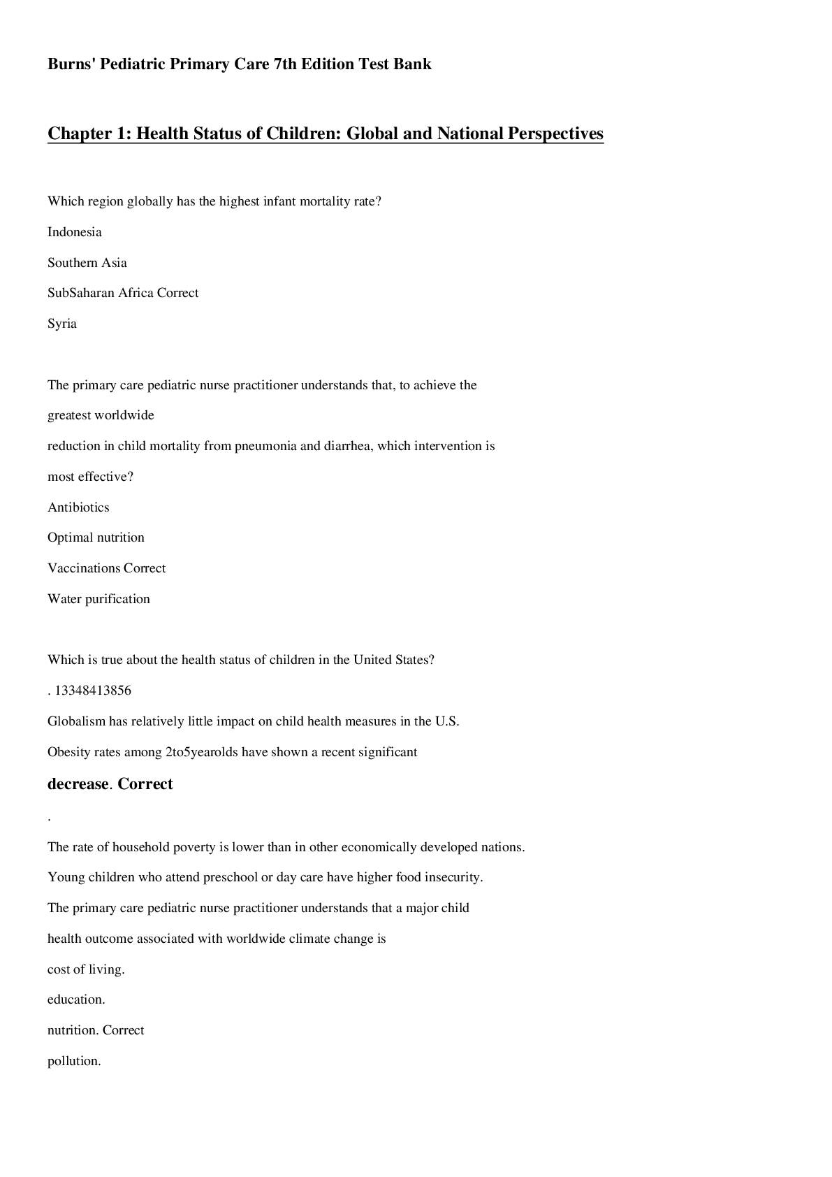
Burns Pediatric Primary Care 7th Edition All Chapters Complete...
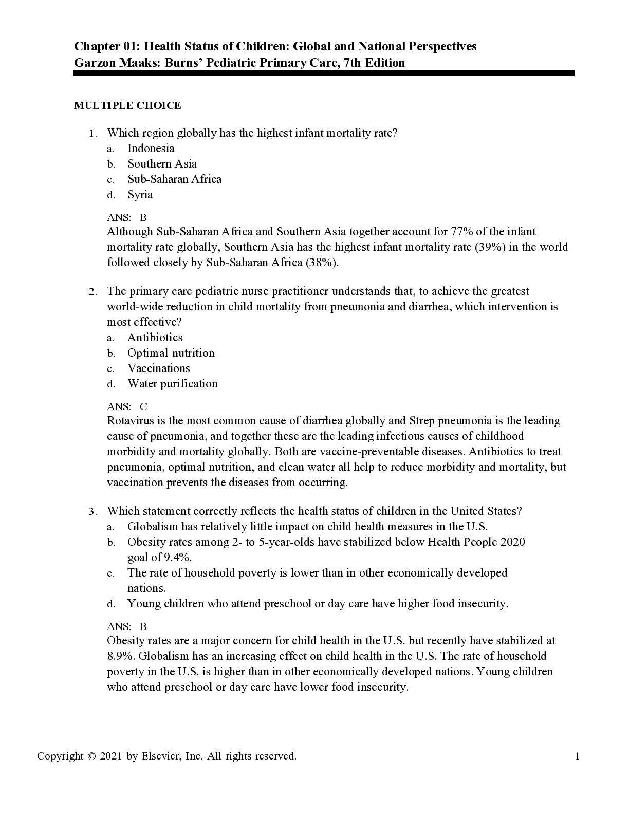
Burns' Pediatric Primary Care, 7th Edition by Garzon, Barber S...
Complete Test Bank Emergency Care 14th Edition Daniel Limmer Q...
Test Bank Emergency Care 14th Edition Daniel Limmer Questions...

![Preview of eBook [PDF] AACN Essentials of Progressive Care Nursing 5th Edition By Sarah Delgado](https://browseimages.nyc3.digitaloceanspaces.com/paper-images/2025/Aug/06/lAjhFV4A2025-08-06-09-286892f5fd83090.png)
eBook [PDF] AACN Essentials of Progressive Care Nursing 5th Ed...
![Preview of [eTextbook] [PDF] Labor Economics 9th Edition By George Borjas](https://browseimages.nyc3.digitaloceanspaces.com/paper-images/2025/Jan/13/JcsHZtUb2025-01-13-07-42678542822cb77.png)
[eTextbook] [PDF] Labor Economics 9th Edition By George Borja...
AACN Essentials of Progressive Care Nursing, 5th Edition by Sa...
![Preview of [eTextbook] [PDF] Our Sexuality Enhanced Edition, Loose-leaf Version 14 Edition By Robert](https://browseimages.nyc3.digitaloceanspaces.com/paper-images/2025/Jan/11/dWqGQ6Ng2025-01-11-11-566782db1b759b0.png)