Surgery > QUESTIONS & ANSWERS > Surgery Week Quiz Series - Part 3 [GRADED A+] (All)
Surgery Week Quiz Series - Part 3 [GRADED A+]
Document Content and Description Below
Last updated: 3 years ago
Preview 1 out of 29 pages
Instant download
![Preview of Surgery Week Quiz Series - Part 3 [GRADED A+]](https://browseimages.nyc3.digitaloceanspaces.com/paper-images/2023/May/08/p5eBDU5s2023-05-08-03-286458eb059216e.png)
Buy this Document to get the Full Access Instantly
Provided by Students Who Aced it
We Verify Document Content to Gurantee Accuracy
Also available in bundle (1)
Click Below to Access Bundle(s)

Surgery Week Quiz Series - Part 1 to 10. [GRADED A+]
Surgery week 10 quiz Q # 1 of 39- 1 Points USMLE Step 2 CK Board Preparation 1. A sexually active woman with multiple partners presents with a 2-day history of red swollen left knee. On examinatio...
By geeks4geeks 3 years ago
$35
10
Reviews( 0 )
Document information
Connected school, study & course
About the document
Uploaded On
May 08, 2023
Number of pages
29
Written in
All

Additional information
This document has been written for:
Uploaded
May 08, 2023
Downloads
0
Views
134
Document Keyword Tags
Recommended For You
Get more on QUESTIONS & ANSWERS »![Preview of Surgery Week Quiz Series - Part 10 [GRADED A+]](https://browseimages.nyc3.digitaloceanspaces.com/paper-images/2023/May/08/zYyq8wl72023-05-08-03-386458ed5846f08.png)
![Preview of Surgery Week Quiz Series - Part 9 [GRADED A+]](https://browseimages.nyc3.digitaloceanspaces.com/paper-images/2023/May/08/ogTzGzKW2023-05-08-03-376458ed074b482.png)
![Preview of Surgery Week Quiz Series - Part 8 [GRADED A+]](https://browseimages.nyc3.digitaloceanspaces.com/paper-images/2023/May/08/lzaPhOBf2023-05-08-03-366458ecb438266.png)
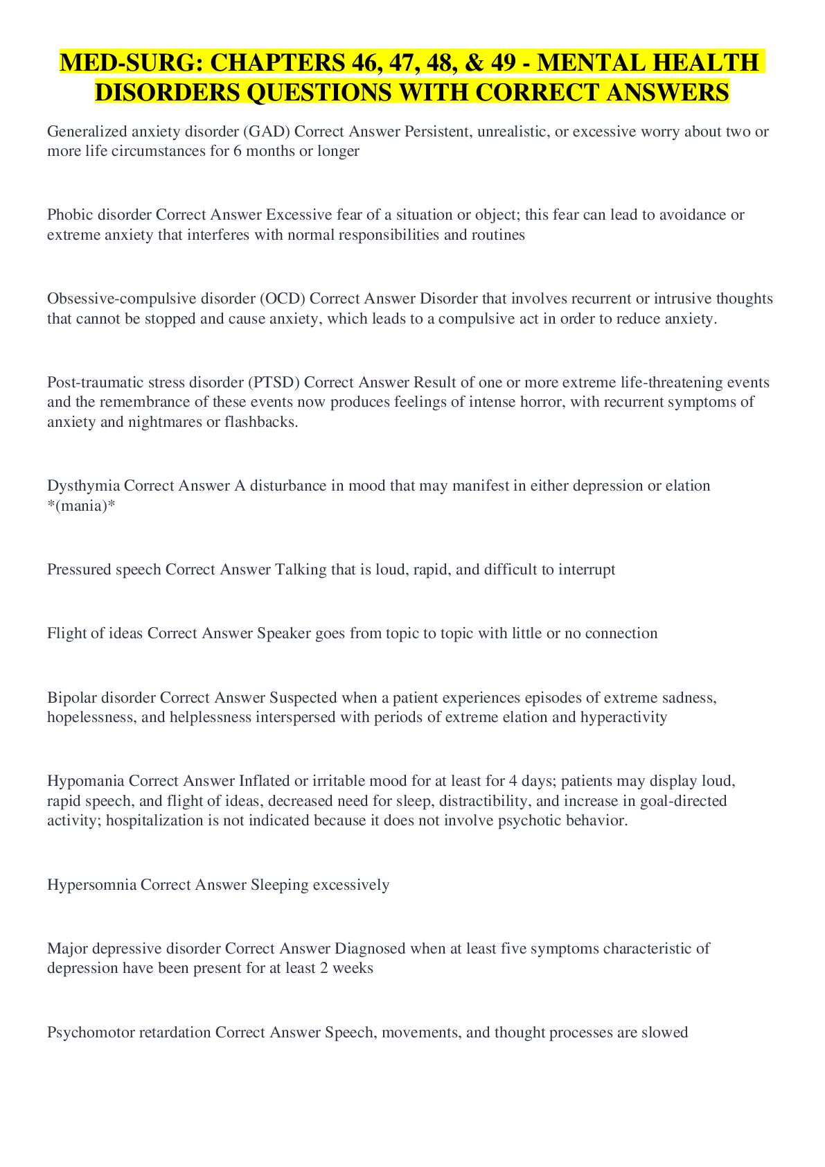
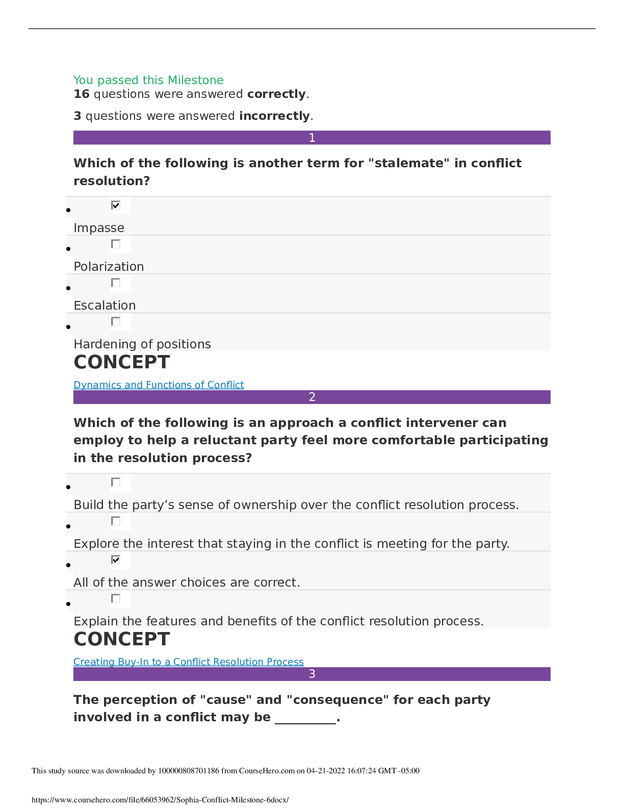
MED-SURG: CHAPTERS 46, 47, 48, & 49 - MENTAL HEALTH DISORDERS...

MED SURG 2022 HESI PRACTICE QUESTIONS WITH CORRECT ANSWERS|GRA...

MED-SURG II HESI TEST BANK|133 QUESTIONS WITH 100% CORRECT ANS...
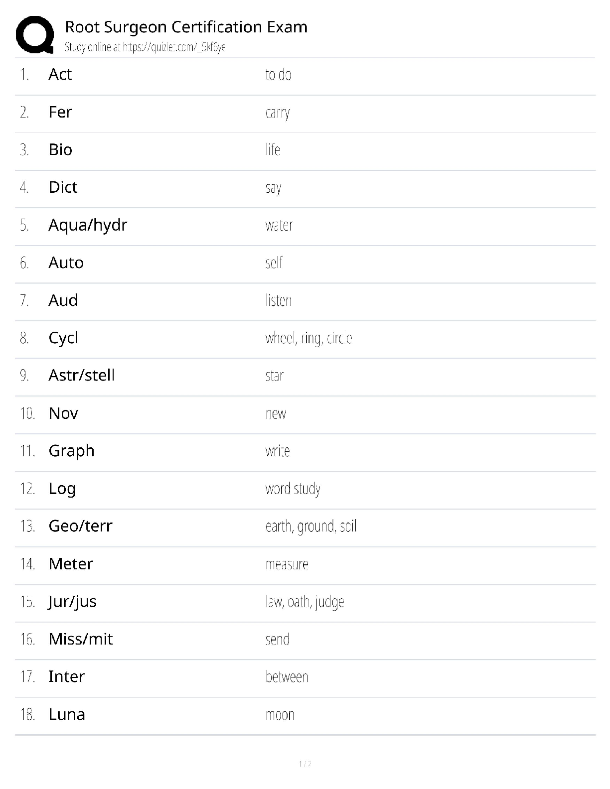
Root Surgeon Certification Exam / RSI Practice Test / 2025 Upd...
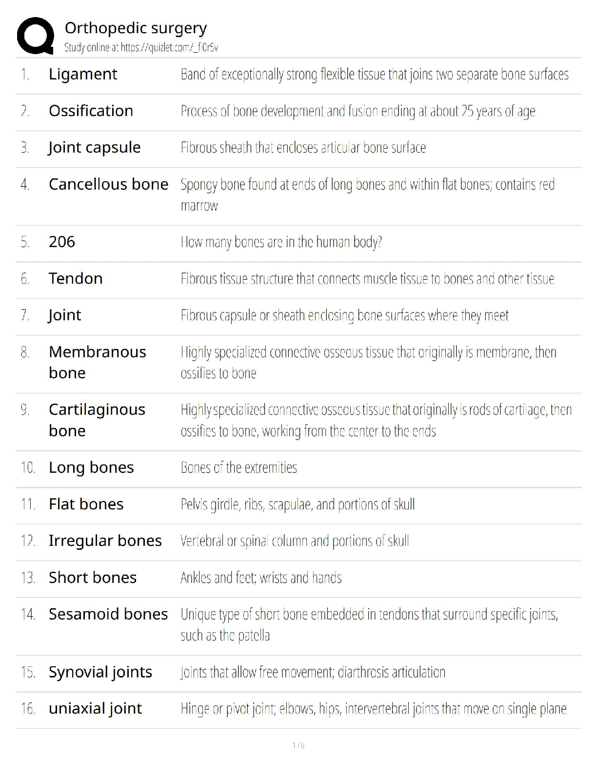
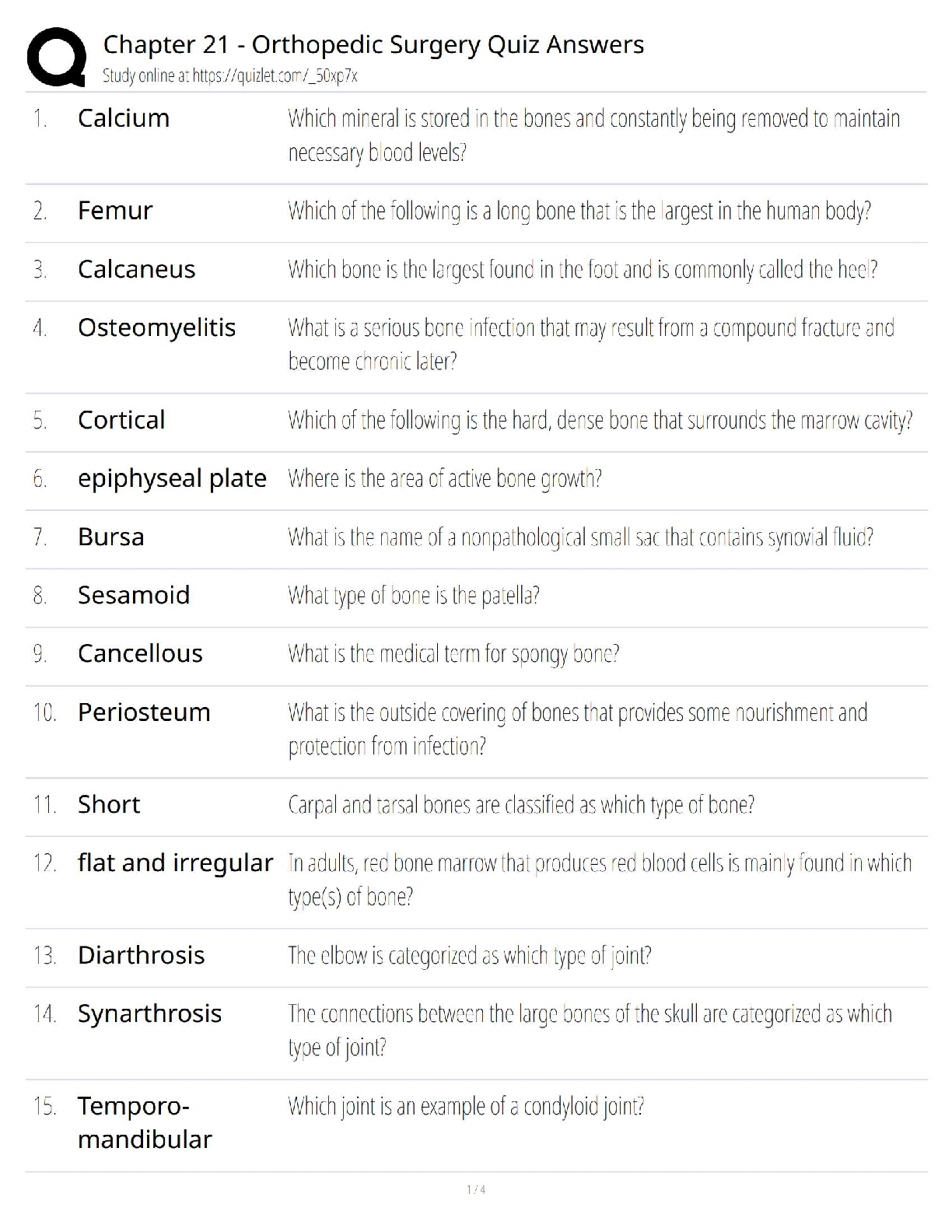
Chapter 21 Orthopedic Surgery Quiz Answers / NCLEX Review / 20...
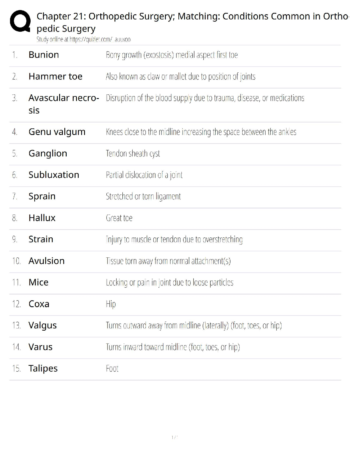
Chapter 21 Orthopedic Surgery / Matching Conditions Common in...
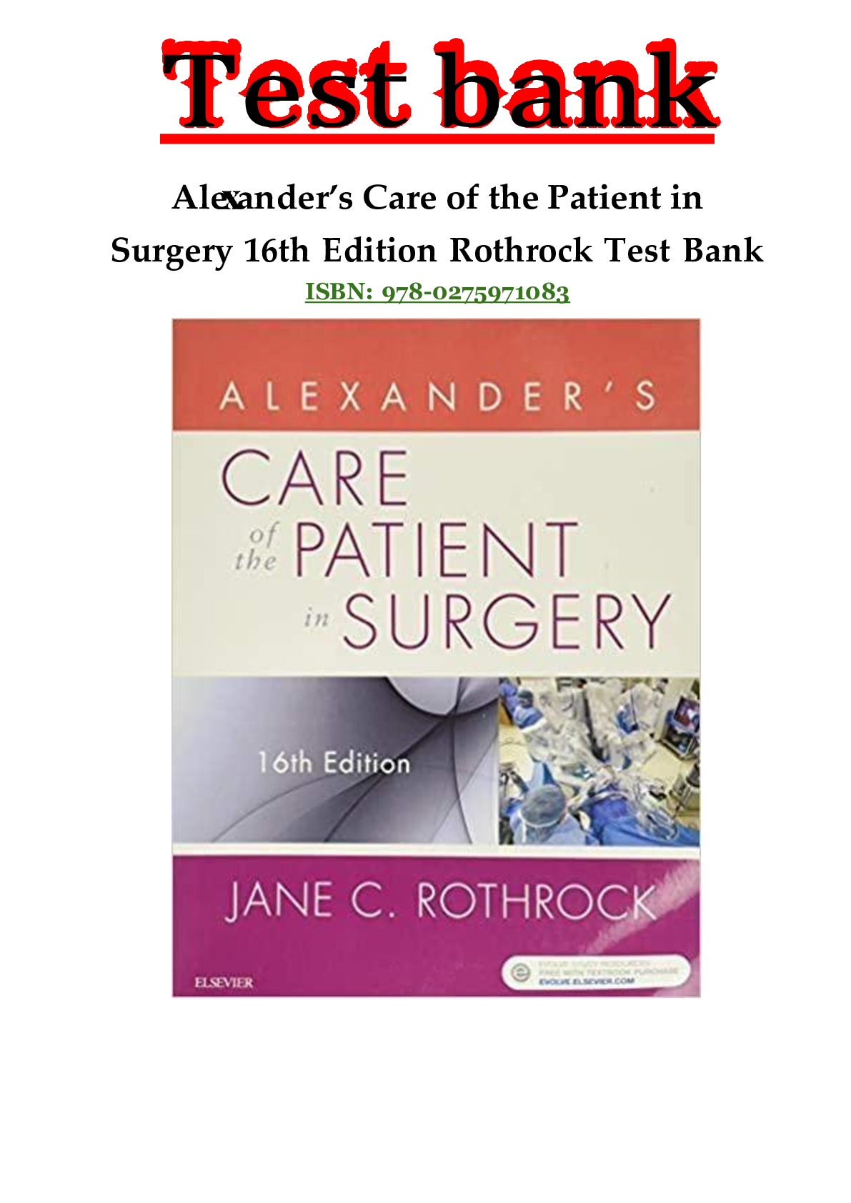
Complete Test Bank Alexander’s Care of the Patient in Surgery...
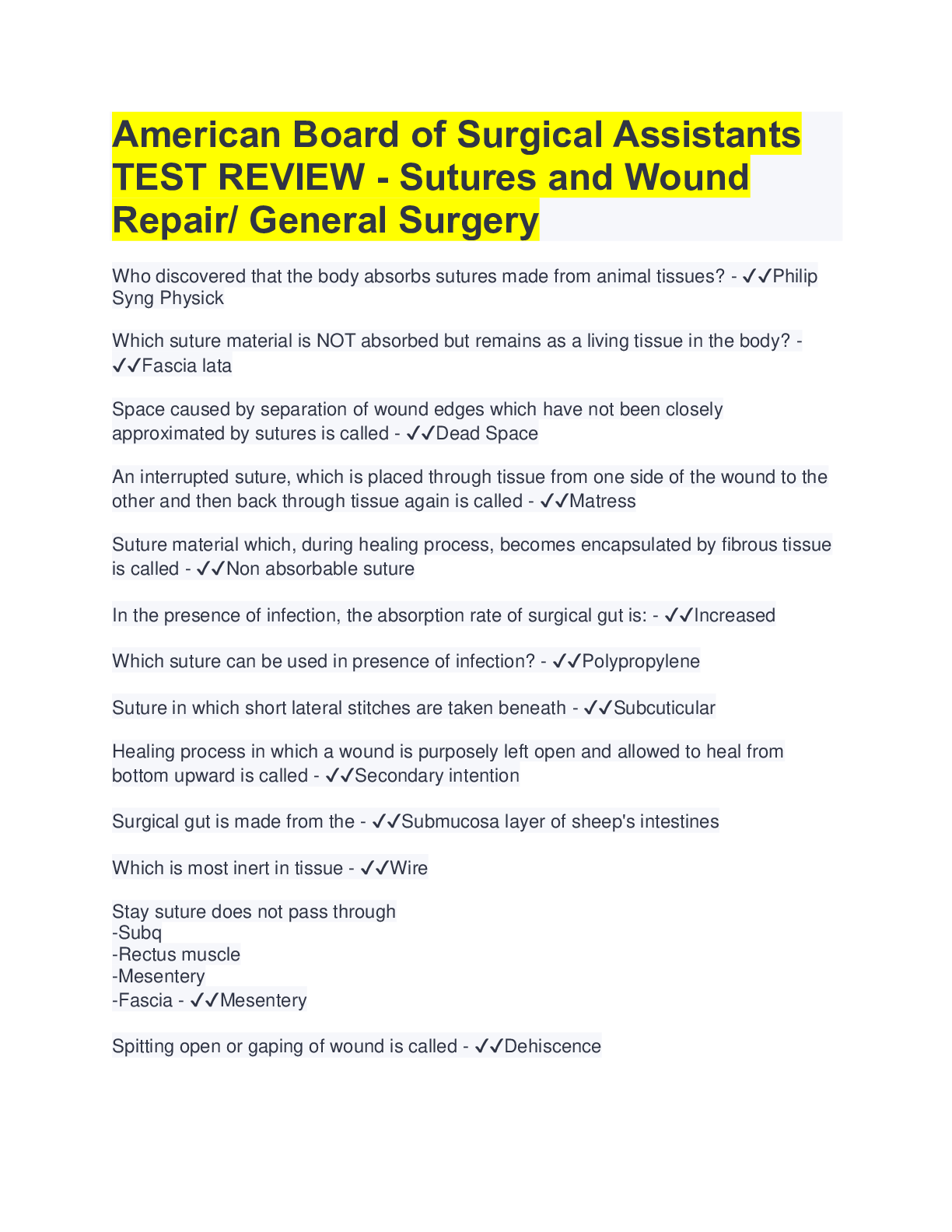
American Board of Surgical Assistants TEST REVIEW - Sutures an...

CST Certification Surgical Specialties / General Surgery Guide...

Cardiology/Cardiac Surgery/Thoracic Surgery Q&A 2022/2023 (Dis...
Test Bank for Surgical Technology 7th Edition by Joanna Kotche...

Complete e-book PDF: Master Techniques in Orthopedic Surgery:...


![Preview of The Genesis of Creation WEEK 1 QUESTIONS with ALL CORRECT SOLUTIONS [GRADED A+]](https://scholarfriends.com/storage/Wk_1_Study_Questions.png)
![Preview of The Creation and Fall of Man WEEK 2 QUESTIONS with ALL CORRECT SOLUTIONS [graded A+]](https://scholarfriends.com/storage/Weekly_Study_Questions_2.png)

![Preview of Essentials in Nutrition EXAM QUESTION AND ANSWERS [ALL CORRECT SOLUTIONS]](https://scholarfriends.com/storage/Case_Study_Module_4.png)
![Preview of NURSING TEST BANK [GRADED A+] [CORRECT SOLUTIONS]](https://scholarfriends.com/storage/Board_Exam_Nursing_Questions_and_Answers_Test_5.png)
![Preview of NURSING TEST BANK [GRADED A+] [CORRECT SOLUTIONS]](https://scholarfriends.com/storage/Board_Exam_Nursing_Questions_and_Answers_Test_4.png)
![Preview of NURSING TEST BANK [GRADED A+] [CORRECT SOLUTIONS]](https://scholarfriends.com/storage/Board_Exam_Nursing_Questions_and_Answers_Test_3.png)
![Preview of NURSING TEST BANK [GRADED A+] [CORRECT SOLUTIONS]](https://scholarfriends.com/storage/Board_Exam_Nursing_Questions_and_Answers_Test_2.png)
![Preview of NURSING TEST BANK [GRADED A+] [CORRECT SOLUTIONS]](https://scholarfriends.com/storage/Board_Exam_Nursing_Questions_and_Answers_Test_1.png)
![Preview of GIZMOs : Archimedes’ Principle - Answer Key [GRADED A+] [TOP RATED]](https://scholarfriends.com/storage/GIZMOs Archimedes’ Principle - Answer Key [TOP RATED].png)
![Preview of GIZMO Coral Reefs 2 Biotic Factors [ALL ANSWERS CORRECT] [GRADED A+]](https://scholarfriends.com/storage/Crump_13.png)