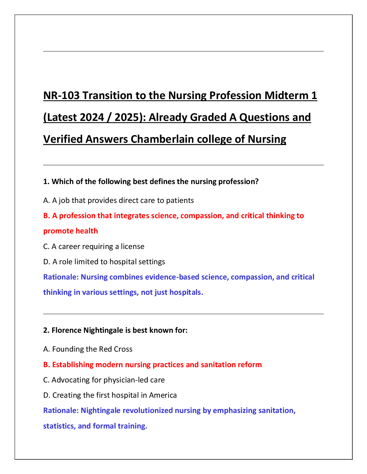
NURSING 265 Adv MedSurg Exam2-1 Questions with Answers,100% CORRECT
Document Content and Description Below
NURSING 265 Adv MedSurg Exam2-1 Questions with Answers
CARE OF PATIENTS WITH COMPLEX RESPIRATORY PROBLEMS
Structures of the lungs
- Trachea, left/right bronchus, segmental bronchus, subsegmental
...
bronchus, alveoli
- Visceral pleura/parietal pleura lubrication
- Right side of lung (3 lobes)
o Usually aspirate on this side r/t longer/straighter airway Gas Exchange Structures
- Bronchiole, terminal bronchiole, respiratory bronchioles, alveoli The Alveoli
- Have about 290 million
- Type 2 pneumocytes secrete surfactant (fatty protein) to keep the alveoli open and keep fluid away from alveoli
Gas Exchange
- Breath O2 in O2 goes into blood stream CO2 releases from blood stream blow CO2 out COPD: effect on lungs
- Healthy alveoli expand and contract giving adequate perfusion
- COPD alveoli have lost elasticity and rely on the impulse from the brain when the CO2 in their blood is too high causing their drive to breath to happen automatically (like kussmaul respirations)
o COPD consists of Emphysema and Chronic Bronchitis
▪ Causing bronchial spasms and dyspnea Bronchitis and Emphysema
- Chronic Bronchitis
o Caused by smoking, characterized by inflammation and structural changes
o Causes excessive secretions (mucous plug)
- Emphysema
o Elastic fibers destroyed leading to hyperinflation
Acute Respiratory Failure
- Progressive or sudden
- Deterioration of gas exchange function in the lungs
o Hypoxemia – PaO2 of less than 50 mmHg (normal 80-100)
o Hypercapnia – PaCO2 greater than 50 mmHg
▪ Decreased LOC if this happens call the Doc to get blood gas
o Acidosis – pH less than 7.35 (normal 7.35 – 7.45)
- Ventilatory failure – Can’t get O2 in
o Asthma, sleep apnea, myasthemia gravis
- Oxygen failure – O2 getting in but it isn’t getting picked up
o Pneumonia, ARDS, PE, shock
CORRECT
respiratory excursion. Clients with cervical and thoracic spinal cord injuries are at high risk for respiratory failure because spinal nerves that affect intercostal muscles are affected. Opiates, which can depress the brainstem, present risk factors for respiratory failure. All of these clients should be monitored closely for respiratory failure. Cocaine is a stimulant, which would not cause respiratory failure unless a stroke ensued.
CORRECT
• Client with acute pancreatitis
Pressure on the brainstem may depress respiratory function. Acute pancreatitis is a risk factor for acute respiratory distress syndrome; abdominal distention also ensues, which can limit respiratory excursion. Clients with cervical and thoracic spinal cord injuries are at high risk for respiratory failure because spinal nerves that affect intercostal muscles are affected. Opiates, which can depress the brainstem, present risk factors for respiratory failure. All of these clients should be monitored closely for respiratory failure. Cocaine is a stimulant, which would not cause respiratory failure unless a stroke ensued.
CORRECT
• Client with a T3 spinal cord injury
Pressure on the brainstem may depress respiratory function. Acute pancreatitis is a risk factor for acute respiratory distress syndrome; abdominal distention also ensues, which can limit respiratory excursion. Clients with cervical and thoracic spinal cord injuries are at high risk for respiratory failure because spinal nerves that affect intercostal muscles are affected. Opiates, which can depress the brainstem, present risk factors for respiratory failure. All of these clients should be monitored closely for respiratory failure. Cocaine is a stimulant, which would not cause respiratory failure unless a stroke ensued.
CORRECT
• Client using patient-controlled analgesia
Pressure on the brainstem may depress respiratory function. Acute pancreatitis is a risk factor for acute respiratory distress syndrome; abdominal distention also ensues, which can limit respiratory excursion. Clients with cervical and thoracic spinal cord injuries are at high risk for respiratory failure because spinal nerves that affect intercostal muscles are affected. Opiates, which can depress the brainstem, present risk factors for respiratory failure. All of these clients should be monitored closely for respiratory failure. Cocaine is a stimulant, which would not cause respiratory failure unless a stroke ensued.
-
Blood Gas Values
- pH = 7.35 – 7.45, pCO2 = 35 – 45 (respiratory), HCO3 = 22 – 28 (metabolic)
- Increased CO2 = acid build-up, acidosis; Increased HCO3 = alkaline build-up, alkalosis
- ROME (Respiratory Opposite, Metabolic Equal) Pathophysiology of Respiration
- Occurs at the alveolar capillary units exchange of oxygen and carbon dioxide oxygen attaches to the circulating hemoglobin molecules 2 processes occur, ventilation and perfusion
- V/Q scan measures how well the alveoli are being ventilated and perfused
o Radioactive dye used to find PE
o Ventilation – perfusion mismatch = PE Causes of Acute Respiratory Failure
- Decreased respiratory drive (narcotics, COPD w/ too much O2)
- Obstruction of the airways (Bronchitis, sleep apnea, asthma)
- Trauma (Injury to the lung tissue or chest wall)
- Dysfunction of the chest wall (spinal cord injury, any condition that affects breathing)
- Disorders (sleep apnea, PE, overdose of opiods/alcohol) Clinical Manifestations of Acute Respiratory Failure
- Early: Impaired O2 (give O2), restlessness, fatigue (promote rest), headache, dyspnea, air hunger, tachycardia, increased BP
o Use interventions
- Progressive: Confusion, lethargy, tachycardia, tachypnea, central cyanosis, diaphoresis, respiratory arrest
o Call rapid response
- Intervention Rapid response ICU Medical Management
- Increased oxygenation, intubation, mechanical ventilation, ICU, bronchodilators, antibiotics, anti- inflammatories
Nursing Management
- Anticipate and assist with intubation
- Monitor (assess): LOC, RR, O2, ABGs continuous pulse oximetry
- Prevent ventilator associated pneumonia
Acute Respiratory Distress Syndrome (ARDS)
- Severe form of acute lung injury, usually results in death
- Starts with Acute Respiratory Failure sudden, progressive pulmonary edema with increasing bilateral infiltrates in lungs
- Refractory hypoxemia – giving pt 100% FiO2 but it isn’t making a difference in O2 stat
- Reduced lung compliance
Causes of Acute Respiratory Distress Syndrome (ARDS)
- Aspiration – acid destroys alveoli/surfactant leads to inflammation
- Drug ingestion and overdose
- Hematologic disorders (DIC massive transfusions)
o TRALI – Transfusion related acute lung injury
- Prolonged inhalation of smoke or corrosive substances, near drowning
- Infection (pneumonia)
- Metabolic disorders
- Shock, trauma, major surgery Clinical Manifestations
- Rapid onset of severe dyspnea
- Arterial hypoxemia
o ABG’s show respiratory acidosis
- Bilateral infiltrates in lungs white out on x-ray
- Intercostal retractions
- Persistent, severe hypoxemia
- Increased alveolar dead space Alveoli collapsed (Atelectasis)
- Hallmark sign refractory hypoxemia 100% O2 no change on ABGs
Medical Management
- Intubation ALWAYS!
o Aggressive supportive care
o If pt declines intubation give Bipap
- Mechanical ventilation
o PEEP – Positive End Expiratory Pressure
▪ Pushes air in to open up alveoli, helps push fluid out of alveoli
▪ Watch for pneumothorax (too much pressure can push air into chest wall causing air to push up against your lungs causing a collapsed lung)
o Prone position (on stomach) to improve ventilation perfusion
- Circulatory support
o Adequate fluid volume
▪ Fluid and diuretics to maintain proper fluid volume
- Nutritional support
o 35-45 kcal/kg per day
o Enteral feeding feeding through gut keeps everything working so you don’t have peristalsis
Pharmacologic Therapy
- Pulmonary specific vasodilators (Nitric Oxide)
- Corticosteroids (to decrease inflammation)
- Anti-inflammatories, Antibiotics
- Surfactant replacement (replace type 2 pneumocytes) Understanding Acute Respiratory Distress Syndrome (ARDS)
- Starts with lung injury
o Antibiotics, anti- inflammatory agents
- Release of vasoactive substances (lungs fill up with fluid)
- Increased permeability of alveoli
- Fluid and protein move into alveoli
o Crackles, dyspnea, tachypnea, hypoxemia r/t fluid build up, possible mechanical ventilation
- Altered ventilation, perfusion
o Use prone position
- Early on CO2 may be in normal range until fluid builds up, O2 goes down early
- Damage to alveolar epithelium
o Causes hemoptysis (coughing up blood)
- Decreased surfactant production
o Decreases compliance of lungs, may need to administer surfactant
- Atelectasis occurs (collapse of alveoli)
o Mechanical ventilation with PEEP (keeps alveoli open, pushes fluid out of alveoli)
- Eventually scarring occurs with loss of functional lung tissue once fibrotic highland membrane is formed, lung damage is permanent
- Interdisciplinary team for this pt consists of social work, resp therapist, pulmonologist, palliative care, dietary
Pulmonary Hypertension
- Can lead to right sided HF Cor Pulmonale
- Increased blood pressure and vasoconstriction in the pulmonary vasculature
- May be arterial, venous, hypoxic, or thromboembolic
- Causes: lung disease, obesity, sleep apnea, narcosis
- Leads to non-ischemic hypertensive cardiomyopathy and right-sided heart failure
Clinical Manifestations
- Signs of right-sided heart failure (Cor Pulmonale)
o SOB (especially with exertion), chest pain, JVD, peripheral edema
- Decreased O2 saturation
- Loud S2 heart sound
Medical Management
- Oxygen, vasodilators, diuretics for edema
- HTN control
o ACE inhibitors, Calcium Channel Blockers
- DVT prophylaxis
- Pain management
- Treat the underlying problem Nursing Management
- Monitor VS every 4 hours, and EKG
- I+O to prevent Cor Pulmonale
- Pain assessment
- Monitor electrolyte levels
- Titration of critical care infusions
- Activity, allow for rest periods, cluster care if possible
Cor Pulmonale
- Pulmonary heart disease
- Originates in the lungs and eventually damages the right side of heart from increased pressure
- Acute – rapid dilation of right ventricle; caused by too much right ventricular stretch
- Chronic – progressive; results in right ventricular hypertrophy
- Can lead to left sided heart failure and death
Causes:
- COPD – Most common
- Deformities of thoracic cage
- Injuries to chest wall; tissue damage
- Chemical agents
- Massive obesity
- PE
- Disorders in the nervous system, respiratory muscles, chest wall, and pulmonary tree Clinical Manifestations
- Peripheral edema, JVD, enlarged liver, ascites, pleural effusion (buildup of fluid between the tissues that line the lungs and the chest), cough, SOB, wheezing, crackles, heart murmurs, fatigue, headache, confusion, Ruddy (greyish/reddish, blotchy skin r/t hypoxemia)
Medical Management
- Treat underlying cause (Supplemental O2, Intubation if necessary, sodium restriction, diuretic therapy, bed rest to promote diuresis)
Pharmacologic Therapy
- Nitrates, diuretics, digitalis, bronchodilators, calcium channel blockers, anticoagulants, inotropes (dobutamine, dopamine, digitalis)
Nursing management
- Client teaching, fluid balance, daily weights, edema, nutrition, sodium restrictions, diuretics, smoking cessation
Pulmonary Embolism (PE) Respiratory Alkalosis
- Thrombus, air, fat (from breaking femur), amniotic fluid
- Clot travels into pulmonary circulation
- Complete or partial obstruction of pulmonary blood vessels
- Increased risk with:
o Prolonged bed rest, after surgery, smokers, cancer (r/t immobilization, meds), venous stasis (make sure pt wearing TED hose, possible SubQ Heparin, do ROM exercises), A fib (blood just sitting in the atrium), obesity
- Potentially lethal, depends on amount of occlusion and location
Clinical Manifestations
- Dyspnea, tachypnea, Sudden SOB, cough, (inspiratory)chest pain, anxiety, petechiae (across chest and upper body), fever, diaphoresis, syncope, hemoptysis (coughing up blood, indicates alveolar damage), crackles, tachycardia, (hypotension dizziness and fainting) low CO2
o If pt has JVD, syncope, cyanosis, and hypotension or dyspnea and chest pain call rapid responses, elevate HOB, and put on O2
o It is important to remember that many pts with PE do not have the “classic”
manifestations but instead have vague symptoms resembling the flu, such as nausea, vomiting, and general malaise.
Diagnostic Tests for PE:
- Ct scan and ABGs
-
- V/Q scan (for definite diagnosis)
o Radioactive compound inhaled into airspace of lung (normal lung distributes equally)
o Radioactive compound injected into vein. Travels to lung tissues in blood vessels
▪ If there is a clot in an artery the dye will stop short of the end of the artery
o Mismatch of inhaled and injected compounds on the lung scan images = PE
- D-dimer (tells you if pt has any blood clots, but not specific to PE) Prevention is KEY!
- Anticoagulants (heparin/lovenox), SCD’s/TED hose, early ambulation after surgery, frequent leg exercises, teach pt not to massage or cross legs
- Client with a diagnosed pulmonary embolism who is receiving IV heparin and has bright-red
hemoptysis
-
Medical Management
- Emergency management
o Pt may be unstable and in shock with hypotension
- Oxygen
- Pharmacologic therap
o Heparin (monitor ptt/appt)
▪ Wait 6 hrs after first dose, get ptt, titrate, wait 6 hours, get ptt
▪ Pt needs 2 normal ptt’s to be switched to maintenance therapy
▪ Occult test to monitor for bleeding
▪ Antidote for heparin is protamine sulfate
▪ Therapy with heparin and warfarin continues until INR is 2.0-3.0
o Warfarin
▪ Antidote is Vitamin K
o Thrombolysis – tPA
▪ Used with massive PE obstructing blood flow to lobe or more than one segment and when BP cannot be maintained with supportive measures
- Other considerations
o Early on: alkalosis r/t hyperventilation
o Progresses into acidosis r/t lactic acid buildup Nursing Management
- Monitor thrombolytic therapy (bleeding no tPA within 1 week of surgery or with head bleed)
- Assess and manage pain, monitor O2 levels, relieve anxiety
- Post op care
o Assess sit for hematoma or infections, use incentive spirometer, VS (to monitor for bleeding and fluid volume), dressing changes, pain (RR), increased HR
- Home care and d/c intructions
o Warfarin - INR (2-3)
▪ Use soft tooth brush and electric razor
Heparin monitor platelet count daily due to side effects of thrombocytopenia Venous Air Embolism
- Entry of air into the venous system above the right atrium
- Can occur with trauma, central line insertion, surgical procedures of head and neck
o Pulling central line – Pt laying down, use Vaseline gauze, hold pressure. Instruct pt to take deep breath and hold it, then pull line. If on ventilator, pull line on inspiration
- Manifestations develop immediately, severity depends on amount of air
Manifestations of Air Emboli
- Dyspnea, chest pain, tachycardia, heart murmur, hypotension, decreased LOC, circulatory shock, sudden death, pt may turn blue
*Reduce the risk of air emboli by carefully priming all IV tubing. Secure all connections in central lines and protect them from becoming dislodged when the client moves around*
Pleural Effusion
- Accumulation of fluid in the pleural space
- Types of fluid:
o Transudate – substances that pass through the membrane
o Exudate – substances that have escaped from blood vessels Clinical Manifestations
- Dyspnea, decreased or absent lung sounds, dull flat sound on percussion, pleural friction rub Treatment
- Fluid is removed from the pleural space by a thoracentesis
o Serous fluid (hydrothorax), Blood (hemothorax), Chyle/lymph (chylothorax), Pus (pyothorax/empyema)
Medical Management
- Thoracentesis
o Be careful how much fluid is pulled
o Tell pt to avoid coughing or deep breathing and not to make any sudden movements Nursing Management
- Assist with thoracentesis
o Send specimens to lab, gather sterile gown, gloves, mask, and hair net, monitor breath sounds (no breath sounds call Dr. for chest tube) VS, assess the site, chest x-ray after procedure to check for pneumothorax
- Assist with chest tube insertion
o Set up chest drainage system, monitor chest drainage, monitor for subcutaneous emphysema (crackles under skin)
Pulmonary Edema
- Classifications
o Cardiogenic – caused by L ventricular failure, mitral valve stenosis, hypertension, cardiomyopathy
o Non cardiogenic – caused by aspiration, sepsis, inhalation of toxic chemicals, near
drowning, smoke inhalation, pneumonia
o Neurogenic – caused by injury to CNS from head trauma
- Abnormal cardiac function
o Orthopnea – trouble breathing when lying down
- Backup of blood into pulmonary vasculature
o Pink, frothy sputum
- Increased microvascular pressure
- Fluid leak into interstitial space, alveoli
- Feeling of impending doom Clinical Manifestations
- Increased dyspnea, air hunger, central cyanosis, agitation, frothy blood tinged sputum, confused, decreased LOC
- Assessment: crackles in bases of lungs, tachycardia, falling pulse ox readings, hypoxemia Nursing Management
- Oxygen, assist with intubation, mechanical ventilation, diuretics, morphine, foley catheter
Pneumothorax
- Any chest injury that allows air into pleural space
o Usually caused by blunt chest trauma Clinical Manifestations
- Reduced breath sounds, affected side moves poorly, tracheal deviation (deviates to the good side unless lungs is punctured somehow then it deviates toward the side of the injury r/t pressure), pleuritic pain, tachypnea, SubQ emphysema/crepitus (skin will crackle when you press on it r/t gas or air under the skin)
Diagnosis
- Ultrasound or x-ray Treatment
- Chest tube may be needed to allow the air to escape and the lung to re-inflate
Tension Pneumothorax
- Results from an air leak in the lung or chest wall. Air forces into the chest cavity causes complete collapse of the affected lung. Air that enters the pleural space during inspiration does not exit during expiration
o PEEP, chest tube, trauma
- If not promptly detected and treated, it is quickly fatal (can develop shock) Clinical Manifestations
- Asymmetry of thorax (one side smaller than the other), tracheal deviation, respiratory distress, absence of breath sounds on one side, JVD (pressure built up on the heart causes neck veins to distend), cyanosis
Diagnosis
- Detectable on chest x-ray, ABGs will show hypoxia and respiratory alkalosis Treatment
- A large-bore needle is inserted into affected side initially, then a chest tube is placed until lung re- inflates
Hemothorax
- Occurs after blunt chest trauma
o Simple hemothorax is blood loss <1500 mL in the chest cavity
o Massive hemothorax is blood loss >1500 mL in the chest cavity Clinical Manifestation
- Vary with size
o Small, pt may have no symptoms
o Large, pt may have respiratory distress
- Breath sounds reduced Diagnosis
- Blood in the pleural space is visible on a chest x-ray and confirmed by thoracentesis Interventions
- Focus on removing blood in the pleural space to normalize breathing and prevent infection
- Anterior and posterior chest tubes are placed to empty pleural space
o Closely monitor chest tube drainage, chest x-rays are used to determine effectiveness
- An open thoracotomy is needed when there is an initial blood loss of 1500 – 2000 mL from the chest, or a persistent bleeding at the rate of 200 mL/hr over 3 hours
- Monitor VS, blood loss, and I+O’s
- Assess pt response to chest tubes, infuse IV fluids and blood as prescribed. Blood lost through chest drainage can be infused back into pt if needed
Pulmonary Contusion
- Rib fractures (auto accidents)
o Decrease pt pain, make sure they turn, cough, and deep breath
Flail Chest (Paradoxyl chest movement)
- CPR, Rib fractures
- Chest moves in on inspiration, out with expiration
- Treatment: turn, cough, and deep breath
- Respiratory failure Ventilator Manifestations
- Anxious, SOB, coughing, decreased ability to clear secretions
MECHANICAL VENTILATION
Indications for Mechanical Ventilation
- Air compromise
o Airway patency is in doubt or pt may be at risk for losing patency
- Need to protect airway
o Worried about airway intubation
▪ Burns, asthma, auto peeping, ARDS, COPD
▪ Extubate if stridor develops reintubate
- Respiratory failure
o Hypoxemic: PaO2 <60 mmHg
o Hypercapnic: PaCO2 >50 mmHg Contraindication for Mechanical Venilation
- When a pts desire to not be resuscitated has been expressed and it is documented in the pts medical record
o DNI (Do not intubate), DNR (Do no resuscitate) Intubation procedure
- Oxygen flowmeter and O2 tubing, Suction apparatus and tubing, Suction catheter and yankauer (suction tip)
- Ambu bag and mask
o If intubation is taking too long you may need to bag pt to bring O2 up
- Laryngoscope with assorted blades, 3 sizes of ET tubes
- Towels for positioning neck
- Stethoscope (check breath sounds to make sure tube is not down into R lung)
o Lung sounds in only one lung means tube is too far in
- Syringe
o Inflate the cuff (to block airway and keep tube in place, pt cannot talk when the cuff is inflated, if pt can talk cuff probably not inflated right
- Restraints to keep in the right position
- If there is color change on the CO2 detector that is GOOD!
- EMERGENCY INTUBATION
o 2 nurses, 1 to record
- Chest x-ray to confirm placement
- Meds used while intubating
o Phentanyl, versed, atomidate
- Pre-oxygenate with 100% oxygen to provide pt with reserve oxygen while attempting to intubate
o Do not allow more than 30 seconds for intubation attempt
o If intubation is unsuccessful, ventilate with 100% oxygen for 3-5 minutes before reattempt
- Confirm tube position
o By auscultation of the chest (confirm breath sounds on both sides)
o Observe bilateral chest rise
o Tube location at the teeth (make sure tube is not moving and document)
o CO2 detector (color change is GOOD!)
- Self extubation
o Assess pt
▪ Good stats: put on nasal cannula
▪ Bad stats: Bag pt until intubation is redone
- Rapid Response
o If pt declining
- Call Code
o If pt is pulseless/not breathing
Types of Ventilators
Pressure-cycled ventilator
- The ventilator pushes air into the lungs until an airway pressure is reached (preset pressure)
- The ventilator is used for short periods, as in the PACU and for RT (same day surgery, short amount of time)
Time-cycles ventilator
- The ventilator pushes air into the lungs until a preset time has elapsed
- The ventilator primarily is used in the pediatric or neonatal client Volume-cycled ventilator
- The ventilator pushes air into the lungs until a preset volume is delivered (to avoid damaging the lungs)
- A constant tidal volume is delivered regardless of the changing compliance of the lungs and chest well or the airway resistance in the client
Ventilator Setting
- Tidal volume: the volume of air that the client receives with each breath
- Rate: number of ventilator breaths delivered per minute
o If you see respiratory acidosis at 12 RR bump up RR to blow off more CO2
- Fraction of Inspired Oxygen (FIO2): O2 concentration delivered to the client, which is determined by the clients condition and the ABGs
o % of oxygen being delivered
o 100% until blood gas is done, then titrate, get ABG 1 hour after change is made to check effectiveness, make 1 change at a time to be able to tell what fixed the issue
▪ You don’t want to give pt 100% O2 for too long, pt can develop O2 toxicity
Continuous Mechanical Ventilation
- Positive pressure (PEEP) is used to force air into lungs and keep alveoli open for gas exchange
- Goals:
o Maintain adequate ventilation, deliver precise FIO2, deliver adequate tidal volume, decrease the work of breathing for the patient
Control Mode (CMV)
- Delivers preset volumes at a preset rate and a preset flow rate (time of breaths)
- The pt CANNOT generate spontaneous breaths, volumes, or flow rates in this mode Assist/Control Mode (A/C)
- Delivers preset volumes at a preset rate and a preset flow rate
- The pt CAN generate spontaneous breaths
o If they don’t the machine will set preset pattern
o If pt starts stacking breaths can lead to alkalosis
- The pt CANNOT generate spontaneous volumes or flow rates in this mode
o Can start auto peeping taking own breath but not own volume leads to alkalosis
- Each pt generated breath over and above the set rate is delivered at the set volume and flow rate Synchronized Intermittent Mandatory Ventilation (SIMV)
- Usually used when pt is being weaned off of ventilator
o Do not use PEEP when weaning the pt off, if already using PEEP decrease gradually until PEEP is no longer on board
- Delivers a preset number of breaths at a set volume and flow rate
- Allows pt to generate spontaneous breaths, volumes, and flow rates between the set breaths
- Detects the pts spontaneous breath and does not initiate a ventilator breath
Positive End Expiratory Pressure (PEEP)
- Prevents atelectasis and keeps alveoli open, decreases need for supplemental O2
- Exerted during expiratory phase of ventilation
- Improves oxygenation by enhancing gas exchange and preventing atelectatsis
- The need for PEEP indicates gas exchange disturbances
- Higher amounts of PEEP increase the chance of complications such as barotrauma or tension pneumothorax
o Crepitus/SubQ Emphysema = crackles under skin
o Hypotension from air squeezing the heart from too much pressure Continuous Positive Airway Pressure (CPAP)
- Application of positive airway pressure throughout the entire respiratory cycle for spontaneously breathing clients
- Keeps alveoli open during inspiration and prevents alveolar collapse
- During CPAP, no ventilator breaths are delivered, but the ventilator delivers O2 and provides monitoring and an alarm system
- Respiratory effort is determined by clients efforts
- Used primarily during weaning efforts Ventilator Alarms
- High pressure alarm (vent is using increased pressure)
o Increased secretions in airway (suction)
o Wheezing or bronchospasm causes decreased airway size (breathing treatment)
o ET tube displaced
o ET tube is obstructed as a result of water or kink in the tubing (unkink tubing)
o Client coughs, gags, or bites on the ET tube (use bite block, may need to sedate pt)
o Client is anxious or fights with the ventilator (may need to sedate pt)
- Low pressure alarm
o Disconnection or leak in the ventilator or in the pts airway cuff occurs
o The pt stops spontaneous breathing
o Pneumothorax
- Assessment starts with patient ex) O2 sats, lung sounds then ventilator Ventilator Questions and Concerns
- What can RN’s do with ventilators?
o RN’s can silence alarms and hyperoxygenate
- Anytime you have questions or concerns about your pts ventilator changes, the alarms, or any issues with your ventilated pt….
o Call the respiratory therapist (RT)
o Make sure you know what the alarm is before silencing it
o Never attempt to change any ventilator settings yourself!
o When Dr. orders any increase or decrease call RT to make changes, out of RN scope Ventilator Associated Pneumonia
- Develops 48 hours after intubation, increases mortality rate, increases healthcare costs, national patient safety goal, monitored by JCAHO, prevention is key!
Priority assessment:
• Assess the client’s color and respirations.
The first priority when caring for a critically ill client is to assess airway and breathing. Alarm settings should be confirmed each shift, more frequently if necessary. Confirming that the client cannot speak ensures that air is going through the endotracheal tube and not around it.
Auscultating for equal bilateral breath sounds assists in confirming that the tube is above the carina. Having visitors remain with the client may promote comfort and prevent confusion.
Routine tracheostomy care is performed according to schedule, not necessarily as part of an initial assessment.
CORRECT
• Confirm alarms and ventilator settings.
The first priority when caring for a critically ill client is to assess airway and breathing. Alarm settings should be confirmed each shift, more frequently if necessary. Confirming that the client cannot speak ensures that air is going through the endotracheal tube and not around it.
Auscultating for equal bilateral breath sounds assists in confirming that the tube is above the carina. Having visitors remain with the client may promote comfort and prevent confusion.
Routine tracheostomy care is performed according to schedule, not necessarily as part of an initial assessment.
CORRECT
• Ensure that the tube cuff is inflated and is in the proper position.
The first priority when caring for a critically ill client is to assess airway and breathing. Alarm settings should be confirmed each shift, more frequently if necessary. Confirming that the client cannot speak ensures that air is going through the endotracheal tube and not around it.
Auscultating for equal bilateral breath sounds assists in confirming that the tube is above the carina. Having visitors remain with the client may promote comfort and prevent confusion.
Routine tracheostomy care is performed according to schedule, not necessarily as part of an initial assessment.
CORRECT
• Listen for bilateral breath sounds.
The first priority when caring for a critically ill client is to assess airway and breathing. Alarm settings should be confirmed each shift, more frequently if necessary. Confirming that the client cannot speak ensures that air is going through the endotracheal tube and not around it.
Auscultating for equal bilateral breath sounds assists in confirming that the tube is above the carina. Having visitors remain with the client may promote comfort and prevent confusion.
Routine tracheostomy care is performed according to schedule, not necessarily as part of an initial assessment.
The Ventilator Bundle
- Institute for Healthcare Improvement (IHI) guidelines:
o Elevate HOB 30-45 degrees
o Daily “sedation vacations” best time for neuro checks
o Daily assessment of readiness to extubate (done by RT)
o Oral care every 2-4 hours, teeth brushing every 12 hours, vigilant hand washing
o Peptic ulcer prophylaxis (protonix), DVT prophylaxis (r/t immobility)
o Continuous suction of subglottic secretions
o Use of Hi-Lo evacuation ET tube
o No routine ventilator circuit changes
o Vent associated pneumonia is leading cause of death with hospital acquired infections Nursing Interventions
- Assess pts ABGs daily
o Morning abnormal look at trend from day to day, may be normal, abnormal call Dr.
- Assess skin around ET tube (move from one side of mouth to other to prevent skin breakdown)
- Suction secretions as needed, provide regular oral care to prevent VAP, responds to vent alarms
- If pt develops respiratory distress on vent
o Remove from vent and bag until problem is detected and resolved
o
Family teaching: “Paralysis and sedatives help decrease the demand for oxygen.” Paralytics and sedation decrease oxygen demand. Sedation is needed more for its effects on oxygenation than to prevent the client from ripping out the endotracheal tube. Suctioning is performed to maintain airway patency.
Minimizing fluids while administering diuretics leads to better outcomes
o
-
Extubation
- Explain to pt what is happening and gather supplies
o Nasal cannula tubing, hook up O2, have suction ready
- RT will deflate cuff and pull ET tube
o RT pulls ET tube on cough
o Nurse helps suction during procedure
- Pt may experience hoarseness (this is expected)
- Pulmonary toilet turn, cough, and deep breath
- Watch for complications
o Dyspnea, stridor, excessive coughing w/o clearing secretions, respiratory fatigue (if RR is too high), may need to be reintubated
PROBLEMS OF METABOLISM
The Endocrine System
- Excretes hormones, travels to target area
- Affects every cell, organ, and function of the body
- Closely linked with the nervous system and the immune system
- Relies on negative feedback mechanism (something is wrong, releases hormone to fix it) 3 Main problems with the endocrine system
- Excess of specific hormone
- Deficiency of specific hormone
- Problem with target tissue receptor site Hypothalmus
- Hormones secreted by the hypothalamus
o CRH, TRH, GHRH, GnRH, Somatostatin
- Hypothalmus controls the release of hormones from the pituitary gland Thyroid Gland
- Protein and Iodine stimulate thyroid hormone
- Cold temp
o Speeds up conversion of T4 T3 to increase heat
- T3 is most active form, T4 turns into T3
- T3/T4 increases metabolism
- Things that inhibit T4 T3
o PTU, Beta Blockers, stress, starvation, amiodorone, corticosteroids
- Calcitonin
o Regulates calcium levels in the blood, helps excrete extra calcium, decreases absorption level, pulls calcium into bone
Thyroid Hormones
- T3 and T4
o Control the metabolic rate of all cells, regulate protein, carb, and fat metabolism, can affect heart rate and contractility, can affect respiratory rate and drive
- Calcitonin
o Secreted from the thyroid gland in response to high plasma calcium levels to increase reabsorption of calcium into bones
Thyroid Diagnostic Tests
- TRH – Thyroid releasing hormone, TSH – Thyroid stimulating hormone, serum levels of T3 and T4, thyroid antibodies (TA)
- Radioactive iodine uptake (RIA)
o No iodine medications 1 week prior to getting RIA
o No procedures with iodine 4 weeks prior to this
o Given orally, uptake in thyroid measured, half-life short no special precautions
o If pt is pregnant, cannot be done
o Normal uptake is 5-35% @ 24 hour mark
Goiter
- Enlargement of the thyroid gland
- May be present with both hypo (r/t the body constantly trying to get the thyroid to work) and hyperthyroidism
- Causes:
o Lack of iodine, inflammation, grave’s disease (causes excess of T4)
Hypothyroidism
- Deficiency of thyroid hormone resulting in decreased metabolism
- Causes:
o Thyroid surgery, radioactive iodine treatment of hyperthyroidism (different than uptake test, destroys thyroid), autoimmune thyroiditis, congenital thyroid problems, thyroid cancer
Manifestations
- Fatigue, hair, skin, and nail changes, numbness and tingling of fingers, cardiac and respiratory complications (decreased RR, HR, BP), subnormal body temp and pulse (cool), weight gain, subdued emotional and mental responses, slow speech, tongue enlarges, hands and feet may enlarge, personality and cognitive changes impaired memory, poor wound healing, periorbital edema
Myxedema (severe life threatening complication of hypothyroidism)
- Severe form of hypothyroidism characterized by an accumulation of mucopolysaccharides in the in the interstitial tissue
o Dry, waxy type of swelling
o The edema is non-pitting and common in the facial and periorbital areas
▪ Puffy eyelids, loss of eyebrow hair, think lips, broad tongue
Myxedema Coma (Medical Emergency)
- Drastic decrease in metabolic rate
- Can be triggered by stress such as surgery or infection
- Hypoventilation causing respiratory acidosis
- Hypotension (Give bolus of fluid and levophed)
- Hypothermia (check temp)
- Mortality rate 100%
- Check patient hourly
Medical Management of Hypothyroidism
- Synthetic levothyroxine replacement therapy (levothroid, synthroid)
o Need to take every day, don’t abruptly stop taking (can cause myxedema)
o Lifelong therapy
o Start on low dose and titrate up to avoid hyperthyroidism
o Don’t switch back and forth between brands, different compounds used
o Can cause heart failure, MI (ask pt about pain, watch RR) can cause angina in elderly
o Don’t give with food, give it after they have eaten a meal
o Watch for heart palpitations, tachycardia, intolerance to heat, unexplained weight loss
- Watch for signs and symptoms of shock
o Decreased urine output Medical Interactions
- May decrease effects of Beta Blockers, increase anticoagulant effects, may alter glucose (closely monitor diabetic pt), do not take multivitamins or calcium supplements with thyroid medication (causes decreased absorption of all)
Nursing Management
- Patient teaching
o Lifelong disease
▪ Client needs to understand manifestations of hypo and hyperthyroidism r/t too much or not enough meds
o Medication management
▪ Take thyroid hormone daily
o Nutrition
▪ High fiber, increase fluid, iodized salt
o Increase physical activity (even though they are tired)
o Follow-up for TH levels
o Heat/cold disturbances call Dr.
o Sleep pattern disturbances, fatigue, dizziness
Hyperthyroidism
- Excessive thyroid hormone secretion from the thyroid gland (increased TSH, Goiter (grave’s)
- Increased metabolism in all body organs
- Grave’s disease
o Diffuse, toxic goiter causing increased stimulation of the thyroid gland
o Most common cause of hyperthyroidism
Manifestations
- Nervousness, palpitations, tremors, thinning hair, poor heat tolerance, excessive sweating, skin (warm/moist), exophthalamus (big protruding eyeballs edema behind optic nerve), increased appetite and dietary intake, weight loss, elevated BP, cardiac dysrhythmias, heart failure
Thyroid Storm (Life threatening)
- Severe form of hyperthyroidism, onset is usually sudden and potentially fatal (usually r/t not knowing they have thyroid problem)
- Major signs of thyroid storm include:
o Marked elevation of body temp
▪ Even 1 degree increase can indicate thyroid storm
o Tachycardia, dehydration, extreme irritability
- Infection, trauma, goiters, exposure to iodine can make it worse
- Thyroid storm is unusual, but when it occurs it is a life threatening emergency
o Can lead to seizures, coma, shock, and death
- Treatment:
o Methimazole
Medical management of Hyperthyroidism (treatment may result in hypothyroidism)
- BP and temp q4hrs
- Minimize stress, decrease noise, promote comfort
- Medications
o PTU and methimazole (blocks conversion of T4 T3)
▪ Associated with liver failure (check liver enzymes, watch for jaundice, dark urine)
o Sodium and potassium iodine solutions (Inhibits TSH release)
o Lithium, Beta Blockers (propranolol)
o Radioactive 131 I therapy (destroys portion of thyroid, pt cannot be pregnant)
▪ Excreted within a month, urine/body fluid will be radioactive – will be in radiation isolation, outpatient procedure, drink through straw to minimize exposure to mouth, can take 6-8 weeks to work, may remain on thyroid meds until the thyroid activity decreases
- Surgery – total or subtotal thyroidectomy
- Diagnosis – TSH levels, T3 and T4, antibodies (grave’s disease) Thyroidectomy
- Total or subtotal
o Don’t give meds after
o Done for goiter that isn’t responding to meds
- Treatment of choice for thyroid cancer
- Cancer surgery may also include modified of radical neck dissection and treatment with radioactive iodine to minimize metastasis
- Preoperative goals include the reduction of stress and anxiety to avoid precipitation of thyroid storm
Preoperative Management
- Administration of euthyroid to bring down to near normal thyroid function
- Dietary guidelines
o Avoid caffeine and other stimulants
- Explanation of tests and procedures
o After surgery, how they will cough, put hands behind neck to cough, support neck with both hands when moving
- Weight is normal, cardiac problems are under control, demonstrate proper post op head support,
decrease size of thyroid give radioactive iodine Postoperative Care
- Monitor for hypocalcemia
o Assesses for possible damage of parathyroid, If signs develop give IV calcium gluconate with Vitamin D supplement
- Monitor dressing for potential bleeding and hematoma formation
o Check posterior of dressing, if dressing saturated call Dr.
- Monitor respirations and protect airway
o Have suction and emergency trach kit in case they develop stridor
- Assess pain and provide pain relief
- Use semi-fowlers position and support head
o No extra stress on suture line
- Assess voice but discourage talking
o Pt may only be able to whisper after procedure, that is still good
- Monitor VS q15mins for 1 hour then every hour after that Post-op supplies
- O2, suction equipment and trach kit (in case pt develops stridor), BP cuff and stethoscope, extra pillows, intubation equipment, calcium gluconate (for hypocalcemia r/t damaged parathyroid (tetany, muscle twitching)
Thyroiditis
- 3 types of thyroiditis
o Acute – bacterial invasion
o Subacute – viral infection; usually follows respiratory tract infections
o Chronic – Hashimoto’s disease; autoimmune destruction of the thyroid gland
Parathyroid Gland
- Parathormone (PTH) regulates calcium and phosphate balance
o Inversely related, calcium high then phos is low, calcium low then phos is high
o Increased PTH acts on kidneys and increases reabsorptions of calcium and increases secretion of phos
- Excessive PTH causes bone damage, hypercalcemia, and kidney damage
Hyperparathyroidism
- Increased levels of PTH act directly on the kidneys
o Increases reabsorption of calcium
o Increases excretion of phosphate
- These processes cause hypercalcemia and hypophosphatemia Manifestations
- Elevated serum calcium level, bone decalcification, renal calculi, fatigue, lethargy, muscle weakness, n+v, constipation, HTN, cardiac dysrhythmias, depression, confusion, personality changes, memory loss
Diagnosis
- PTH, calcium and phos levels, cAMP Treatment
- Parathyroidectomy
- Hydration therapy to dilute calcium
- Encourage mobility and reduce calcium excretion
- Diet: encourage fluids and avoid excess or restricted calcium
- Heart monitor r/t increased calcium levels
- Lasix
- Safety falls risk for fractures!
- Calcitonin and glucocorticoids = synergistic effect
Hypercalcemic Crisis (Emergency)
- Serum calcium level >14 mg/dL, acute s/s of hypercalcemia
- Treatment:
o Large amounts of IV hydration and Lasix IV
- Monitor closely after thyroid and parathyroid surgery
Hypoparathyroidism
- Causes:
o Removal of parathyroid tissue during thyroidectomy or radial neck dissection
o Low serum magnesium levels
- Results in hypocalcemia and hyperphosphatemia Manifestations
- Muscle tetany, numbness and tingling in extremities, stiffness in hands and feet, bronchospasms, laryngeal spasms, anxiety irritability, depression, ECG changes, Chvostak’s sign, Trousseau’s sign
Diagnosis
- Decreased PTH, magnesium, calcium. Increased phosphate Management of Hypoparathyroidism
- Increase serum calcium level, administer calcium gluconate IV
- Maintain environment free from noise, drafts, bright lights, and sudden movement
- Provide diet high in calcium and low in phosphorus
o Greens, beans, spinach
- Initiate Vitamin D supplementation
Acute Hypoparathyroidism (Life threatening)
- Elevate serum calcium levels as rapidly as possible IVP!
- Goal is to elevate the calcium level and prevent the following:
o Seizures, laryngeal spasms, bronchospasms, and respiratory obstruction
ADRENAL GLANDS
- Adrenal medulla
o Functions as part of the autonomic nervous system
o Catecholamines: epinephrine and norepinephrine
- Adrenal Cortex
o Glucocorticoids
▪ Cortisol, increases liver glycogen (preventing hypoglycemia), stimulates digestive enzymes (carb, fat, protein metabolism)
o Mineralocorticoids
▪ Steroid hormones, includes aldosterone (regulated by K+ and ACTH, main mineralocorticoid, watch K+ level), regulates Na and K+ by retaining Na and excreting K+
o Androgens
Adrenal Insufficiency
- Insufficient levels of cortisol, thought to be caused by TB, autoimmune, fungal infections, AIDS, adrenalectomy, tumors of the pituitary gland
- Hypofunction of the adrenal glands
- Atrophy and destruction of the adrenal glands
- Decreased levels of Glucocorticoids, mineralocorticoids, and androgens
- High plasma concentration of ACTH
- Decreased cortisol, increased ACTH Manifestations
- Muscle weakness, fatigue, anorexia, confusion r/t low serum sodium (brain does not like changes in sodium), increased K+ (watch cardiac rhythm), low blood glucose r/t cortisol level, bronzing to their skin year round, always look tan
Diagnostic Tests
- Cortisol levels (decreased), ACTH levels (increased) because it is not being used to excrete cortisol, K+ (increased), sodium (decreased) r/t decreased aldosterone, BUN elevated r/t dehydration
Assessment of pt with Adrenocortical Insufficiency
- Level of stress and any illness or stressors that may precipitate problems
- Fluid and electrolyte status (increased K+, decreased sodium)
- VS and orthostatic BP’s
Addison’s Crisis (life-threatening)
- Severe hypotension, circulatory collapse, shock, coma
- Rapid infusion of fluids to treat
- Levophed, vasopressin, steroids (to elevate level of cortisol), sodium succinate
- Treat K+ (K+ restrictions, kayexelate, insulin w/ D5W, monitor glucose levels) Nursing focus for the pt with Adrenal Insufficiency
- Fluid volume deficit, electrolyte imbalance (sodium, K+), activity intolerance and fatigue, knowledge deficit
Nursing Interventions
- Monitor for signs and symptoms of fluid volume deficit (decreased BP, increased HR, elevated BUN, elevated creatinine, dry mucous membranes, tenting, decreased LOC)
- Encourage fluids; select foods high in sodium
- Avoid stress activity until stable, perform all activities for pt when in crisis, maintain a quiet non- stressful environment, implement measures to reduce anxiety
- Monitor I+O’s, monitor cardiac rhythms
- Cortisol
o Hydrocortisone, prednisone
▪ Divide dose to give it the way the body releases it
o Fludrocortisone imitates aldosterone to reabsorb water/sodium Patient teaching
- Explicit instructions regarding life time replacement of adrenal hormones
- May need to modify drug dose and increase salt intake in hot weather r/t increased sweating
- Modify diet and fluid intake to control fluid and electrolyte balance
- Pt needs to carry injection kit with Dexamethasone at all times for crisis situations
Cushing’s Syndrome
- Causes:
o Excessive adrenocortical activity or corticosteroid medications (ACTH decreased)
o ACTH producing tumor (ACTH increased)
- Cortisol increased Manifestations
- Hyperglycemia (r/t increased cortisol), weight gain (r/t increased sodium), central obesity, heavy trunk, thin extremities, buffalo hump (extra fat around the neck and upper part of back), fragile skin, ecchymosis (discoloration of skin resulting from bleeding underneath), sleep disturbances, mood changes, osteoporosis (r/t increased cortisol), muscle wasting, weakness, hypertension, slow healing, women may grow extra hair, men will develop gynecomastia (man boobs), decreased immune system
Assessment of the pt with Cushing’s syndrome
- Activity level and ability to carry out self-care
- Skin assessment (pt skin is thin, use paper tape, turn frequently)
- Changes in physical appearance and pt responses to these changes
- Mental function, emotional status
- Medications, compliance, and administration
- Nutrition
o Increased protein, decreased sodium, fluid restrictions, Vit B/C (help with immune function), potassium supplement (r/t too much aldosterone)
- Monitor for fluid overload
o Edema, JVD, crackles in lungs, decreased urine output Nursing Focus for the pt with Cushing’s syndrome
- Risk for injury, risk for infection, self-care deficit, impaired skin integrity, disturbed body image, hypervolemia
Planning care for pt with Cushing’s syndrome
- Decrease risk for injury, decrease risk for infection, assess ability to carry out self-care activities, monitor skin integrity, monitor response to body image, provide teaching and resources for follow- up
o Goal=Absence of complications Nursing interventions
- Establish a protective environment; assist with ADL’s, encourage a diet high in protein, calcium, vitamin D, K+. Fluid and sodium restrictions.
- Avoid exposure to infections (decreased immune system)
- Plan rest and activity periods on a regular schedule
- Provide meticulous skin care and frequent, careful skin assessment
- Explain the causes of emotional instability to the pt and his family; need to avoid stress (stress will increase the release of cortisol making the pts condition worse)
ADRENOMEDULLARY DISORDERS
Pheochromocytoma
- Found in the adrenal medulla, rare tumors, most are benign
- Causes hyperactivity of gland; excessive release of catecholamines (epinephrine, norepinephrine)
o Avoid stimulants, don’t change positions quickly, don’t lay pt flat (if you have to check BP frequently!)
- HTN is the main manifestation (intermittent episodes), may have pounding headache, or
may feel impending doom
- If diagnosed early, tumor can be surgically removed
o Monitor BP after surgery
- If left untreated it can lead to cardiomyopathy, stroke, MI, renal failure, death
- Diagnostics
o VMA (Vanillylmandelic Acid)
▪ 24 hour urine collection looking for biproducts of catecholamines
PITUITARY DISORDERS
Hyperpituitarism
- Over secretion of hormones
- Usually affects anterior pituitary (pituitary tumor or injury)
- Most common hormones effected are prolactin and growth hormone
o Causes acromegaly (abnormal growth of hands, feet, and face) and gigantism (excessive growth, unusual or abnormal largeness)
Manifestations
- Visual field disturbances, headaches Surgical Management
- Tumor resection
- Transsphenoidal hypophysectomy (removal of pituitary gland and tumor)
- Post op care similar to craniotomy
o Assess for cerebral swelling, increased ICP, altered LOC
o Nasal packing 2-3 weeks
▪ Avoid sneezing, coughing, bending, don’t blow nose
▪ If dressing saturated reinforce and call Dr.
▪ No brushing teeth for 2 weeks, use mouth wash
▪ Monitor urine output
▪ HOB elevated
o Teach the pt they will be on lifelong hormones
o Pt can develop diabetes insipidus after surgery
ADH deficiency – major disorder
- Diabetes insipidus
o Can be caused by shock, malnutrition, surgery, head trauma/ tumors
o Pt may be on vasopressin (enhances reabsorption of water, decreasing urine output)
- Excretion of large volumes of fluid
o Causes dehydration, decreased specific gravity, increased HR, decreased BP, increased BUN, hypernatremia, hypovolemia, shock (strict I+O, foley cath, daily weights)
ADH excess (SIADH)
- SIADH – syndrome of inappropriate antidiuretic hormone fluid volume overload)
o Hyponatremia, decreased HR, increased BP, decreased BUN, bounding pulse, increased specific gravity
- Can also occur with head injuries, lung cancer, neurosurgery
- Water retention and hyponatremia
o Fluid restriction
▪ Encourage gatorade instead of free water
▪ I+O’s, daily weights
o 3% normal saline (has specific guidelines for titrating up and down) with diuretics
o Declomycin
▪ Blocks action of ADH
▪ Watch kidneys (nephrotoxic) ACUTE COMPLICATIONS OF DIABETES Hypoglycemia
- Abnormally low blood glucose (below 70)
- Causes include too much insulin or oral hypoglycemic agents, too little food, and/or excessive physical activity
Manifestations
- Inability to concentrate, headache, confusion, memory lapses, slurred speech, numbness of lips and tongue, irrational or combative behavior, double vision, and drowsiness
- Dizziness, syncope
- Sweating, tremors, tachycardia, palpitations, nervousness, and hunger
- Disorientation, seizures, and loss of consciousness Assessment
- Onset is abrupt and may be unexpected
- Symptoms vary from person to person
- Decreased adrenergic response may affect symptoms in person who have had diabetes for years Management
- Treatment must be immediate
- Give 15 g of fast-acting, concentrated carbohydrate
o 3-4 glucose tablets
o 4-6 ounces of juice or regular soda (no diet soda)
o 5-6 hard candies
o 2-3 teaspoons of honey
- Retest BS in 15 minutes, retreat if <70 mg/dL
- Provide a snack with protein and carbohydrate unless the patient plans to eat meal within 30-60 minutes
- EMERGENCY MEASURES: If pt cannot swallow or is unconscious give 1 mg SubQ or IM of glucagon, or 25-50 mL 50% dextrose solution IV
DKA (Diabetic Ketoacidosis)
- Caused by absence of or inadequate amount of insulin resulting in abnormal metabolism of carbs, protein, and fat
- Clinical features
o Hyperglycemia, dehydration, acidosis (ABGs, ketones in urine) Manifestations
- Polyuria, polydipsia, polyphagia, n+v, abdominal pain, headache, blurred vision, hyperventilation (kussmaul respirations, compensation for acidosis), acetone breath, mental status changes, hypovolemic, increased HR, dry mucous membranes, decreased LOC, ketones in urine
- Watch for signs of progression, if pt begins to progress you need to see them immediately!
Assessment of DKA
- Blood glucose levels
- Severity not related to blood glucose level
- Low levels of CO2, pH, PCO2 (Metabolic acidosis with respiratory compensation, kussmaul respiration, causing low CO2 levels because pt is blowing it off. pH, HCO3, PCO2, will all be low)
- Ketones in blood and urine
- Electrolytes vary according to water loss and level of hydration (sodium, K+) Prevention
- “SICK DAY RULES”
o Self-monitoring blood glucose levels monitor more often, if 240-250 call Dr.
o Monitor urine ketones dip sticks
o Do not stop taking insulin
o Adequate nutrition and fluids 8-12 oz. of water per hour even if vomiting Treatment of DKA
- Rehydration with IV fluid
o Switch to D5W when pt BS at 250 to prevent hypoglycemia
o Give lantus when you are ready to take off D5W, turn off 2 hours after giving lantus
- Continuous IV infusion of regular insulin
o Don’t drop blood glucose more than 50-70 an hour to avoid cerebral edema
- Reverse acidosis and restore electrolyte imbalance
- NaHCO3 is given for the acid-base imbalance of diabetic ketoacidosis, not the hyperglycemic- hyperosmolar state, which presents with hyperglycemia and absence of ketosis/acidosis. KCl 20
mEq for each liter of IV fluid will correct hypokalemia from diuresis. IV regular insulin at 2 units/hr will correct hyperglycemia. IV normal saline at 100 mL/hr will correct dehydration.
-
- Monitor
o Blood glucose and renal function
o EKG and electrolyte levels: potassium
▪ May need to give K+ during therapy because of dilution of blood with fluids, watch for signs and symptoms of hypokalemia (PVC’s)
▪ 10 mEq peripheral line
▪ 20 mEq central line
- Client with type 1 diabetes whose insulin pump is beeping “occlusion”
Because glucose levels will increase quickly in clients who use continuous insulin pumps, the nurse should assess this client and the insulin pump first to avoid diabetic ketoacidosis. Thirst is a symptom of hyperglycemia and, although important, is not a priority; the nurse could delegate a fingerstick blood glucose to unlicensed assistive personnel while assessing the client whose insulin pump is beeping. Although a blood glucose reading of 150 mg/dL is mildly elevated, this is not an emergency. Mild hypertension is also not an emergency.
-
▪
HHNS (Hyperglycemic Hyperosmolar Nonketotic Syndrome)
- Hyperglycemia occurs due to lack of effective insulin; ketosis is minimal or short
- Hyperglycemia causes osmotic diuresis with loss of water and electrolytes; hypernatremia and increased osmolality occur
- Manifestation include hypotension, profound dehydration, tachycardia, and variable neurologic signs due to cerebral dehydration
- High mortality rate
- No ketones in urine, no kussmauls/acidosis
- BS >600
Treatment of HHNS
- Rehydration, Insulin administration, monitor fluid and electrolytes Prevention of HHNS
- BGSM
- Diagnosis and management of diabetes
- Assess and promote self-care management Teaching pt self-care
- Assess knowledge and adherence to plan of care
- Provide basic information about diabetes, its cause, and symptoms, and acute/chronic complications and their treatment
- Teach self-care activities to prevent long term complications including foot care, eye care, and risk-factor management
- Include family in plan of care
- Provide information, encourage health promotion activities, and recommend health screenings Diabetic patients: PLAY IT SAFE!
S: Signs and symptoms of hypo/hyperglycemia A: Administration of oral hypoglycemic agents F: Finger stick blood glucose monitoring
E: Emergency numbers to call and outpatient diabetes education
[Show More]
Last updated: 2 years ago
Preview 1 out of 30 pages

.png)