6/12/2019 HOL - Instance
https://myhol.holscience.com/instances/5862#/experimentation/77844/final 1/19
Home (https://myhol.holscience.com) My Account (https://myhol.holscience.com/users/53676)
Contact Us (https://myho
...
6/12/2019 HOL - Instance
https://myhol.holscience.com/instances/5862#/experimentation/77844/final 1/19
Home (https://myhol.holscience.com) My Account (https://myhol.holscience.com/users/53676)
Contact Us (https://myhol.holscience.com/contactUs) Logout (https://myhol.holscience.com/logout)
(/#/home)
BIOL 1020L Microbiology
Microscopy for Microbiology – Use and Function
Student Name
Student ID
Lesson
Institution
Session
Course
Instructor
Final Report
Microscopy for
Microbiology – Use and
Function
University of New England
SUMMER 2019
BIOL 1020L Microbiology
Michael Shea Dustin Kunz
53676
EVALUATION
Closely review your work. You will NOT be able to change your answers in the
experimentation section once you click “Submit Your Data” below.
This Final Report page displays the answers and data you recorded throughout the lesson.
Review the entire page to ensure your work is accurate and nothing is missing. To finalize the
report, click “Submit Your Data” below. Turn in the Final Report using your instructor’s
preferred method.
6/12/2019 HOL - Instance
https://myhol.holscience.com/instances/5862#/experimentation/77844/final 2/19
Eye piece that is typically 10X or 15X
magnification
Calculated by multiplying the
magnification of the ocular lens with
the objective lens 2 Total magnification factor 2
Visible area of a slide that can be
observed
Distance range that is in focus at a
given time
Held by the turret closest to the stage,
and range in power from 4x to 100x
magnification 5 Objective lens 2
1. Match the term with the definition.
1 Ocular lens 2
3 Field of view 2
4 Depth of field 2
6/12/2019 HOL - Instance
https://myhol.holscience.com/instances/5862#/experimentation/77844/final 3/19
Exploration
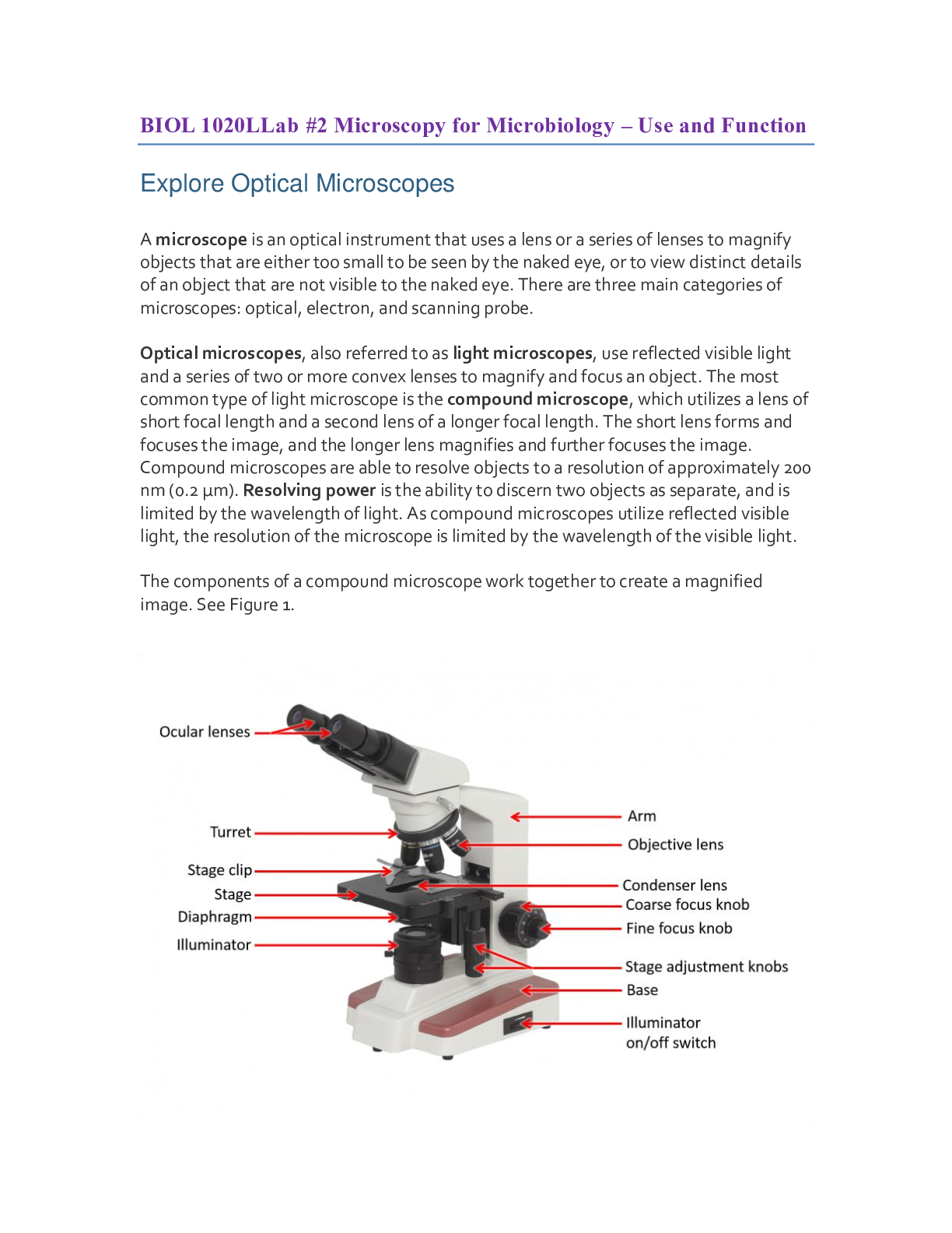
2. Label the parts of the microscope.
1. A compound microscope is a type of optical microscope.
True 2
False
Exercise 1
2. The _____ sits below the stage and varies the field for viewing the specimen.
diaphragm 2
coarse adjustment knob
base
turret
3. The depth of field is independent of the total magnification factor.
True
False 2
1. Describe the details in the slides "Letter e" that become visible as the power changed
from low power, to medium power, to high power.
2. Why is it important to calculate the diameter of the field when first using the
microscope?
Letter Component Name Component Function
A Ocular Lens Usually 10x or 15x, this is
the first lens through
which we look, and it
provides the first
ifi ti f t
B Tube The tube connects the
ocular and objective
l
C Turret The turret allows for
switching between
objective lenses.
D Objective Lenses This lens provides our
second magnification
factor, usually
4x,10x,40x, or 100x.
E Condenser Lens This lens focuses light
from the illuminator under
the slide.
F Stage The stage holds the slide
to be viewed.
G Diaphragm The diaphragm works as
an iris, allowing
manipulation of the size
of the cone of light
allowed to pass through
th lid
H Illuminator The illuminator takes
place of the older mirror,
providing its own steady
source of light.
I Coarse Focus Knob Allows major adjustment
of focus on the slide.
J Fine Focus Knob Allows minor adjustment
of focus on the slide.
K Stage Adjustment Knob This moves the stage
towards and away from
the viewer.
L Stage Adjustment Knob This moves the stage to
the left and right
M Base The base provides a
stable surface for the
microscope to sit upon.
6/12/2019 HOL - Instance
https://myhol.holscience.com/instances/5862#/experimentation/77844/final 7/19
Data Table 3: Field of View
Data Table 4: Letter e Viewing Results
Lens Ocular Magnification Objective Magnification Total Magnification
Low Power 15x 4x 60x
Medium Power 15x 10x 150x
High Power 15x 40x 600x
Oil Immersion n/a n/a n/a
Lens Total Magnification Field of View (mm) Field of View (µM)
Low Power 60x 4.5 4500
Medium Power 150x 1.8 1800
High Power 600x .45 450
Oil Immersion n/a Since HOL's V-Scope
does not have a method
of determining FOV, I
have used sample data
for this question. Since HOL's V-Scope
does not have a method
of determining FOV, I
have used sample data
for this question.
6/12/2019 HOL - Instance
https://myhol.holscience.com/instances/5862#/experimentation/77844/final 8/19
Photo 1: Letter e - Low Power
Photo 2: Letter e - Medium Power
Lens Observations
Low Power At low power we can see
the entire letter (a
broader FOV), some
speckling around the
Medium Power At medium power, the
lines appear more blurry,
though the horizontal line
is still quite sharp. The
diff t
High Power At high power the edges
of the line appear rough--
almost furry--and we can
see how the paper
medium absorbs some of
the ink in every direction,
creating a spreading
effect. We can even
differentiate between the
speckles on the
background quite clearly.
Our field is now so
limited, though, that we
can't even see that it's an
Oil Immersion n/a
1. Using the field of view calculated in Exercise 1 for the high power lens, approximately
how far across are each of the cells in the Bacteria Coccus Form slide in Photo 10? Show
your calculations.
2. Detail techniques you found helpful for focusing on the various slides in this exercise.
Slide Magnification
Amoeba 600x - While the image is
good at 150x, and we
can see the overall
shape, at 600x we can
see features of the
surface and inside of the
amoeba not otherwise
i ibl
Penicillium 600x
Yeast 600x
Spirillium 600x
Bacillus 600x
Coccus 600x
6/12/2019 HOL - Instance
https://myhol.holscience.com/instances/5862#/experimentation/77844/final 11/19
Photo 6: Penicillium Slide
Photo 7: Yeast Slide
6/12/2019 HOL - Instance
https://myhol.holscience.com/instances/5862#/experimentation/77844/final 12/19
Photo 8: Spirillium Slide
Photo 9: Bacillus Slide
6/12/2019 HOL - Instance
https://myhol.holscience.com/instances/5862#/experimentation/77844/final 13/19
Photo 10: Coccus Slide
Exercise 3
The cheek wet mount had a clearly defined cell structure and details could be seen at 600x. The
dental plaque mount seems more like a smear than individual structures, and may be more
comprehensible at a higher magnification. Both were invisible to the naked eye.
1. Describe the similarities and differences between the cheek cell wet mount and dental
plaque wet mount.
6/12/2019 HOL - Instance
https://myhol.holscience.com/instances/5862#/experimentation/77844/final 14/19
Data Table 6: Wet-Mount Viewing Results
Photo 11: Prepared Slide - Cheek Cell Smear
The process was easier the second time because I had already done it, so not needing to repeatedly
reread instructions allowed me to be smoother and more confident as I pressed the cover down.
2. How did the process of preparing wet-mount slides become easier as you prepared the
second wet-mount slide of this exercise?
Slide Magnification
Cheek Cell Smear 600x
Dental Tartar Smear 600x
Competency Review
1. The main component of an optical microscope that is used to focus an image at a high
power of magnification is the _____.
coarse adjustment knob
turret
fine adjustment knob 2
diaphragm
microscope.
2. The slide is placed on the _____ of an optical microscope.
objective lens
turret
base
stage 2
microscope.
6/12/2019 HOL - Instance
https://myhol.holscience.com/instances/5862#/experimentation/77844/final 18/19
3. Depth of field refers to the distance range that is in focus at a given time.
True 2
False
FEEDBACK
This question focuses on Learning Objective 2: Describe field of view and depth of field.
4. When calculating total magnification of a microscope, _____ should be considered.
the ocular lens
the objective lens
both the ocular lens and the objective lens 2
neither the ocular lens nor the objective lens
5. The oil immersion lens provides optimal viewing of large specimens.
True
False
6. The field of view _____ as magnification increases.
7. When creating a wet-mount slide, an individual should drop the cover slip on the top of
a water droplet placed on the slide without touching it to the slide first.
1. The optical microscope is regularly used to identify pathogenic microbes. In 1918 the
Spanish Flu infected almost one third of the world and was thought to be caused by
bacteria. However, a virulent virus was the cause of the Spanish Flu.
From what we have learned throughout this lab on microscopy, why weren’t scientists able
to identify the Spanish Flu with an optical microscope?
[Show More]