NPTE Questions Exam 2
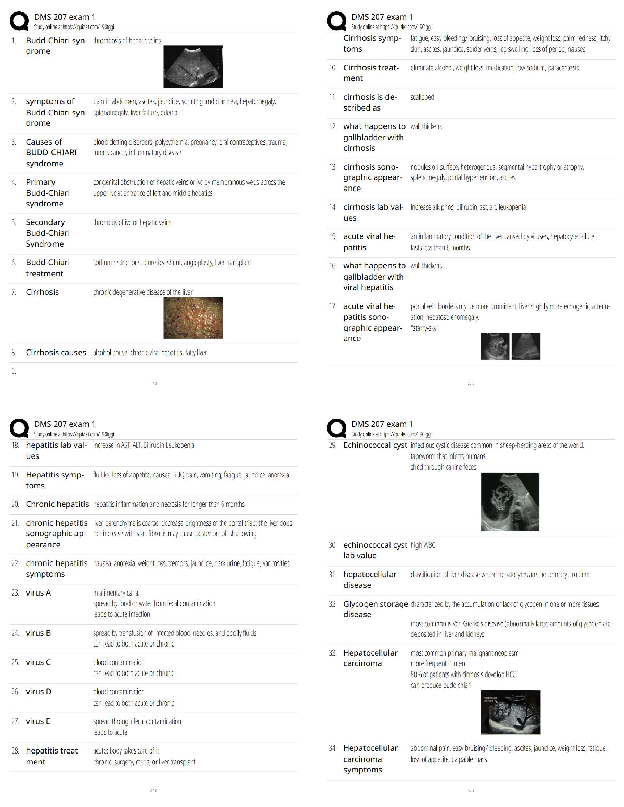
1. During a cervical spine examination, the PT observes restricted left rotation of the C7-T1
spinal level. After stabilizing the thoracic spine, the therapist's hand placement for mobilization
t
...
NPTE Questions Exam 2
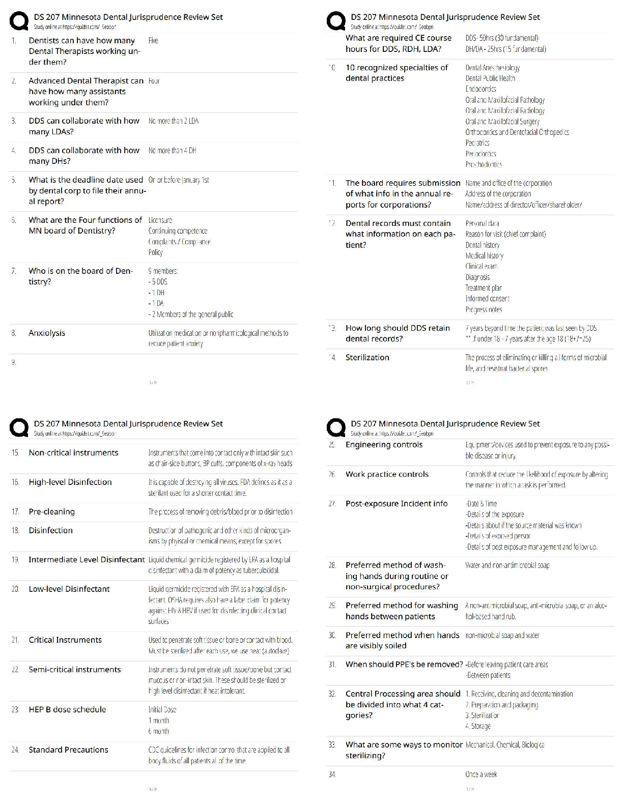
1. During a cervical spine examination, the PT observes restricted left rotation of the C7-T1
spinal level. After stabilizing the thoracic spine, the therapist's hand placement for mobilization
to improve left rotation should be at the: (A-10)
a. Posterior right C7 articular pillar
b. Posterior left C7 articular pillar
c. Tip of T1 spinous process
d. Posterior left C6 articular pillar - ✔✔a. Posterior right C7 articular pillar
The most effective hand placement for mobilization into greater left rotation is at the posterior
aspect of the right C7 articular pillar because it rotates the C7 vertebra to the left
2. A patient was referred to physical therapy complaining of loss of cervical AROM. X-rays
showed degenerative joint disease (DJD) at the uncinated processes in the cervical spine. The
motion that would be MOST restricted? (A-88)
a. Flexion
b. Extension
c. Rotation
d. Side-bending - ✔✔d. Side-bending
The uncinated processes (joints of Luschka) are located at the inferolateral aspect of the lower
cervical vertebrae. Side bending is lost with degenerative changes at the joint that the uncinated
process makes with the vertebra below.
3. A patient demonstrates weakness when rotating the head to one side as well as weakness
flexing the head laterally and forward to the same side. The therapist recognizes these symptoms
of the lesion of the: (A-98)
a. Spinal nerve root of the accessory nerve of the same side
b. Motor portion of the facial nerve
c. Spinal nerve root of the accessory nerve on the contralateral side
d. Motor portion of the hypoglossal nerve - ✔✔c. Spinal nerve root of the accessory nerve on the
contralateral side
The spinal nerve root of the accessory nerve (CN XI) innervates the SCM. This allows for head
rotation and flexion of the head laterally and forward, both to the contralateral side.
4. A client with RA presents at the PT clinic with severe whiplash from a motor vehicle accident
1 week ago. Initial cervical radiograph results revealed osseous structures appeared intact. The
client's chief complaints are of cervical pain and sudden falls with loss of consciousness.
Examination reveals a (+) Romberg sign and hyperreflexia. The PT's INITIAL action is to: (A173)
a. Fit this client with a hard cervical collar and contact the referring physician recommending a
CT scan
b. Immediately inform the referring physician and recommend a MRI scan
c. Immediately inform the referring physician and recommend another series of radiographs
d. Perform a test for transverse ligamental laxity
6. To reduce an elderly individual's chronic forward head posturing in standing and sitting the
therapist should consider stretching exercises to: (B-73)
a. Middle trapezius and rhomboids
b. Longus capitiis and longus colli muscles
c. Rectus capitis posterior minor and rectus capitis posterior major muscles
d. Rectus capitis anterior muscles
Forward head posturing or forward translation of the occiput in relation to the neck and trunk is
associated with extension of the occipital and axial joint flexion of the lower and mid cervical
spines. Chronic extension of the occipital axial joint will lead to shortening of the suboccipital
extensor muscles (rectus capitis posterior major and minor) and localized stretching of these
muscles would be indicated as part of the therapeutic intervention to reduce forward head
posturing - ✔✔a. Fit this client with a hard cervical collar and contact the referring physician
recommending a CT scan
This patient is exhibiting signs and symptoms of spinal cord compression with UMN signs
(hyperreflexia), a (+) Romberg sign, and sudden falls with loss of consciousness. This requires
immediate immobilization and contact with the physician for further imaging. Some cervical
lesions (non-displaced dens fracture, rupture of the transverse ligament) require greater imaging
detail than radiographs provide. This individual also has RA, which often accompanied by
erosion of the dens and facets and ligamental laxity (transverse). Immediately informing the
physician is important, and if the client is exhibiting spinal cord compression, immediate
stabilization is required.
5. A PT is performing the maximal cervical quadrant test to the right with a patient with right
C5-6 facet syndrome. The patient would most likely complain of: (A-181)
a. Pain in the right cervical region
b. Tightness in the right upper trapezius
c. Radicular pain in the right upper limb
d. Referred pain to the left midscapular region - ✔✔a. Pain in the right cervical region
The test position would consist of right cervical side bending with extension. This shortens the
upper trapezius and stresses the right cervical facets. When a pathological cervical facet is
provoked, the result will cause pain in the ipsilateral cervical region, with referred pain to the
ipsilateral scapular region. The test might also compress the nerve root, creating radicular signs,
but only on the right side.
6. To reduce an elderly individual's chronic forward head posturing in standing and sitting the
therapist should consider stretching exercises to: (B-73)
a. Middle trapezius and rhomboids
b. Longus capitiis and longus colli muscles
c. Rectus capitis posterior minor and rectus capitis posterior major muscles
d. Rectus capitis anterior muscles - ✔✔c. Rectus capitis posterior minor and rectus capitis
posterior major muscles
Forward head posturing or forward translation of the occiput in relation to the neck and trunk is
associated with extension of the occipital and axial joint flexion of the lower and mid cervical
spines. Chronic extension of the occipital axial joint will lead to shortening of the suboccipital
extensor muscles (rectus capitis posterior major and minor) and localized stretching of these
muscles would be indicated as part of the therapeutic intervention to reduce forward head
posturing
7. A patient with traumatic onset (Motor vehicle accident) of neck pain presents with subjective
complaints of frank upper cervical spine instability. One test that would safely assist in
identifying the integrity of the C1-C2 articulation would be: (B-121)
a. Vertebral artery test
b. Maximum cervical compression test
c. Transverse ligament stress test
d. Hautant's test
The transverse ligament stress test is specifically designed to assess the integrity of the transverse
ligament, which maintains the position of the dens of C2 with the anterior arch of C1 - ✔✔c.
Transverse ligament stress test
The transverse ligament stress test is specifically designed to assess the integrity of the transverse
ligament, which maintains the position of the dens of C2 with the anterior arch of C1
8. A patient presents with neck pain, which is a result of a motor vehicle accident (hit from
behind while the car was at rest). To determine the function of the deep cervical flexors, the PT
decides to perform a muscle function test utilizing the cranio-cervical flexion test. Findings of a
normal test would be: (B-136)
a. During active chin tuck, the patient is able to hold the head 1 inch above the table for 30
seconds
b. During active chin tuck, the pressure in the stabilizer cuff increases to 22 and the patient can
hold this position for 10 seconds
c. When palpating the anterior cervical musculature during the active chin tuck, the SCM
activates prior to the longus colli muscle
d. During active chin tuck, the patient is able to maintain the normal cervical lordosis for 10
seconds - ✔✔b. During active chin tuck, the pressure in the stabilizer cuff increases to 22 and the
patient can hold this position for 10 seconds
During an active chin tuck, the pressure in the stabilizer cuff increases to 22 and the patient can
hold this position for 10 seconds.
9. A patient presents with complaint of neck pain on the right. During the AROM examination,
the PT observes the following osteokinematic neck motions-full side bending left, full rotation to
the left, full forward flexion, limited and painful extension, limited and painful right side bending
and limited and painful right rotation. Based on this pattern, what is the arthrokinematic
restriction? (B-173)
a. Restriction with downglide of a facet on the right
b. Restriction with upglide of a facet on the right
c. Restriction with downglide of a facet on the left
d. Restriction with upglide of a facet on the left - ✔✔a. Restriction with downglide of a facet on
the right
If the facet on the right was restricted with downgliding (arthokinematic restriction), then the
osteokinematic motions that would be limited would be rotation and side bending to the right
with limited extension. The fact that there is pain on the right supports that the restriction is on
the right.
10. A patient with long-term postural changes exhibits an excessive forward head, and complains
of pain and dizziness when looking upward. The MOST effective physical therapy intervention
is: (B-175)
a. Manual therapy techniques to provide pain relief and postural reeducation
b. Anterior cervical muscle stretching and postural reeducation to relieve vertebral artery
compression
c. Strengthening exercise to the posterior cervical musculature
d. Postural reeducation to reduce compression of the cervical sympathetic ganglia - ✔✔a.
Manual therapy techniques to provide pain relief and postural reeducation
Long-term postural changes with forward head posture include shortening of the posterior
muscles, potential joint restrictions, with possible vertebral artery compromise at the occiput.
Restoration of normal movement throughout the cervical region and postural reeducation is the
best choice for this condition
11. A patient with a confirmed left C6 nerve root compression due to foraminal encroachment
complains of pain in the left thumb and index finger. The MOST effective cervical position to
alleviate this radicular pain in WB is: (C-103)
a. Lower cervical flexion
b. Left side-bending
c. Lower cervical rotation
d. Right rotation - ✔✔a. Lower cervical flexion
Flexion increases the space at the intervertebral foramen, allowing the C6 nerve root to
decompress and reduce or alleviate radicular pain
12. A patient presents with decrease motion at the occiptoatlantal joint (OA). The PT wants to
use the principles of coupled motions that occur in that area of the spine during manual therapy
techniques. In order to improve OA mobility, when the occiput is side-bent to the right, the
therapist should mobilize C1 into: (C-116)
a. Flexion
b. Extension
c. Rotation to the right
d. Rotation to the left - ✔✔d. Rotation to the left
Given the rules of coupled movement in the upper cervical spine, when the occiput is side-bent
into one direction, C1 rotates into the opposite direction. Side bending and rotation occur in the
same direction from C2-C7 regardless if the spine is in flexion or extension.
13. A patient is referred for physical therapy with a diagnosis of DJD affecting C2 and C3. The
patient complains of pain and stiffness in the cervical region and transient dizziness with some
cervical motions. The BEST initial examination procedure is: (C-152)
a. Lhermitte's test
b. Vertebral artery test
c. Oppenheim's test
d. Adson's maneuver - ✔✔b. Vertebral artery test
The vertebral artery test checks the integrity of the blood flow through the artery in the cervical
region. Because the patient is experiencing symptoms of circulatory disturbance and a unilateral
pull could compress the left cervical structures, the vertebral artery test is an appropriate
screening test. The test consists of passively placing the patient's head in extension and side
flexion in supine position. Then the head and neck are slowly rotated to the laterally flexed side
and held for 30 sec. Some of the (+) signs may be syncope, lightheadedness, nystagmus or visual
disturbances. Though there are some doubts about the sensitivity/specificity of this test, the
patient's initial complain of dizziness with come cervical movements would indicate that it be
applied in this case.
14. A patient presents with a complaint of severe neck and shoulder pain of 2 days duration. The
patient reports falling asleep on the couch watching TV, has been stiff, and sore since. There is
tenderness of the cervical muscles on the right, with increased pain upon palpation. Passive
ROM is most limited in flexion, then side bending left, and then rotation left and active
extension. Side bending right and rotation right are also painful. Based on these examination
findings, the patient's diagnosis is: (C-190)
a. Cervical radiculopathy
b. Facet syndrome
c. Cervical strain
d. Herniated disc
A facet syndrome presents with localized pain - ✔✔b. Facet syndrome
A facet syndrome presents with localized pain
[Show More]


.png)