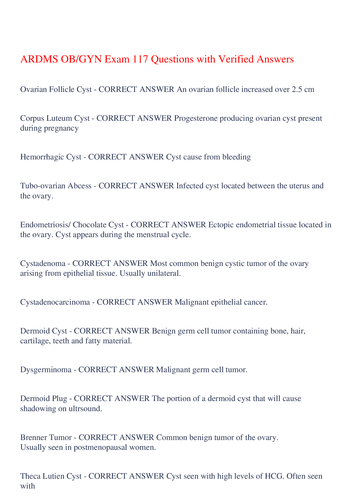
ARDMS OB/GYN Exam 117 Questions with Verified Answers
Ovarian Follicle Cyst - CORRECT ANSWER An ovarian follicle increased over 2.5 cm
Corpus Luteum Cyst - CORRECT ANSWER Progesterone producing ovarian cyst present
...
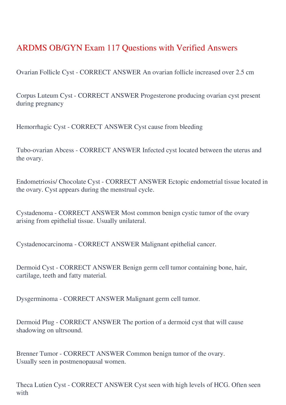
ARDMS OB/GYN Exam 117 Questions with Verified Answers
Ovarian Follicle Cyst - CORRECT ANSWER An ovarian follicle increased over 2.5 cm
Corpus Luteum Cyst - CORRECT ANSWER Progesterone producing ovarian cyst present during pregnancy
Hemorrhagic Cyst - CORRECT ANSWER Cyst cause from bleeding
Tubo-ovarian Abcess - CORRECT ANSWER Infected cyst located between the uterus and the ovary.
Endometriosis/ Chocolate Cyst - CORRECT ANSWER Ectopic endometrial tissue located in the ovary. Cyst appears during the menstrual cycle.
Cystadenoma - CORRECT ANSWER Most common benign cystic tumor of the ovary arising from epithelial tissue. Usually unilateral.
Cystadenocarcinoma - CORRECT ANSWER Malignant epithelial cancer.
Dermoid Cyst - CORRECT ANSWER Benign germ cell tumor containing bone, hair, cartilage, teeth and fatty material.
Dysgerminoma - CORRECT ANSWER Malignant germ cell tumor.
Dermoid Plug - CORRECT ANSWER The portion of a dermoid cyst that will cause shadowing on ultrsound.
Brenner Tumor - CORRECT ANSWER Common benign tumor of the ovary. Usually seen in postmenopausal women.
Theca Lutien Cyst - CORRECT ANSWER Cyst seen with high levels of HCG. Often seen with hydadiform mole.
Ovarian Torsion - CORRECT ANSWER Complete or partial rotation of the ovarian pedicle on its axis. The right ovary is three times more likely to torse than the left.
Ectopic Pregnancy - CORRECT ANSWER Gestational sac located anywhere outside of the fundus of the uterus.
Heterotopic Pregnancy - CORRECT ANSWER Intrauterine pregnancy and correlating ectopic pregnancy in the same patient.
Polycystic Ovarian Disease - CORRECT ANSWER Ovarian disease characterized by anovulation due to an endocrine imbalance.
Stein-Leventhal Syndrome - CORRECT ANSWER Another name for polycystic ovarian disease.
Hyperstimulated Ovaries - CORRECT ANSWER Overstimulated follicles increased in size due to infertility treatments.
Thecoma - CORRECT ANSWER Benign ovarian tumor usually seen in postmenopausal women. Produces estrogen.
Fibroma - CORRECT ANSWER Benign ovarian tumor comprised of muscle.
Sertoli-Leydig Cell Tumor - CORRECT ANSWER An androgen producing ovarian tumor that causes masculinization.
Granulosa Tumor - CORRECT ANSWER Estrogen producing ovarian tumor.
Krukenberg Tumor - CORRECT ANSWER Malignant tumor of the ovary that metastasized from the GI tract such as the stomach or colon.
Fibromyoma - CORRECT ANSWER Uterine mass made of muscle.
Adenomyosis - CORRECT ANSWER Ectopic endometrial tissue within the myoma. Causes enlargement of the uterus with abnormal contours.
Uterine Polyps - CORRECT ANSWER Overgrowth of uterine tissue into the endometrial cavity.
Endometritis - CORRECT ANSWER Infection of the endometrium seen after sexually transmitted disease.
T-shaped Uterus - CORRECT ANSWER T-shaped endometrium. Associated with the use of DES medication for infertility.
Uterine Adenocarcinoma - CORRECT ANSWER Uterine cancer usually seen in postmenopausal women. Causes abnormal bleeding.
Threatened Abortion - CORRECT ANSWER First trimester bleeding with a closed cervix and normal GS position.
Incomplete Abortion - CORRECT ANSWER Miscarriage with either fetal or placental components left behind.
Complete Abortion - CORRECT ANSWER Full expostulation of fetus and placenta from the uterus before term.
Septic Abortion - CORRECT ANSWER Miscarriage with either fetal or placental components left behind that causes infection in the mother.
Inevitable Abortion - CORRECT ANSWER In the first trimester when the GS is seen to move close to the cervix and the cervix dilates.
Incompetent Cervix - CORRECT ANSWER Cervix dilates too early for delivery. Measures less that 3cm in length.
Placenta Previa - CORRECT ANSWER When the placenta grows so that it covers the cervix.
Circumvallate Placenta - CORRECT ANSWER The placenta doubles back causing a ring-like shape.
Hydatidiform Mole Placenta - CORRECT ANSWER Overgrowth of placental tissue that forms a mass.
Placenta Hydrops - CORRECT ANSWER Enlarged placental thickness >4cm with cystic changes. Usually seen with hydrops fetalis.
Abruptio Placentae - CORRECT ANSWER Abnormal detachment of the placenta from the uterine wall.
Choriogangioangioma - CORRECT ANSWER Tumor made up of blood vessels.
Trophoblastic Disease - CORRECT ANSWER Another name for molar pregnancy.
2 Vessels Cord - CORRECT ANSWER Umbilical cord containing only one artery. Associated with trisomy 18 and 21.
Cord Previa - CORRECT ANSWER Umbilical cord covering the cervical os.
Cord Prolapse - CORRECT ANSWER Umbilical cord presents first during delivery and is seen within the cervix.
Nuchal Cord - CORRECT ANSWER More than one umbilical cord loop around the fetal neck.
Velamentous Cord Insertion - CORRECT ANSWER Umbilical cord inserts into the side of the placenta.
Choriod Plexus Cyst - CORRECT ANSWER Normal cyst in the choriod plexus until the 26th week.
Encephalocele - CORRECT ANSWER Bony defect in the skull with the brain tissue protruding.
Agenesis of Corpus Callosum - CORRECT ANSWER Dilation of the posterior horns of the lateral ventricles (tear drop). Upward displacement of the CSP and the 3rd ventricle.
Hydrocephalus - CORRECT ANSWER Excess fluid in the lateral ventricles measuring greater than 10 mm. Dangling choriod plexus.
Hydraencephaly - CORRECT ANSWER Excess fluid fills the head. Falx is absent or incomplete.
Cystic Hygroma - CORRECT ANSWER Bilateral cysts at the back of the head. Seen with turners syndrome. Only present in females.
Arnold Chiari Type II Malformation - CORRECT ANSWER Group of cranial abnormalities associated with spina bifida. Absent cisterna magna, banana sign, and lemon sign.
Micrognathia - CORRECT ANSWER Small chin.
Hyperglossia - CORRECT ANSWER Large tongue.
Anencephaly - CORRECT ANSWER Absence of a large part of the brain or skull.
Holoprosencephaly - CORRECT ANSWER Failure of the forebrain to divide into hemispheres.
Semilobar Holoprosencephaly - CORRECT ANSWER A partial fissure between two hemispheres of the brain. Associated with facial anomalies.
Lobar Holoprosencephaly - CORRECT ANSWER A fissure exists between the two hemispheres of the brain without communication.
Alobar Holoprosencephaly - CORRECT ANSWER One large ventricle. No fissure present. Associated with severe facial anomalies.
Cleft Lip - CORRECT ANSWER Fissure or gap in the lip caused by abnormal development.
Proboscis - CORRECT ANSWER Abnormality of the nose.
Iniencephaly - CORRECT ANSWER Extreme extension of the neck so that the back of the skull and the top of the shoulders are touching.
Ectopia Cordis - CORRECT ANSWER Fetal heart located outside the thorax. Associated with pentalogy of cantrell.
Atrial Septal Defect - CORRECT ANSWER Abnormal opening of the wall between the atria in the fetal heart.
Ventricular Septal Defect - CORRECT ANSWER Abnormal opening of the wall between the ventricles in the fetal heart.
Endocardial Cushion Defect - CORRECT ANSWER Abnormality in the walls between all four heart chambers. Associated with down syndrome.
Ebstein Anomaly - CORRECT ANSWER Atrial displacement of the septal and posterior tricuspid valves leaflets.
Moderator Band - CORRECT ANSWER Thickened papillary muscle in the fetal heart. Usually seen in the right ventricle. Associated with down syndrome.
Transposition of Great Vessels - CORRECT ANSWER Switching of the aorta and the pulmonary artery.
Dextrocardia - CORRECT ANSWER Axis of the heart is switched so the right atrium is closest to the spine.
Hypoplastic Left Heart Syndrome - CORRECT ANSWER Small left atrium and left ventricle.
Extralobar Lung Sequesteration - CORRECT ANSWER One segment of the lung is sepreated from the other lung tissue and has it's own blood supply from the aorta.
Diaphragmatic Hernia - CORRECT ANSWER Deviation in the diaphragm allowing abdominal contents into the thorax.
Hydrothorax - CORRECT ANSWER Fluid in the thorax cavity
Double Bubble Sign - CORRECT ANSWER Normal abdomen and dilated duodenum caused by duodenal atresia. Associated with down syndrome.
Omphalocele - CORRECT ANSWER Mass within the umbilical cord covered by the umbilical membrane. Seen with trisomys 21 and 18.
Gastroschisis - CORRECT ANSWER Mass protruding from the stomach not covered by a membrane. Seen with trisomys 21 and 18.
Esophageal Atresia - CORRECT ANSWER Blockage of the esophagus leading to polyhydraminos and an absent stomach on sonogram.
Meconium Peritonitis - CORRECT ANSWER Rupture of the fetal bowl leading to meconium leaking into the surrounding areas.
Hirschsuprung's Disease - CORRECT ANSWER Disease characterized by a lack of nerves in the fetal bowl.
Infantile Polycystic Kidney - CORRECT ANSWER AKA Potter 1. Bilateral renal microcysts. Is a recessive disease.
Mulitcystic Dysplastic Kindney - CORRECT ANSWER AKA Potter 2. Unilateral renal cyst of varriable size.
Hydronephrosis - CORRECT ANSWER Excess fluid in the kidneys.
Posterior Urethral Valve Obstruction - CORRECT ANSWER Urethra is block causing enlarged bladder and hydronephrosis. Only seen in male fetus.
Bladder Extrophy - CORRECT ANSWER Urinary bladder is located outside the fetal body.
Spina Bifida Occulta - CORRECT ANSWER Splaying of the lamina in the vertebrae with no tissue outside the spine.
Spina Bifida Apperta - CORRECT ANSWER Splaying of the lamina in the vertebrae with spinal tissue or meninges protruding outward.
Meningocele - CORRECT ANSWER An abnormality associated with spina bifida where the meninges only protrude outside the body.
Myelomeningocele - CORRECT ANSWER An abnormality associated with spina bifida where the spinal cord and nerves develop outside the body.
Lumbar-Sacral Teratoma - CORRECT ANSWER A benign cystic tumor arising out of the lower back.
Achondrogenesis - CORRECT ANSWER Failure of the fetus to produce cartilage.
Osteogenesis Imperfecta - CORRECT ANSWER Abnormal bone formation characterized by many fractures especially in the fetal ribs.
Rhizomelia - CORRECT ANSWER Shortening of the distal limbs.
Micromelia - CORRECT ANSWER Shortening of the limbs.
Campomelia - CORRECT ANSWER Bowing of the long bones.
Mesomelia - CORRECT ANSWER Shortening of the proximal limbs.
Sirenomeila - CORRECT ANSWER Fusion of the lower legs.
Polydactyly - CORRECT ANSWER More than the normal amount of digits.
Syndactyly - CORRECT ANSWER Fusion of digits.
Talipes Equinovarius - CORRECT ANSWER Club foot.
Clinodactyly - CORRECT ANSWER Clenched hand and curved digits.
Gestational Diabetes - CORRECT ANSWER Increases risk of heart anomalies, caudal regression, and macrosomia.
Twin to Twin Transfusion Syndrome - CORRECT ANSWER Syndrome in which one twin receives the blood and nutrients from the other twin causing the regression of one.
Acardiac-Acephalic Twin - CORRECT ANSWER Syndrome in which one twin has no heart and no head.
Amniotic Band Syndrome - CORRECT ANSWER Syndrome in which the amniotic membrane breaks free and attaches in places to the fetus. Causes amputations.
Trisomy 13 - CORRECT ANSWER Patau's syndrome. Associated with holoprosencephaly, IUGR, polyhydraminos, micrognathia, agenesis of the corpus callosum, and proboscis.
Trisomy 18 - CORRECT ANSWER Edwards Syndrome. Associated with clenched hand, polyhydraminos, choriod plexus cyst, diaphragmatic hernia, omphalocele.
Trisomy 21 - CORRECT ANSWER Down syndrome. Associated with abnormal nuchal fold, abnormal nuchal translucency, cushion defect, club foot, cleft lip, sandal toe, moderator band, and abnormal nasal bone.
Fetal Alcohol Syndrome - CORRECT ANSWER Syndrome caused by maternal alcohol intake. Associated with IUGR, microcephaly, microphthalmos.
Beckwith-Weideman Syndrome - CORRECT ANSWER Syndrome associated with omphalocele, macroglossia, and giantism.
Pentalogy of Cantrell - CORRECT ANSWER Group of anomalies including omphalocele, ectopia cordis and 2 vessels cord.
Meckel Gruber Syndrome - CORRECT ANSWER Group of anomalies including encephalocele, polycystic kindey, cleft lip, and polydactyly.
Treacher Collins Syndrome - CORRECT ANSWER Associated with micrognathia.
VACTEL - CORRECT ANSWER Vacters syndromes. Vertebral, anal, cardial, trachea atresia, esophageal atresia, and limb.
Toxoplasmosis - CORRECT ANSWER Toxin carried by cats that causes intracranial calcifications in the fetus.
[Show More]