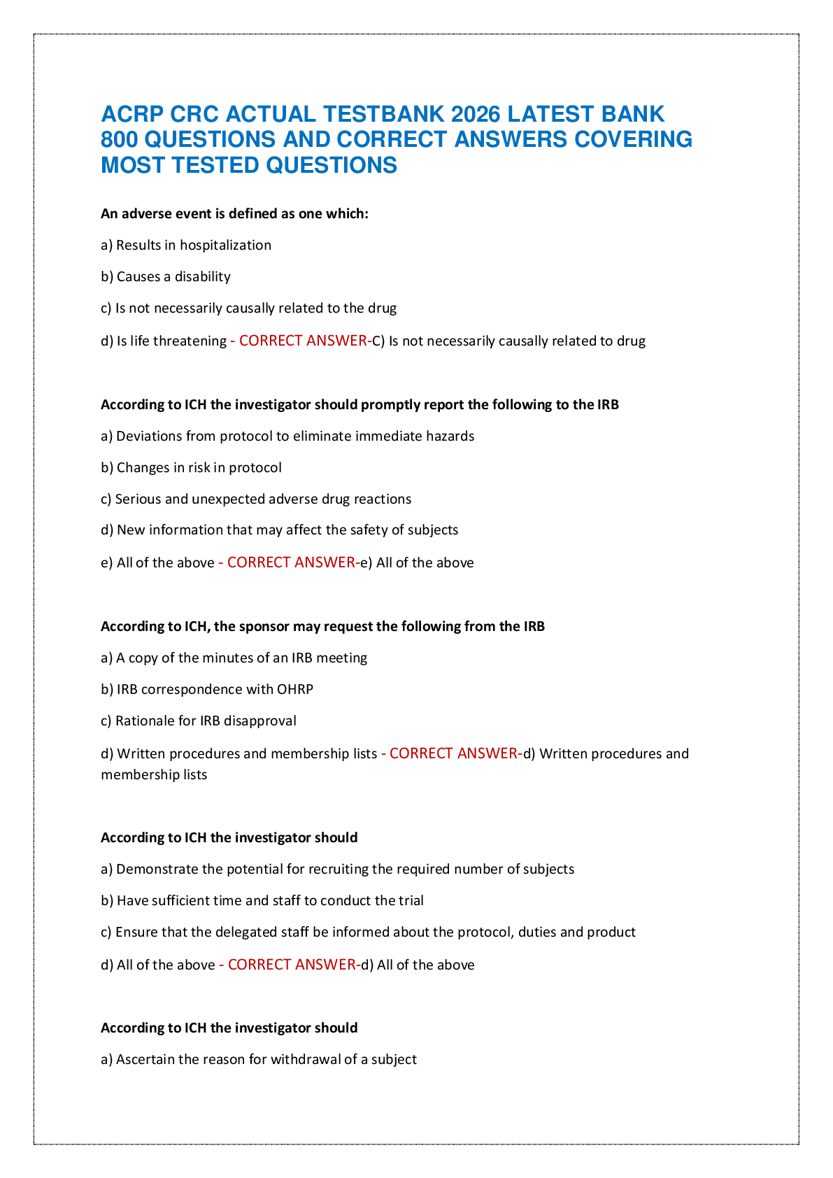
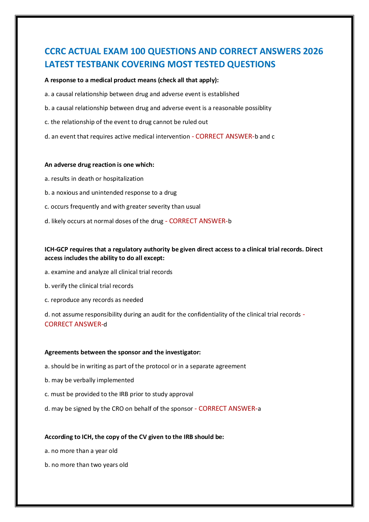
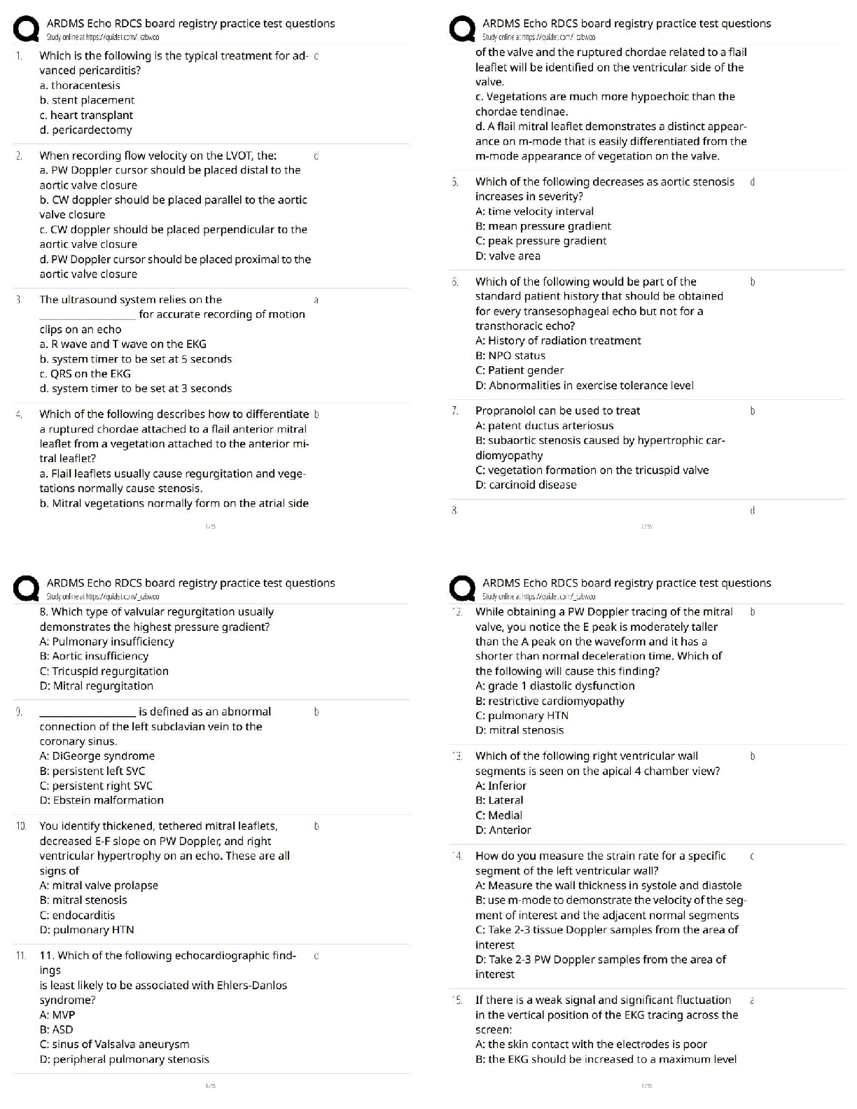
ARDMS Echo RDCS Board Registry practice Test 200 Questions with Verified Answers
Which is the following is the typical treatment for advanced pericarditis?
a. thoracentesis
b. stent placement
c. heart transplant
...
ARDMS Echo RDCS Board Registry practice Test 200 Questions with Verified Answers
Which is the following is the typical treatment for advanced pericarditis?
a. thoracentesis
b. stent placement
c. heart transplant
d. pericardectomy - CORRECT ANSWER d
When recording flow velocity on the LVOT, the:
a. PW Doppler cursor should be placed distal to the aortic valve closure
b. CW doppler should be placed parallel to the aortic valve closure
c. CW doppler should be placed perpendicular to the aortic valve closure
d. PW Doppler cursor should be placed proximal to the aortic valve closure - CORRECT ANSWER d
The ultrasound system relies on the ____________________ for accurate recording of motion clips on an echo
a. R wave and T wave on the EKG
b. system timer to be set at 5 seconds
c. QRS on the EKG
d. system timer to be set at 3 seconds - CORRECT ANSWER a
Which of the following describes how to differentiate a ruptured chordae attached to a flail anterior mitral leaflet from a vegetation attached to the anterior mitral leaflet?
a. Flail leaflets usually cause regurgitation and vegetations normally cause stenosis.
b. Mitral vegetations normally form on the atrial side of the valve and the ruptured chordae related to a flail leaflet will be identified on the ventricular side of the valve.
c. Vegetations are much more hypoechoic than the chordae tendinae.
d. A flail mitral leaflet demonstrates a distinct appearance on m-mode that is easily differentiated from the m-mode appearance of vegetation on the valve. - CORRECT ANSWER b
Which of the following decreases as aortic stenosis
increases in severity?
A: time velocity interval
B: mean pressure gradient
C: peak pressure gradient
D: valve area - CORRECT ANSWER d
Which of the following would be part of the
standard patient history that should be obtained
for every transesophageal echo but not for a
transthoracic echo?
A: History of radiation treatment
B: NPO status
C: Patient gender
D: Abnormalities in exercise tolerance level - CORRECT ANSWER b
Propranolol can be used to treat
A: patent ductus arteriosus
B: subaortic stenosis caused by hypertrophic cardiomyopathy
C: vegetation formation on the tricuspid valve
D: carcinoid disease - CORRECT ANSWER b
8. Which type of valvular regurgitation usually
demonstrates the highest pressure gradient?
A: Pulmonary insufficiency
B: Aortic insufficiency
C: Tricuspid regurgitation
D: Mitral regurgitation - CORRECT ANSWER d
____________________ is defined as an abnormal
connection of the left subclavian vein to the
coronary sinus.
A: DiGeorge syndrome
B: persistent left SVC
C: persistent right SVC
D: Ebstein malformation - CORRECT ANSWER b
You identify thickened, tethered mitral leaflets,
decreased E-F slope on PW Doppler, and right
ventricular hypertrophy on an echo. These are all
signs of
A: mitral valve prolapse
B: mitral stenosis
C: endocarditis
D: pulmonary HTN - CORRECT ANSWER b
11. Which of the following echocardiographic findings
is least likely to be associated with Ehlers-Danlos
syndrome?
A: MVP
B: ASD
C: sinus of Valsalva aneurysm
D: peripheral pulmonary stenosis - CORRECT ANSWER d
While obtaining a PW Doppler tracing of the mitral
valve, you notice the E peak is moderately taller
than the A peak on the waveform and it has a
shorter than normal deceleration time. Which of
the following will cause this finding?
A: grade 1 diastolic dysfunction
B: restrictive cardiomyopathy
C: pulmonary HTN
D: mitral stenosis - CORRECT ANSWER b
Which of the following right ventricular wall
segments is seen on the apical 4 chamber view?
A: Inferior
B: Lateral
C: Medial
D: Anterior - CORRECT ANSWER b
How do you measure the strain rate for a specific
segment of the left ventricular wall?
A: Measure the wall thickness in systole and diastole
B: use m-mode to demonstrate the velocity of the segment of interest and the adjacent normal segments
C: Take 2-3 tissue Doppler samples from the area of interest
D: Take 2-3 PW Doppler samples from the area of interest - CORRECT ANSWER c
If there is a weak signal and significant fluctuation
in the vertical position of the EKG tracing across the
screen:
A: the skin contact with the electrodes is poor
B: the EKG should be increased to a maximum level
C: the alcohol used to clean the skin is causing artifact
D: a cell phone or other nearby electrical device is causing interference - CORRECT ANSWER a
A patient currently undergoing chemotherapy for
breast cancer is referred for an echo. A small mass
is noted within the right ventricular wall and a mild
pericardial effusion. There is debris and fibrous
strands throughout the effusion. The mass is in the
right ventricle is most likely
A: primary cardiac malignancy
B: rhabdomyoma
C: secondary cardiac malignancy
D: myxoma - CORRECT ANSWER c
Global longitudinal strain is measured from:
A: parasternal views
B: subcostal views
C: apical views
D: more than one of the above - CORRECT ANSWER c
Which of the following is the most common cause of tricuspid regurgitation?
A: pulmonary HTN
B: atherosclerosis
C: prolapse
D: rheumatic fever complication - CORRECT ANSWER a
When measuring the left ventricular end systolic
volume, you should use the cine function to:
A: find the frame just after the MV closes
B: find the frame just before the MV opens
C: find the frame just before the AV opens
D: find the frame at the peak of the R wave - CORRECT ANSWER b
Which of the following statements is correct
regarding tricuspid valve prolapse(TVP)?
A: TVP rarely leads to significant tricuspid regurgitation.
B: TVP most commonly occurs as an isolated finding.
C: TVP refers to abnormal diastolic displacement of the valve leaflet into the RA.
D: The anterior and septal leaflets are most commonly involved. - CORRECT ANSWER d
Dobutamine is used to assess suspected
hibernating myocardium. If the affected wall
motion improves at low doses but declines at
higher doses,
A: the myocardium has irreversible damage and reperfusion would not be recommended
B: the patient will be scheduled for a transesophageal echo to better evaluate the endocardium
C: the patient will be scheduled for a reperfusion procedure
D: the patient will be scheduled for a treadmill stress echo - CORRECT ANSWER c
The echocardiogram demonstrates akinesis of the
left ventricular apex. Which coronary artery is most
likely affected by coronary artery disease?
A: obtuse artery
B: right coronary artery
C: left anterior descending artery
D: left circumflex artery - CORRECT ANSWER c
Aortic and pulmonic insufficiency will cause
A: a mid-diastolic murmur
B: a crescendo-decrescendo murmur
C: an early diastolic murmur
D: an early systolic murmur - CORRECT ANSWER c
Multiple intrapericardial tumors are identified with
a moderate pericardial effusion. These findings are
most suggestive of:
A: Rhabdomyoma
B: Metastatic tumors
C: Invasive sarcoma
D: Fibroelastomas - CORRECT ANSWER b
A patient is referred for an echo for pre-surgical
clearance for a hip replacement. The only pertinent
cardiac history is a 6 year history of systemic HTN.
What will be the most likely findings on the exam?
A: dilated aortic root and aortic insufficiency
B: interventricular septum and inferior wall = 1.1cm thickness
C: interventricular and interatrial septum = 1.1cm thickness
D: right ventricular free wall =0.5cm thickness - CORRECT ANSWER b
When measuring left atrial volume;
A: LA length is measured from the center of the mitral annulus to the inner edge of the posterior LA wall
B: the same view used for LV volumes should be used for measurement of LA volumes
C: the proximal pulmonary veins should be included in the tracing
D: place a caliper at the center of the MV annular plane, trace the cavity to create a full circle and return to the center of the MV annulus - CORRECT ANSWER a
28. A vena contracta width greater than or equal
to _____ indicates severe mitral
regurgitation.
A: 3mm
B: Smm
C:. 7mm
D: 9mm - CORRECT ANSWER c
The best view to evaluate the left atrial appendage
is:
A: apical 4 chamber
B: parasternal long axis
C: apical 2 chamber
D: subcostal - CORRECT ANSWER c
If the systemic BP is 120/80, LVOT velocity is 1m/s
and the aortic valve velocity is 5m/s, what is the
peak pressure gradient at the valve?
A: 100mmHg
B: 64mmHg
C: 4mmHg
D: 25mmHg - CORRECT ANSWER a
Severe dilatation of the aortic root is diagnosed at _____
or greater
A: 5.0cm
B: 6.0cm
C: 4.5cm
D. 3.8cm - CORRECT ANSWER a
A Sonographer should instruct the patient on the
use of the Borg Scale during:
A: a stress echo
B: a saline contrast exam
C: a transesophageal echo
D: a microbubble contrast exam - CORRECT ANSWER a
Diastolic flow reversal in the hepatic veins and a
mitral E/A ratio of 1.9 correlates with which of the
following?
A: constrictive pericarditis
B: right upper and lower pulmonary vein stenosis
C: moderate mitral stenosis
D: 2mm perimembranous VSD - CORRECT ANSWER a
The early heart tube normally:
A: loops medially then laterally
B: loops laterally then anterior
C: loops anterior and to the right
D: loops anterior and to the left - CORRECT ANSWER c
Severe aortic regurgitation demonstrates
A: a flat slope
B: a very faint waveform pattern due to turbulence
C: a peak velocity =5m/s
D: a significant difference between the peak velocity at the onset and the end of the regurgitation - CORRECT ANSWER d
What classic characteristic of hypertrophic
cardiomyopathy is not identified in patients with focal mid-cavity hypertrophy?
A: dagger shaped waveform at the narrowed area of the ventricle
B: systolic crescendo-decrescendo murmur
C: systolic anterior motion of the mitral valve
D: turbulent flow at the area of narrowing in the ventricle - CORRECT ANSWER c
Which pulmonary vein is able to be evaluated by
PW Doppler in the apical 4 chamber view in most
patients?
A: left upper
B: right lower
C: left lower
D: right upper - CORRECT ANSWER d
Leaflet redundancy and myxomatous degeneration
are common signs of:
A: subvalvular aortic stenosis
B: mitral valve prolapse
C: carcinoid disease
D: supravalvular aortic stenosis - CORRECT ANSWER b
You are performing an apical view during a
microbubble contrast echo on a patient with
suspected wall motion abnormalities in the basal
anterolateral left ventricular segment. You notice
significant swirling of the contrast in the area of the
apex. How can you adjust the controls to reduce
this artifactual swirling?
A: move the focal zone to the apex
B: inject contrast at a faster rate
C: turn off harmonic imaging
D: decrease the mechanical index setting - CORRECT ANSWER d
Which of the following is true regarding an Austin
Flint murmur?
A: It is associated with significant mitral regurgitation causing mitral valve fluttering seen on the m-mode tracing.
B: It is associated with a large VSD causing paradoxical interventricular septal motion seen on the m-mode.
C It is associated with severe aortic regurgitation causing compression/vibration of the anterior mitral valve leaflet in diastole
D: It is associated with severe aortic stenosis causing vibration of the aortic root in systole; Seen on the m-mode tracing of the aortic root. - CORRECT ANSWER c
Atrial depolarization is represented by what part of
the EKG tracing?
A: P wave
B: QRS wave
C: R wave
D: T wave - CORRECT ANSWER a
You identify a left atrial mass with an irregular shape and a "grape cluster" appearance. It is heterogeneous with areas of calcification. These
findings are most suggestive of:
A: rhabdomyoma
B: hypernephroma
C: fibroma
D: myxoma - CORRECT ANSWER d
Which of the following will decrease after a mitral
commissurotomy in a patient with isolated mitral
stenosis?
A: pressure half time
B: mitral valve area
C: A wave velocity
D: mitral regurgitation - CORRECT ANSWER a
What is the average normal aortic valve size?
A: 3-4cm
B: 2-3cm ;
C: 4-5cm 2
D: 3.5-4.5cm - CORRECT ANSWER a
Doppler evaluation of the aortic valve
demonstrates ________________ with atrial
fibrillation.
A: beat to beat variation in aortic velocity
B: loss of the A wave
C: E to A reversal
D: dagger shaped waveform - CORRECT ANSWER a
Renal cell carcinoma most commonly metastasizes
to the heart through the
A: IVC
B: portal venous system
C: lymphatic system
D: aorta - CORRECT ANSWER a
Which type of cardiomyopathy is related to fatty
and fibrous tissue replacement of the myocardium
of the right ventricle?
A: right ventricular hemochromatosis
B: lipomatous septal hypertrophy
C: arrhythmogenic right ventricular cardiomyopathy
D: endomyocardial fibrosis - CORRECT ANSWER c
The pattern of wall hypertrophy in hypertensive
heart disease is ______________, and in hypertrophic cardiomyopathy the pattern is usually ______________
A: symmetric, the same
B: symmetric, asymmetric
C: asymmetric, symmetric
D: focal, diffuse - CORRECT ANSWER b
A 22yr old patient presents for an echo and you
identify biventricular wall thickening, mild
pericardial effusion and thickening of the leaflets of
all four valves. Which of the following is the most
likely cause for these findings?
A: amyloidosis
B: hypertrophic cardiomyopathy
C: carcinoid disease
D: rheumatic fever - CORRECT ANSWER a
A patient presents for an echo due to a recent
stroke. The order states that they are looking for an
embolic source for the stroke. She has been
admitted to the hospital and is currently on her
second day of Heparin treatment due to a recent
diagnosis of DVT in her left leg. A recent VQ scan
was normal with no evidence of pulmonary
embolism. Which of the following is a potential
reason for why the study was ordered?
A: to evaluate the patient for CHF
B: to evaluate the patient for a possible vegetation on the tricuspid valve
C: to evaluate the patient for a possible patent foramen ovale
D: to evaluate the patient for a possible VSD - CORRECT ANSWER c
Which of the following is a normal response seen
with stress echocardiography?
A: decrease in left ventricular size in diastole and reduced fractional shortening
B: hyperdynamic wall motion and increase of EF%
C: ST segment depression on EKG
D: ST segment elevation on EKG - CORRECT ANSWER b
A new atrial septal defect is a complication most
commonly seen with what procedure?
A: transcatheter aortic valve implantation
B: coronary artery stent placement
C: septal ablation for hypertrophic cardiomyopathy
D: balloon mitral valvotomy - CORRECT ANSWER d
Which of the following adjustments would improve
the appearance of contrast on the echo image?
A: Turn off harmonic imaging
B: Use multiple focal zones
C: Decrease output power
D: Increase the thermal index - CORRECT ANSWER c
What effect will a mechanical index(Ml) of 1.0 have
on a contrast echo exam?
A: An MI setting of 1.0 or greater is preferred for contrast exams.
B: The increased beam intensity will burst the contrast bubbles quickly.
C: Mechanical index has no effect on contrast exams. The thermal index must be properly set for an optimal contrast exam.
D: The decreased beam intensity will reduce the appearance of the bubbles on the image. - CORRECT ANSWER b
When placing the electrodes on a patient for a 12
lead EKG for to a stress echo, the electrode for left
arm is placed:
A: 2cm below the left clavicle
B: at the anterior axillary line between the last rib and the iliac crest
C: left forearm
D: right forearm - CORRECT ANSWER a
How is the contraction velocity of the left atrial
appendage evaluated?
A: M-mode sample line positioned approximately 1 cm proximal to the entry of the appendage into the body of the LA
B: M-mode sample line positioned approximately across the left atrium and the body of the left atrial appendage
C: PW Doppler sample volume positioned 1cm proximal to the entry of the appendage into the body of the LA
D: PW Doppler sample volume positioned in the center of the left atrial appendage - CORRECT ANSWER c
Systole is defined as:
A: from the end of the isovolumic contraction period to the start of the isovolumic relaxation period
B: from mitral valve closure to aortic valve closure
C: from the end of the isovolumic relaxation period to the start of the isovolumic contraction period
D: from the opening of the aortic valve to aortic valve closure - CORRECT ANSWER b
If the heart rate is 80bpm and the stroke volume is
50mL, what is the cardiac output?
A: 4000mL
B: 400 L/min
C: 4 mL/min
D: 4 L/min - CORRECT ANSWER d
Which of the following should be suspected on the
echo in the presence of a diastolic murmur?
A: aortic stenosis
B: MVP
C: ASD
D: mitral stenosis - CORRECT ANSWER d
Which of the following adjustments should be
made to the Doppler settings when evaluating left
ventricular inflow in a patient with constrictive
pericarditis?
A: Decrease the PRF to demonstrate the expected low velocity peaks of the mitral waveform
B: Decrease the sweep speed
C: Increase the PRF to demonstrate the expected E peak velocities greater than 2.0m/s
D: Increase the sweep speed - CORRECT ANSWER b
The motion of which of the following wall segments
can be evaluated on the apical four chamber view?
A: basal anteroseptal wall
B: mid anteroseptal wall
C: lateral right ventricular wall
D: basal inferolateral wall - CORRECT ANSWER c
Which of the following is the most common type of
infiltrative cardiomyopathy to develop restrictive
cardiomyopathy?
A: endomyocardial fibrosis
B: amyloidosis
C: sarcoidosis
D: Fabry disease - CORRECT ANSWER b
Which of the following lab tests should be
performed regularly for a patient with a Starr-
Edwards valve?
A: staphylococcus aureus levels
B: prothrombin time
C: white blood cell count
D: eosinophil count - CORRECT ANSWER b
Which of the following is the most likely EF% on a
patient with dilated cardiomyopathy?
A: 45%
B: 85%
C: 15%
D: 60% - CORRECT ANSWER c
A patient presents for a follow up scan due to a
history of moderate aortic insufficiency noted on
an exam 2 years ago. Which of the following would
be an expected finding on the exam?
A: hypercontractile left ventricle
B: hypertrophic left ventricle
C: atrophied left ventricle
D: pulmonary artery dilatation - CORRECT ANSWER a
A patient with a history of Marfan syndrome
presents for his annual echo. What measurement
is most important for you to obtain during today's
exam?
A: dp/dt
B: peak velocity across the pulmonic valve
C: LV wall thickness in diastole
D: aortic root diameter - CORRECT ANSWER d
What is the difference between an aortic dissection and an intramural hematoma?
A: An intramural hematoma has no intimal flap or false lumen like a dissection does
B: An intramural hematoma only affects the abdominal aorta and a dissection only affects the ascending segment
C An intramural hematoma is always spontaneous and a dissection is always caused by trauma
D: An intramural hematoma forms on top of the intima and a dissection has thrombus formation posterior to the intima - CORRECT ANSWER a
Which of the following is a responsibility of the
Sonographer assisting with a pericardiocentesis?
A: measure the volume of the fluid on the preliminary echo
B: cover the highest frequency linear probe with a sterile probe cover
C: instruct the patient breathing techniques necessary during the procedure
D: send the needle to be sterilized after the procedure - CORRECT ANSWER c
After the ductus arteriosus closes and atrophies it
is called the:
A: supracardiac ligament
B: ligamentum arteriosum
C: infracardiac ligament
D: ligamentum venosus - CORRECT ANSWER b
Which of the following describes a technique used
to differentiate constrictive pericarditis from
restrictive cardiomyopathy?
A: Ask the patient to perform the Valsalva maneuver while using Doppler to evaluate aortic outflow.
B: Measure the deceleration time of the mitral valve
C: Evaluate the respiratory collapse of the IVC
D: Use tissue Doppler to assess the mitral annulus - CORRECT ANSWER d
You identify mitral valve prolapse on an echo.
Which of the following describes what you should
do to evaluate this patient for a commonly
associated structural defect?
A: Evaluate the aortic valve for prolapse of the right coronary cusp.
B: Evaluate the atrial septum for a secundum defect.
C: Evaluate the ventricular septum for an apical muscular defect.
D: Evaluate the pulmonary veins for stenosis. - CORRECT ANSWER b
A color Doppler jet of aortic insufficiency that fills
approximately 1/2 of the LVOT width indicates
what level of aortic insufficiency?
A: trace
B: mild
C: moderate
D: severe - CORRECT ANSWER c
<25% suggests mild regurgitation
25% to 65% suggests moderate regurgitation
=65% suggests severe regurgitation
All the following are caused by tricuspid
regurgitation, except?
A: IVC dilatation
B: pulmonary HTN
C: increased right atrial pressures
D: right heart volume overload - CORRECT ANSWER b
Where is the oblique sinus?
A: at the LV apex
B: posterior to the left atrium
C: between the aortic root and left pulmonary artery
D: between the aortic root and right pulmonary artery - CORRECT ANSWER b
A 20yr old male presents for an echo with visible
distension of the jugular veins. Which of the
following is the least likely cause for this symptom?
A: Aortic coarctation
B: Pulmonary HTN
C: Tricuspid stenosis
D: Constrictive pericarditis - CORRECT ANSWER a
81. Which the following is commonly demonstrated
with right ventricular infarction?
A: hypertrophied right ventricle
B: dilated pulmonary artery
C: dilated right ventricle
D: high velocity tricuspid regurgitation - CORRECT ANSWER c
A thin layer of muscle provides
support for the atrial walls.
A: pectinate
B: precordial
C: pectoralis
D: papillary - CORRECT ANSWER a
The McConnell sign refers to hyperdynamic motion
of the apex of the right ventricle with akinesis of
the free wall that is usually seen with:
A: cortriatriatum
B: congenital pulmonary stenosis
C: acute pulmonary embolism
D: chronic pulmonary HTN - CORRECT ANSWER c
A peak emptying velocity of 15cm/s in the left atrial
appendage indicates:
A: normal flow
B: the presence of an ASD
C: increased risk of thrombus formation
D: stenosis of the outlet - CORRECT ANSWER c
Average emptying flow velocity of the LAA is around 40cm/s. Flow moving
<20cm/s indicates an increased risk of thrombus formation.
The peak velocity of aortic regurgitation indicates
the maximum pressure gradient between:
A: the aorta and the left ventricular in systole
B: the left ventricular and the left atrium in diastole
C: the aorta and the left ventricular in diastole
D: the left ventricle and the left atrium in diastole - CORRECT ANSWER c
At what point in the cardiac cycle is the left
ventricle at its greatest dimension?
A: mid systole
B: early diastole
C: end diastole
D: end systole - CORRECT ANSWER c
Pectus excavatum is most commonly associated
with what cardiac abnormality?
A: Tetralogy of Fallot
B: mitral stenosis
C: MVP
D: VSD - CORRECT ANSWER c
The Doppler tracing is consistent with which of the
following cardiac abnormalities?
A: atrial fibrillation
B: premature ventricular contraction
C: supraventricular tachycardia
D: ventricular fibrillation - CORRECT ANSWER a
Which of the following correctly describes why the
hepatic vein is more commonly evaluated with
Doppler to assess venous inflow into the right
atrium than the inferior vena cava?
A: Respiratory variation in flow is not easily identified in the IVC but is easily evaluated in hepatic vein flow
B: The IVC is commonly affected by extrinsic compression that can change the flow pattern where the hepatic veins do not respond to extrinsic compression
C: The flow in the hepatic veins is perpendicular to the ultrasound beam which provides a better Doppler signal
D: The flow in the hepatic veins is parallel to the ultrasound beam which provides a better Doppler signal - CORRECT ANSWER d
Mild aortic insufficiency will demonstrate a
pressure half time of
A: =500ms
B: 200-500ms
C: <200ms
D: <500ms - CORRECT ANSWER a
The vena contracta measurement for tricuspid
regurgitation should be performed:
A: inthe parasternal RV outflow view
B: only on patients with atrial fibrillation and poor CW Doppler evaluation
C: in the parasternal RV inflow view
D: in the apical 4 chamber view - CORRECT ANSWER c
Which of the following is describing the normal mitral valve?
A: anterior leaflet has no attachment to a papillary muscle
B: tri-leaflet valve
C: anterior leaflet is longer than the posterior leaflet
D: posterior leaflet has no attachment to a papillary muscle - CORRECT ANSWER c
Which of the following is typically associated with
an E/A ratio >2.0?
A: restrictive cardiomyopathy
B: mild aortic regurgitation
C: chronic systemic HTN
D: hypertrophic cardiomyopathy - CORRECT ANSWER a
Restrictive cardiomvopathy, constrictive pericarditis and severe Al typically result in a high velocity E wave and a E/A ratio greater than 2.0.
Placing the transducer at the 5th intercostal space
on the patient's chest can lead to
A: an oval appearance of the left ventricle in short axis.
B: a decreased angle between the interventricular septum and the aortic root in long axis.
C: improved visualization of mitral valve prolapse.
D: better visualization of the pulmonary artery. - CORRECT ANSWER a
If a patient had a prior repair of an aortic
dissection, what are the expected findings on the
echo?
A: thick linear reflection extending from the sinuses to the arch that inhibits all color Doppler evaluation
B: it is not possible to repair an aortic dissection
C: echodense, cylindrical structure with a uniform diameter attached to native sinuses
D: multiple areas of increased echogenicity where the donor aorta was attached to the ventricle - CORRECT ANSWER c
Which of the following is a characteristic of chronic
systemic HTN identified on Doppler evaluation?
A: E/A ratio=1.5
B: Left ventricular hypertrophy
C: Tricuspid regurgitation
D: E to A reversal - CORRECT ANSWER d
A patient presents for an echo due to a recent
stroke. Considering there is a normal septum,
which of the following could be a potential cause?
A: aortic valve stenosis
B: right atrial rhabdomyoma
C: dissection of the descending thoracic aorta
D: tricuspid valve vegetation - CORRECT ANSWER a
A patient presents for an echo and the order does
not list an indication for the exam. You review the
chart for the patient history to locate an
appropriate indication for the echo. Which of the
following is an appropriate indication to perform
an echo?
A: onset of systemic HTN 6 months ago
B: lab work that demonstrates the presence of staphylococcus aureus
C: history of coumadin therapy
D: suspected abdominal aortic aneurysm - CORRECT ANSWER b
Which of the following views is preferred for
measurement of the vena contracta of moderate
aortic insufficiency?
A: Parasternal short axis
B: Apical 5
C: Parasternal long axis
D: Apical 3 - CORRECT ANSWER c
Which orthogonal plane is most commonly used to
obtain a planimetry measurement of the mitral
valve area?
A: long axis
B: short axis
C: apical
D: subcostal - CORRECT ANSWER b
What is the major advantage of using color Doppler
in transesophageal echo (TEE) evaluation of a
prosthetic valve over color Doppler evaluation of
the same valve on a transthoracic echo (TTE) exam?
A: TEE Can identify paravalvular Ieakage which is usually very limited on a TTE.
B: TEE can use color Doppler to assess the PISA radius of MR which is usually limited by valve masking on TTE.
C: TEE can identify turbulence caused by valvular stenosis which cannot successfully be diagnosed on TTE.
D: TEE can identify valvular dehiscence while TTE usually cannot diagnose this abnormality with color Doppler. - CORRECT ANSWER a
The tricuspid valve opens _________ the mitral
valve opens and closes __________ the mitral valve
closes.
A: after, before
B: after, after
C: before, after
D: simultaneously with, 60ms before - CORRECT ANSWER c
You are performing a bubble study to evaluate a
patient with a possible pulmonary arteriovenous
malformation(AVM). The standard echo
demonstrates no shunt flow across the septum.
Which of the following correctly describes what a
positive study would demonstrate?
A: if bubbles are noted in the left atrium within three minutes of the injection, a pulmonary AVM is present
B: if bubbles are noted in the left atrium within four to eight cardiac cycles after the injection, a pulmonary AVM is present
C: if bubbles are noted in the left atrium within 60 seconds of the injection, a pulmonary AVM is present
D: if bubbles are noted in the right atrium within four to eight cardiac cycles after the injection, a pulmonary AVM is present - CORRECT ANSWER b
With systemic blood pressure of 110/70mmHg and
mitral regurgitation velocity of 5m/s, what is the
left atrial pressure?
A: 30mmHg
B: 100mmHg
C: 25mmHg
D: 10mmHg - CORRECT ANSWER d
LA pressure = systolic BP - MR peak pressure gradient
MR pressure gradient = 4 (5 x 5) = 100mmHg; LA pressure = 110mmHg - 100mmHg =
10mmHg
What type of magnification provides the highest
frame rate and best image resolution?
A: partial
B: postprocessing
C: complete
D: preprocessing - CORRECT ANSWER d
Which of the following would cause increased
afterload?
A: aortic stenosis
B: ASD
C: tricuspid regurgitation
D: mitral regurgitation - CORRECT ANSWER a
A patient presents for an echo and the order does
not list an indication for the exam. You review the
patient history in the chart to locate an appropriate
indication for the echo. Which of the following is an
appropriate indication to perform an echo?
A: history of diabetes mellitus
B: history of stroke 6 months ago
C: history of chemotherapy for liver carcinoma
D: onset of systemic HTN 6 months ago - CORRECT ANSWER c
A recent diagnosis of HTN and a history of DM are risk factors for cardiovascular disease but are not appropriate indications for an echo. A recent stroke would be appropriate to look for an embolic source but a stroke from 6 months ago would not be an appropriate indication for the current echo order. Chemotherapy has been shown to cause a decrease in the EF% and lead to heart failure.
After you complete a stress echo, you are creating
the preliminary report and need to score the
systolic motion of the wall segments. If the basal
inferior wall was akinetic, what score will you give
this segment?
A: 0
B: 2
C: 2
D: 4 - CORRECT ANSWER c
Wall Motion Scoring Index
1 = Normal or hyperdynamic
2 = Hypokinesis
3 = Akinesis
4 = Dyskinesis or Aneurysmal
While performing the parasternal long axis views of
the heart, you notice dilatation of the aortic root.
Which 2D echocardiographic view will help to best
evaluate the extent of this aortic abnormality?
A: apical 5 chamber
B: suprasternal long axis
C: subcostal short axis
D: apical 3 chamber - CORRECT ANSWER b
How does Doppler evaluation demonstrate that
the pulmonary vascular resistance is normally
lower than resistance in the systemic vascular
system?
A: The aortic flow pattern is more rounded while the pulmonary artery flow pattern is more peaked
B: the pulmonary valve tracing always demonstrates a lower peak velocity than the aortic valve tracing
C: the flow through aortic valve reaches a peak velocity faster than the flow through the pulmonary valve
D: the flow through pulmonary valve reaches a peak velocity faster than the flow through the aortic valve - CORRECT ANSWER c
Which of the following echo findings is most
consistent with Holt Oram syndrome?
A: global dysfunction of the LV
B: dilated aortic root
C: atrial septal defect
D: left ventricular hypertrophy - CORRECT ANSWER c
What effect does dilated cardiomyopathy have on
the heart rate?
A: increased
B: decreased
C: unable to determine without more information
D: no change - CORRECT ANSWER a
The heart tries to compensate for the decreased output by increasing the heart rate.
Which of the following statements is true regarding
the papillary muscles?
A: The posteromedial muscle is supplied by a single coronary artery and the anterolateral muscle receives blood from two separate coronary arteries.
B: The anterolateral papillary muscle has a higher incidence of rupture than the posteromedial muscle.
C: The anterior leaflet of the mitral valve may sometimes be connected to a third papillary muscle identified on the interventricular septum.
D: There are usually 2 papillary muscles within the left ventricle but a normal variation identified is the presence of 1 or 3 papillary muscles. - CORRECT ANSWER a
How many pulmonary veins empty oxygenated
blood into the left atrium?
A: 3
B: 2
C: 4
D: 6 - CORRECT ANSWER c
Which of the following correctly describes how to
measure the acceleration time of the pulmonary
valve waveform?
A: measure the slope of the line from the onset of systole to the systolic peak
B: trace the waveform from start to finish
C: measure the distance from the onset of systole to the systolic peak
D: measure the time from the onset of systole to the systolic peak - CORRECT ANSWER d
A normal left ventricular response to exercise is
A: reduced ejection fraction
B: reduced end systolic volume
C: reduced LV wall thickness in systole
D: increased end systolic volume - CORRECT ANSWER b
A patient is undergoing a pharmacologic stress
echo and the last dose of dobutamine has been
delivered with little increase in heart rate. Which of
the following would most likely be administered to
this patient to continue to try to increase the heart
rate?
A: Adenosine
B: Persantine
C: Atropine
D: Perfluorocarbon - CORRECT ANSWER c
What portion of the aorta is located between the
innominate artery and the left subclavian artery?
A: ascending
B: infundibulum
C: descending
D: arch - CORRECT ANSWER d
An m-mode tracing of the mitral valve records
normal motion with a shaggy or fuzzy appearance
of the anterior leaflet in all phases of the cardiac
cycle. Which of the following is the most likely
cause for this?
A: eccentric mitral regurgitation jet
B: vegetation formation
C: mitral stenosis
D: eccentric aortic insufficiency jet - CORRECT ANSWER b
Which of the following is a correct statement
describing the guidelines for wearing a mask?
A: Wash your hands, hold the mask over your mouth by touching the front of the mask. tie the upper strings over your ears toward the top of your head and the bottom strings behind your neck
B: Wash your hands, tie the upper strings over your ears toward the top of your head and the bottom strings behind your neck
C: Wash your hands, put on gloves, tie the upper strings over your ears toward the top of your head and the bottom strings behind your neck
D: Wash your hands, put on gloves, hold the mask over your mouth by touching the front of the mask. tie the upper strings over your ears toward the top of your head and the bottom strings behind your neck - CORRECT ANSWER b
Which of the following should be omitted from the
optimal parasternal long axis view of the left
ventricle ?
A: coronary sinus
B: right ventricular outflow tract
C: papillary muscle
D: descending aorta - CORRECT ANSWER c
Pedal edema, anasarca and hepatomegaly are
signs of:
A: severe tricuspid regurgitation
B: severe pulmonic regurgitation
C: severe aortic regurgitation
D: severe mitral regurgitation - CORRECT ANSWER a
What is the first parameter to consider when
assessing a patient for diastolic dysfunction?
A: tricuspid regurgitation velocity
B: mitral valve E velocity
C: left ventricular ejection fraction
D: mitral valve E' velocity - CORRECT ANSWER c
When considering treatment options for an
aneurysm of the left ventricle, the most important
information the surgeon needs to see in the pre-op
echo report is:
A: presence of bypass grafts within affected segment
B: presence/absence of thrombus
C: motion of unaffected wall segments
D: size of the aneurysm - CORRECT ANSWER c
How can a contrast echo help to confirm the diagnosis of constrictive pericarditis?
A: used to better evaluate the flow velocities in the IVC
B: used to better delineate the left ventricular walls to assess for compaction which is a common complication of constrictive pericarditis
C: contrast will increase the "bounce" motion of the interventricular septum
D: used to evaluate the IVC for the appearance of presystolic contrast reversal at the right atrial junction - CORRECT ANSWER d
A patient presents with difficulty breathing and
palpitations when lying on their left side. This is
termed:
A: apnea
B: dyspnea
C: orthopnea
D: trepopnea - CORRECT ANSWER d
Which of the following correctly describes how to
manipulate the transducer from the parasternal
long axis view of the left heart to visualize the right
ventricular inflow tract?
A: angle lateral with slight rotation
B: tilt inferior toward the right hip
C: 60 degree clockwise rotation
D: 60 degree counterclockwise rotation - CORRECT ANSWER b
Which of the following describes how to visualize
all three hepatic veins on the same image?
A: subcostal sagittal view, just to the right of midline
B: subcostal transverse view, angled toward the patient's right shoulder
C: subcostal sagittal view, just to the left of midline
D: subcostal transverse view, angled toward the patient's left shoulder - CORRECT ANSWER b
Which echocardiographic view is best when trying
to identify Ebstein anomaly?
A: Parasternal long axis
B: Subcostal
C: Apical
D: Parasternal short axis - CORRECT ANSWER c
A cardiac murmur is auscultated with the patient
supine. The murmur gets louder when the patient
stands next to the table. These findings are most
suggestive of?
A: Bicuspid aortic valve without stenosis
B: Rheumatic valve disease
C: Aortic insufficiency
D: Hypertrophic cardiomyopathy - CORRECT ANSWER d
Which of the following describes the left ventricle?
A: lowest oxygen saturation of the two ventricles
B: systolic pressure is normally around 120mmHg
C: heavily trabeculated walls
D: diastolic pressure normally near OmmHg - CORRECT ANSWER b
Which of the following measurements is an
accurate predictor of adverse outcomes in LV
diastolic dysfunction?
A: Right atrial pressure
B: Ejection fraction
C: Peak velocity of tricuspid regurgitation
D: MV deceleration time - CORRECT ANSWER d
Which of the following is most likely to cause
holodiastolic flow reversal in the descending
thoracic aorta?
A: significant pulmonary HTN
B: ruptured sinus of Valsalva
C: aortic insufficiency with a pressure half time =500ms
D: LV ejection fraction of 25% - CORRECT ANSWER b
Which of the following patients would benefit from
the placement of an intraaortic balloon pump?
A: patient with dilated cardiomyopathy
B: patient with significant cyanosis
C: patient with Libman Sacks endocarditis
D: patient with suspected patent foramen ovale - CORRECT ANSWER a
The infundibulum is a part of the
A: right atrium
B: proximal LVOT
C: distal pulmonary artery
D: proximal RVOT - CORRECT ANSWER d
2D evaluation demonstrates a dilated left ventricle
with an EF of 80% and a 3.8cm left atrium. What
will the color Doppler evaluation demonstrate to
explain these findings?
A: moderate aortic regurgitation and moderate mitral regurgitation
B: isolated moderate aortic regurgitation
C: aortic stenosis
D: 3mm VSD - CORRECT ANSWER b
A patient presents for an echo following a recent
myocardial infarction and the new onset of a
diastolic murmur with dyspnea. The chart states
that the physician suspects a flail leaflet. Which
valve is most commonly affected by a myocardial
infarction and should be evaluated closely for a
related flail leaflet?
A: aortic valve
B: pulmonic valve
C: tricuspid valve
D: mitral valve - CORRECT ANSWER d
Which chamber listed below is filling with blood
during the Q-T interval in the EKG?
A: Right ventricle
B: Right and left atrium
C: IVC
D: Left ventricle - CORRECT ANSWER b
__________ can cause __________ closure of the mitral valve.
A: Increased left atrial pressure, early
B: Pulmonary HTN, delayed
C: Mitral regurgitation, delayed
D: Increased left ventricular diastolic pressures, early - CORRECT ANSWER d
In the parasternal long axis view, the coronary sinus is located ________ to the _________
A: anterior, descending aorta
B: anterior, left atrium
C: posterior, descending aorta
D: anterior, ascending aorta - CORRECT ANSWER A
Which of the following refers to the left ventricle?
A: contains the moderator band at the apex
B: mitral valve controls left ventricular outflow
C: normally contains two papillary muscles
D: triangular shape - CORRECT ANSWER c
Congestive heart failure, pulmonary HTN, and left
atrial thrombus are complications related to:
A: chronic severe mitral stenosis
B: Ebstein anomaly
C: moderate infundibular stenosis
D: moderate peripheral pulmonary artery stenosis - CORRECT ANSWER a
In a patient with cardiac tamponade, the IVC will
A: respond normally to respiration by varying the vessel diameter
B: be collapsed and difficult to image due to decreased flow through the vessel
C: respond with approximately a 50% respiratory collapse
D: remain dilated with no respiratory changes in diameter - CORRECT ANSWER d
The pericardial space around the heart is found
between what two layers of the cardiac wall?
A: parietal pericardium, myocardium
B: visceral pericardium, myocardium
C: epicardium, parietal pericardium
D: epicardium, visceral pericardium - CORRECT ANSWER c
Which of the following correctly describes the
movement of blood from the left atrium into the
left ventricle in rapid filling phase?
A: atrial pressure increases, mitral valve opens and blood is pushed into the left ventricle due to the atrial contraction
B: atrial pressure increases, mitral valve opens and blood is pulled into the left ventricle due to the rapidly relaxing ventricle
C: atrial pressure drops, mitral valve opens and blood is pulled into the left ventricle due to the rapidly relaxing ventricle
D: atrial pressure drops, mitral valve opens and blood is pushed into the left ventricle due to the atrial contraction - CORRECT ANSWER b
How do you adjust the transducer from the
parasternal long axis view of the left ventricle to
view the long axis of the ascending aorta ?
A: slide up one or two rib spaces
B: slide down one or two rib spaces
C: rotate 90 degrees
D: tilt toward the patient's right hip - CORRECT ANSWER a
Which of the following windows provides the best evaluation of aortic stenosis using the pedoff probe?
A: suprasternal notch
B: right parasternal
C: left parasternal
D: apical - CORRECT ANSWER a
Which of the following correctly describes how to measure the LVOT diameter?
A: leading edge to leading edge at mid systole
B: at the level of the aortic valve insertion points
C: inner edge to inner edge at mid diastole
D: 3-10 mm from the aortic valve insertion points - CORRECT ANSWER d
Which of the following is an advantage of PW
Doppler over CW Doppler?
A: ability to determine velocities at exact locations
B: ability to evaluate deeper flow patterns
C: not limited by PRF
D: no aliasing - CORRECT ANSWER a
You perform an echo and the only abnormal
finding present is mild pulmonic regurgitation.
What do you expect the peak pressure gradient to
be for the regurgitant jet?
A: 1mmHg
B: 5mmHg
C: 10mmHg
D: 15mmHg - CORRECT ANSWER b
A patient presents for an echo through the
emergency room. The order in the chart lists
dyspnea, chest pain, pedal edema and pulsus
paradoxus. What is the doctor expecting to find on
the echo?
A: Dressler syndrome
B: acute pericarditis
C: flail mitral leaflet
D: cardiac tamponade - CORRECT ANSWER d
Pulsus paradoxus - systolic BP measured during expiration is 10mmHg or more than
the same pressure measured during inspiration
A patient presents for an echo because they have
been experiencing positional and respiratory
variation in chest pain. You expect to find which of
the following on the exam?
A: Kawasaki's syndrome
B: pericarditis
C: congestive heart failure
D: hypertrophic cardiomyopathy - CORRECT ANSWER b
Which of the following prosthetic valves carries the
highest risk of failure?
A: St. Jude MVR
B: Starr Edwards MVR
C: Stjude AVR
D: Porcine AVR - CORRECT ANSWER d
Which of the following is true regarding the
stenosis of the tricuspid valve?
A: Tricuspid stenosis is more common than pulmonary stenosis
B: Tricuspid stenosis leads to right ventricular dilatation.
C: Tricuspid stenosis is rarely an isolated abnormality and is usually identified along with mitral stenosis
D: Atherosclerosis is the most common cause of tricuspid stenosis. - CORRECT ANSWER c
Which of the following correctly describes the
isovolumic contraction period?
A: The volume is constant in all 4 chambers but the pressure in the ventricles decreases
B: The volume is constant in all 4 chambers but the pressure in the ventricles increases
C: The pressure is constant in all 4 chambers but the volume in the ventricles increases
D: The pressure is constant in all 4 chambers but the volume in the ventricles decreases - CORRECT ANSWER b
Aside from the evaluation of coronary artery
disease, stress echo can be helpful in the
evaluation of:
A: the change in pulmonary pressures in patients with mitral stenosis
B: the severity of mitral regurgitation in patients with prolapse
C: the change in pressure gradient across coarctation of the aorta
D: more than one of the above - CORRECT ANSWER d
The standard sector width for transthoracic
echocardiography is:
A: 180 degrees
B: 90 degrees
C: 30 degrees
D: 60 degrees - CORRECT ANSWER d
Which of the following has no influence on the
stroke volume and cardiac output?
A: preload
B: myocardial contractility
C: afterload
D: respiration - CORRECT ANSWER d
Which of the following describes the hemodynamic
effect of a large right atrial myxoma on right
ventricular inflow?
A: Increased E wave velocity with absent A wave
B: increased right ventricular inflow mimic tricuspid stenosis
C: decreased right ventricular inflow mimics tricuspid stenosis
D: decreased right ventricular inflow causes pulmonary HTN - CORRECT ANSWER c
For optimal Doppler flow evaluation of the cardiac
valves, a cursor angle of degrees
should be used.
A: 0
B: 45
C: 60
D: 90 - CORRECT ANSWER a
Which of the following patients would NOT be an
ideal candidate for a diagnostic 3D evaluation?
A: patient with suspected tricuspid stenosis
B: patient with suspected ASD
C: patient with atrial fibrillation and suspected atrial thrombus
D: patient with suspected thrombus in left atrial appendage - CORRECT ANSWER c
3D evaluation requires a stable heart rate and transducer position for
artifact free imaging. Atrial fibrillation will cause an irregular rate and degrade the 3D
images.
Left ventricular hypertrophy and a longitudinal
strain measurement of -8 indicates:
A: Dressler syndrome
B: systemic HTN
C: dilated cardiomyopathy or CHF
D: hypertrophic cardiomyopathy or amyloidosis - CORRECT ANSWER d
Which of the following left atrial diameters would
most likely be demonstrated in a patient with
chronic mild mitral regurgitation?
A: 3.8cm
B: 4.6cm
C: 4.9cm
D: >5.0 - CORRECT ANSWER a
A patient enters the ER with a fever of 103 degrees
and admitted history of IV drug use. They also
demonstrate a recent onset of left sided paralysis.
A recent CT scan of the brain demonstrates an
infarct in the right parietal lobe. What is the most
likely source of the emboli that led to the infarct?
A: vegetation on the tricuspid valve
B: vegetation on the mitral valve
C: thrombus formation in the right heart due to decreased cardiac function with dilated cardiomyopathy
D: arteritis causing inflammation of the cerebral arteries - CORRECT ANSWER b
the most likely cardiac source for a cerebral embolism is from the left side of the heart.
Hypertrophic cardiomyopathy will usually cause
what change in brachial blood pressures during
treadmill testing?
A: decrease in systolic and diastolic pressure
B: increased systolic and decreased diastolic pressure
C: decreased systolic and increased diastolic pressure
D: increase in systolic and diastolic pressure - CORRECT ANSWER a
__________ is used to estimate __________
A: Simpson rule, left ventricular EF%
B: Bernoulli equation, right atrial pressure
C: Continuity equation, peak pressure gradient at the aortic valve
D: dp/dt, diastolic function of the left ventricle - CORRECT ANSWER a
Severe pulmonic stenosis is diagnosed when the
peak systolic velocity exceeds a minimum velocity
of
A: 1.0m/s
B: 6.0m/s
C: 2.0m/s
D: 4.0m/s - CORRECT ANSWER d
Which of the following cannot be effectively
evaluated with MRI?
A: left ventricular systolic volume
B: left ventricular mass
C: regurgitant volume of MR
D: MVP - CORRECT ANSWER d
Increasing the imaging depth will
A: decrease the beam intensity
B: decrease frame rate
C: increase the pulse repetition frequency
D: improve temporal resolution - CORRECT ANSWER b
On an apical 4 chamber view, if you move the color
Doppler sample box from the mitral level to the
pulmonary veins,
A: the sample size should be increased for better pulmonary venous flow evaluation
B: temporal resolution will improve
C: the PRF increases
D: the frame rate decreases - CORRECT ANSWER d
Which echocardiographic view provides the best
2D evaluation of aortic stenosis?
A: Suprasternal notch view
B: Apical 5 chamber
C: Parasternal long axis
D: Parasternal short axis - CORRECT ANSWER d
Which of the following is an autosomal recessive
disorder?
A: Pompe disease
B: Patau syndrome
C: Hypertrophic cardiomyopathy
D: Down syndrome - CORRECT ANSWER a
The best view to obtain the Doppler evaluation of
the mitral valve is?
A: subcostal
B: parasternal short axis
C: apical 4 chamber
D: parasternal long axis - CORRECT ANSWER c
Which benign cardiac tumor is a gelatinous growth
most commonly seen in the left atrium in female
patients?
A: lipoma
B: rhabdomyoma
C. myxoma
D: teratoma - CORRECT ANSWER c
Which echo view is used to assess flow propagation
velocity of the mitral valve?
A: apical 4 chamber
B: right parasternal long axis
C: subcostal 4 chamber
D: left parasternal long axis - CORRECT ANSWER a
After a recent stent placement in the left circumflex
artery, the catheterization report states that
anterior lateral walls are now receiving proper
perfusion by coronary blood. On the echo, the
motion of the anterolateral walls remains
unchanged from the echo performed prior to the
heart cath. Which of the following terms can be
used to describe the newly perfused wall
segments?
A: necrotic myocardium
B: hibernating myocardium
C: stunned myocardium
D: scarred myocardium - CORRECT ANSWER c
In utero, a fetus with tricuspid atresia will
demonstrate flow from the right atrium to the left
atrium through the foramen ovale. If the foramen
ovale remains open after birth, what other defects
required for the baby to survive?
A: Eisenmenger syndrome
B: an ASD
C: a patent ductus venosus
D: a VsD - CORRECT ANSWER d
Blood in a True aneurysm or the left ventricle is
contained by the ___________________. Blood in a
pseudoaneurysm of the left ventricle is contained
by the ____________________
A: pericardium, diaphragm
B: endocardium, myocardium
C: myocardium, pericardium
D: pericardium, myocardium - CORRECT ANSWER c
Biatrial enlargement is a normal finding in a patient
that has had a:
A: transcatheter aortic valve replacement
B: mitral commissurotomy
C: cardiac transplant
D: coronary bypass surgery - CORRECT ANSWER c
Which of the following treatments would be
preferred for a patient with an EF% of 45%,
unstable atrial fibrillation and significant mitral
regurgitation?
A: synchronized cardioversion
B: cardiac resynchronization
C: biventricular pacemaker
D: unsynchronized cardioversion - CORRECT ANSWER a
Synchronized cardioversion is used in patients with unstable atrial fibrillation, atrial flutter, atrial tachycardia, and supraventricular tachycardia
Which of the following describes the right ventricle?
A: smooth lateral wall
B: 2 septal papillary muscles
C: located posterolaterally in the chest
D: several chordae tendinae attach directly to the ventricular wall - CORRECT ANSWER d
52. Which of the following statements correctly
describes normal flow in the coronary arteries?
A: resistance to flow in the coronary arteries is usually quite high
B: flow moves into the coronary arteries at a continuous rate throughout the cardiac cycle
C: early in ventricular diastole flow moves into the coronary arteries
D: during the isovolumic relaxation period flow moves into the coronary arteries as it exits
the LV - CORRECT ANSWER c
You are evaluating a patient for LV diastolic
dysfunction. Systolic function and the myocardium
appear normal. Which of the following parameters
is used to grade dysfunction?
A: E-to-A ratio
B: mitral regurgitation peak velocity
C: left atrial volume index
D: peak E velocity of the tricuspid valve - CORRECT ANSWER c
A patient presents for an echocardiogram due to a
murmur found on a routine physical. The echo
demonstrates a small mass on the aortic side of
the AV and a similar mass on the ventricular side of
the anterior mitral valve. These findings are most
suggestive of:
A: lipomatous transformation of the valve leaflets
B: papillary fibroelastomas
C: myxomas
D: bacterial vegetations - CORRECT ANSWER b
Which of the following exam techniques is best for
the quantitative evaluation of left ventricular
diastolic dysfunction?
A: tissue velocity imaging
B: color Doppler
C: myocardial strain imaging
D: stress echo - CORRECT ANSWER a
Frequencies used In transthoracic Imaging are
usually in the range of ______________. Frequencies
used in transesophageal imaging are usually in the
range of ________________
A: 3-6MHz, 6-8MHz
B: 1-3MHz, 6-8MHz
C: 2.5-4MHz, 5-7MHz
D: 3-6MHz, 5-7MHz - CORRECT ANSWER c
Pulsus paradoxus is defined as a >10mmHg
decrease in systolic BP during inspiration and is
commonly seen with which of the following
abnormalities?
A: coarctation of the aorta
B: coarctation
C: medium to large ASD
D: cardiac tamponade - CORRECT ANSWER d
How does acromegaly typically affect the heart?
A: coarctation
B: left ventricular hypertrophy
C: severe mitral and tricuspid regurgitation
D: pulmonary HTN - CORRECT ANSWER b
Acromegaly is a disorder where the pituitary gland produces too much growth hormone. it causes enlargement of the face, hands and feet. Associated
cardiac changes include left ventricular hypertrophy, increased LV mass, and impaired
relaxation.
The peak velocity of tricuspid regurgitation will
increase with:
A: diuretic therapy
B: inspiration
C: expiration
D: congestive heart failure - CORRECT ANSWER b
How is the measurement of the dp/dt of the mitral
regurgitation(MR) tracing performed?
A: measure the velocity of the MR between 1 and 3 seconds from the onset
B: measure the time to peak MR velocity and divide by the total MR time
C: measure the velocity of the MR and divide it by the time to peak MR velocity
D: measure the time it takes for the MR velocity to go from 1m/s to 3m/s - CORRECT ANSWER d
The left atrial volume measurement is performed
during what portion of the cardiac cycle?
A: end diastole
B: end systole
C: mid systole
D: mid diastole - CORRECT ANSWER b
An intra-aortic balloon pump or a heart transplant
are recommended for patients with advanced __________________
A: dilated cardiomyopathy
B: Tetralogy of Fallot
C: hypertrophic cardiomyopathy
D: cor pulmonale - CORRECT ANSWER a
The American Society of Echocardiography
recommends that, whenever possible, all 2D and
3D measurements of the left ventricle be taken at
the interface between:
A: blood and myocardium
B: endocardial lining and blood
C: compacted and noncompacted myocardium
D: epicardial lining and blood - CORRECT ANSWER c
Which of the following describes the correct
change in transducer position from the subcostal
four chamber view in order to view the four
pulmonary veins emptying into the left atrium?
A: rotate 30 - 60 degrees clockwise
B: rotate 30 - 60 degrees counter clockwise
C: rotate 60 - 90 degrees counter clockwise
D: angle slightly anterior - CORRECT ANSWER d
Which of the following statements is/are true
regarding when the mitral valve normally closes as
related to an EKG tracing from a patient in normal
sinus rhythm?
A: 60ms after start of QRS wave
B: 60ms after the end of QRS wave
C: immediately after the QRS wave is complete
D: between the Rand S points on the QRS wave - CORRECT ANSWER a
On the Doppler tracing of a pulmonary vein, the
peak velocity of the __________ indicates
the peak flow during ventricular systole and the
peak velocity of the __________ indicates the
peak flow during atrial systole.
A: wave, A wave
B: D wave, A wave
C: S wave, D wave
D: A wave, S wave - CORRECT ANSWER a
The vena contracta measurements for mitral and
aortic regurgitation are obtained in the:
A: apical 5 chamber view
B: parasternal long axis view
C: parasternal short axis view
D: apical 4 chamber view - CORRECT ANSWER b
______________ is a term used to describe the
discordant motion of a cardiac wall segment.
A: akinesis
B: hypokinesis
C: dyskinesis
D: hyperkinesis - CORRECT ANSWER c
Hypokinesia- decreases contractility
Akinesia- absence of wall motion
Dyskinesia- paradoxical wall motion; as the normal myocardium contracts the
increased pressure in the left ventricular cavity causes distention of the ischemic
segment which passively retracts and moves in a direction opposite to the norma
myocardium
What portion of the aorta is located from the
isthmus to the diaphragm?
A: descending thoracic aorta
B: transverse arch
C: descending abdominal aorta
D: infundibulum - CORRECT ANSWER a
The right coronary artery supplies which of the
following with blood?
A: LV apex
B: LV anterior wall
C: basal ventricular septum
D: interatrial septum - CORRECT ANSWER d
The RCA supplies blood to both atria, the atrial septum, the right
ventricular free wall and right ventricular apex. It also supplies the posterior medial
papillary muscle. It branches into posterior descending artery (PDA) that supplies the
inferior ventricular septum and inferior wall of the left ventricle.
A patients referred for an echo to follow up a
previous diagnosis of restrictive cardiomyopathy
caused by amyloidosis. Which of the following is an
expected finding on the exam?
A: left ventricular wall thinning with increased echogenicity
B: E/A ratio of 2.0 or higher
C: E'>14
D: mitral deceleration time >220ms - CORRECT ANSWER b
When a patient has a depressed EF%, which of the
following parameters must be considered first for
diagnosis of LV diastolic dysfunction?
A: septal E'
B: tricuspid regurgitation velocity
C: lateral E'
D: E/A ratio - CORRECT ANSWER d
During the isovolumic contraction period
A: ventricular pressures increase
B: ventricular volumes increase
C: systole occurs
D: atrial volumes increase - CORRECT ANSWER a
A patient presents for an echo for a suspected
myxoma. Which part of the heart should you
evaluate first?
A: Right ventricle
B: Left atrium
C: Right atrium
D: Left ventricle - CORRECT ANSWER b
A severely decreased ejection fraction is indicated
when the EF% falls below
A: 25%
B: 30%
C: 35%
D: 40% - CORRECT ANSWER b
Lutembacher syndrome refers to the presence of
which of the following cardiac abnormalities?
A: mitral stenosis and secundum ASD
B: inlet VSD and primum ASD
C: bicuspid AV and aortic coarctation
D: pulmonary stenosis and membranous VSD - CORRECT ANSWER a
[Show More]






.png)