NR 602 Midterm Review_2020 – Chamberlain
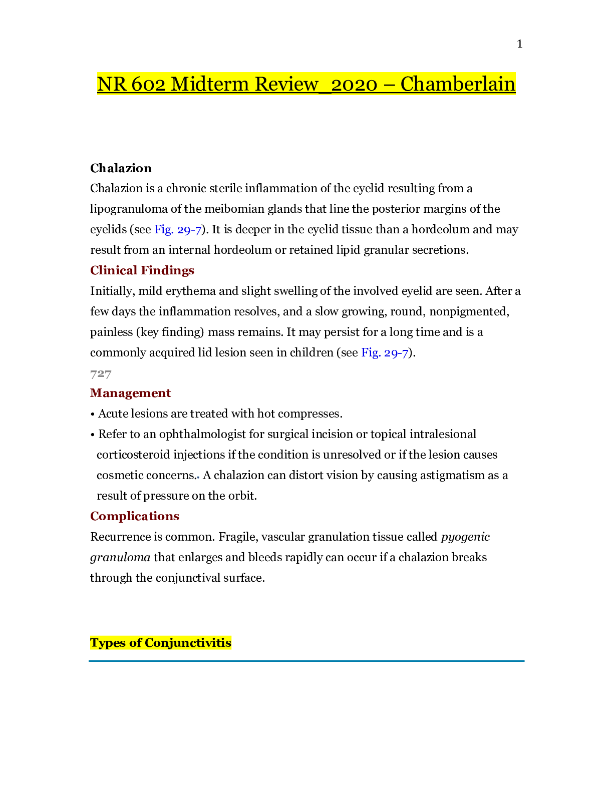
Chalazion
Chalazion is a chronic sterile inflammation of the eyelid resulting from a lipogranuloma of the meibomian glands that line the posterior margins of the eyelids (se
...
NR 602 Midterm Review_2020 – Chamberlain
Chalazion
Chalazion is a chronic sterile inflammation of the eyelid resulting from a lipogranuloma of the meibomian glands that line the posterior margins of the eyelids (see Fig. 29-7). It is deeper in the eyelid tissue than a hordeolum and may result from an internal hordeolum or retained lipid granular secretions.
Clinical Findings
Initially, mild erythema and slight swelling of the involved eyelid are seen. After a few days the inflammation resolves, and a slow growing, round, nonpigmented, painless (key finding) mass remains. It may persist for a long time and is a commonly acquired lid lesion seen in children (see Fig. 29-7).
727
Management
• Acute lesions are treated with hot compresses.
• Refer to an ophthalmologist for surgical incision or topical intralesional corticosteroid injections if the condition is unresolved or if the lesion causes cosmetic concerns. A chalazion can distort vision by causing astigmatism as a result of pressure on the orbit.
Complications
Recurrence is common. Fragile, vascular granulation tissue called pyogenic granuloma that enlarges and bleeds rapidly can occur if a chalazion breaks through the conjunctival surface.
Types of Conjunctivitis
Type Incidence/Etiology Clinical Findings Diagnosis Management*
Ophthalmia neonatorum Neonates: Chlamydia trachomatis, Staphylococcus aureus, Neisseria gonorrhoeae, HSV (silver nitrate reaction occurs in 10% of neonates) Erythema, chemosis, purulent exudate with N. gonorrhoeae; clear to mucoid exudate with chlamydia Culture (ELISA, PCR), Gram stain, R/O N. gonorrhoeae, chlamydia Saline irrigation to eyes until exudate gone; follow with erythromycin ointment
For N. gonorrhoeae:ceftriaxone or IM or IV
For chlamydia: erythromycin or possibly azithromycin PO
For HSV: antivirals IV or PO
Bacterial conjunctivitis In neonates 5 to 14 days old, preschoolers, and sexually active teens: Haemophilus influenzae(nontypeable), Streptococcus pneumoniae, S. aureus, N. gonorrhoeae Erythema, chemosis, itching, burning, mucopurulent exudate, matter in eyelashes; ↑ in winter Cultures (required in neonate); Gram stain (optional); chocolate agar (for N. gonorrhoeae) R/O pharyngitis, N. gonorrhoeae, AOM, URI, seborrhea Neonates: Erythromycin 0.5% ophthalmic ointment
≥1 year old: Fourth-generation fluoroquinolone
For concurrent AOM: Treat accordingly for AOM
Warm soaks to eyes three times a day until clear
No sharing towels, pillows
No school until treatment begins
Chronic bacterial conjunctivitis (unresponsive conjunctivitis previously treated as bacterial in etiology) School-age children and teens: Bacteria, viruses, C. trachomatis Same as above; foreign body sensation Cultures, Gram stain; R/O dacryostenosis, blepharitis, corneal ulcers, trachoma Depends on prior treatment, laboratory results, and differential diagnoses
Review compliance and prior drug choices of conjunctivitis treatment
Consult with ophthalmologist
Inclusion conjunctivitis Neonates 5 to 14 days old and sexually active teens: C. trachomatis Erythema, chemosis, clear or mucoid exudate, palpebral follicles Cultures (ELISA, PCR), R/O sexual activity Neonates: Erythromycin or azithromycin PO
Adolescents: Doxycycline, azithromycin, EES, erythromycin base, levofloxacin PO
Viral conjunctivitis Adenovirus 3, 4, 7; HSV, herpes zoster, varicella Erythema, chemosis, tearing (bilateral); HSV and herpes zoster: unilateral with photophobia, fever; zoster: nose lesion; spring and fall Cultures, R/O corneal infiltration Refer to ophthalmologist if HSV or photophobia present
Cool compresses three or four times a day
Allergic and vernal conjunctivitis Atopy sufferers, seasonal Stringy, mucoid exudate, swollen eyelids and conjunctivae, itching (key finding), tearing, palpebral follicles, headache, rhinitis Eosinophils in conjunctival scrapings Naphazoline/pheniramine, naphazoline/antazoline ophthalmic solution (see text)
Mast cell stabilizer (see text)
Refer to allergist if needed
*See text for dosages.
Blepharitis
Blepharitis is an acute or chronic inflammation of the eyelash follicles or meibomian sebaceous glands of the eyelids (or both). It is usually bilateral. There may be a history of contact lens wear or physical contact with another symptomatic person. It is commonly caused by contaminated makeup or contact lens solution. Poor hygiene, tear deficiency, rosacea, and seborrheic dermatitis of the scalp and face are also possible etiologic factors. The ulcerative form of blepharitis is usually caused by S. aureus. Nonulcerative blepharitis is occasionally seen in children with psoriasis, seborrhea, eczema, allergies, lice infestation, or in children with trisomy 21.
Clinical Findings
• Swelling and erythema of the eyelid margins and palpebral conjunctiva
726
• Flaky, scaly debris over eyelid margins on awakening; presence of lice
• Gritty, burning feeling in eyes
• Mild bulbar conjunctival injection
• Ulcerative form: Hard scales at the base of the lashes (if the crust is removed, ulceration is seen at the hair follicles, the lashes fall out, and an associated conjunctivitis is present)
Differential Diagnosis
Pediculosis of the eyelashes.
Management
Explain to the patient that this may be chronic or relapsing. Instructions for the patient include:
• Scrub the eyelashes and eyelids with a cotton-tipped applicator containing a weak (50%) solution of no-tears shampoo to maintain proper hygiene and debride the scales.
• Use warm compresses for 5 to 10 minutes at a time two to four times a day and wipe away lid debris.
• At times antistaphylococcal antibiotic (e.g., erythromycin 0.5% ophthalmic ointment) is used until symptoms subside and for at least 1 week thereafter. Ointment is preferable to eye drops because of increased duration of contact with the ocular tissue. Azithromycin 1% ophthalmic solution for 4 weeks may also be used (Shtein, 2014).
• Treat associated seborrhea, psoriasis, eczema, or allergies as indicated.
• Remove contact lenses and wear eyeglasses for the duration of the treatment period. Sterilize or clean lenses before reinserting.
• Purchase new eye makeup; minimize use of mascara and eyeliner.
• Use artificial tears for patients with inadequate tear pools.
Chronic staphylococcal blepharitis and meibomian keratoconjunctivitis respond to oral erythromycin. Doxycycline, tetracycline, or minocycline can be used chronically in children older than 8 years old.
Hand-Foot-Mouth Syndrome
Enteroviruses
Nonpolio Enteroviruses
Of the more than 100 serotypes of nonpolio RNA enteroviruses, 10 to 15 serotypes account for most diseases. They are grouped into four genomic classifications: human 495enteroviruses (HEVs) A, B, C, and D. Coxsackieviruses and echoviruses are subgroups of HEVs. Hand-foot-mouth, herpangina, pleurodynia, acute hemorrhagic conjuncti¬vitis, myocarditis, pericarditis, pancreatitis, orchitis, and dermatomyositis-like syndrome are manifestations of infection. These enteroviruses are the most common cause of aseptic meningitis and have also been associated with paralysis, neonatal sepsis, encephalitis, and other respiratory and GI symptoms. The specific serotype may not be unique to any given disease (Abzug, 2011).
As evidenced by the name, enteroviruses concentrate on the GI tract as their primary invasion, replication, and transmission site; they spread by fecal-oral contamination, especially in diapered infants. They are also transmitted via the respiratory route and vertically either prenatally, during parturition, or possibly by way of breastfeeding by an infected mother who lacks antibodies to that particular serotype. Transplacental infection can lead to serious disseminated disease in the neonate that involves multiorgan systems (liver, heart, meninges, and adrenal cortex).
Enteroviruses have worldwide distribution, occurring in temperate climates during the summer and fall and in tropical climates year round. In known cases, infants younger than 12 months old have the highest prevalence rate (>25%), and HEVs account for 55% to 65% of hospitalizations for suspected infant sepsis. Illness occurs more frequently in males; those living in crowded, unsanitary conditions; and in those of lower socioeconomic status (Abzug, 2011). Infection can range from asymptomatic to undifferentiated febrile illness to severe illness. Young children are more likely to be symptomatic. The incubation period is 3 to 6 days (less for hemorrhagic conjunctivitis). After infection, the virus is shed from the respiratory tract for up to 3 weeks and from the GI tract for up to 7 to 11 weeks; it is viable on environmental surfaces for long periods.
Nonpolio enteroviral infection is not a reportable disease, nor is it routinely tested for in the clinical setting, so the overall incidence rate is not known. The CDC administers the National Respiratory and Enteric Virus Surveillance System (NREVSS) and the National Enterovirus 496Surveillance System (NESS) to monitor detection patterns of respiratory and enteric adenoviruses. The 2014 outbreak of an illness in children referred to as acute flaccid myelitis bears some similarity to infections caused by viruses, including enterovirus; epidemiologic studies are ongoing (CDC, 2015f).
-
-
-
-
-
-
-
-
-
-
-
Continued
[Show More]