*NURSING > DISCUSSION POST > BIOS 255 Week 3 Discussion: Blood Vessel Structure – Download To Pass Exams (All)
BIOS 255 Week 3 Discussion: Blood Vessel Structure – Download To Pass Exams
Document Content and Description Below
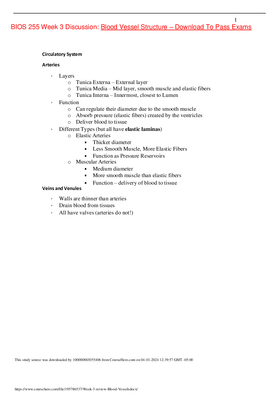
BIOS 255 Week 3 Discussion: Blood Vessel Structure – Download To Pass Exams Circulatory System Arteries • Layers o Tunica Externa – External layer o Tunica Media – Mid layer, smooth ... muscle and elastic fibers o Tunica Interna – Innermost, closest to Lumen • Function o Can regulate their diameter due to the smooth muscle o Absorb pressure (elastic fibers) created by the ventricles o Deliver blood to tissue • Different Types (but all have elastic laminas) o Elastic Arteries ▪ Thicker diameter ▪ Less Smooth Muscle, More Elastic Fibers ▪ Function as Pressure Reservoirs o Muscular Arteries ▪ Medium diameter ▪ More smooth muscle than elastic fibers ▪ Function – delivery of blood to tissue Veins and Venules • Walls are thinner than arteries • Drain blood from tissues • All have valves (arteries do not!) This study source was downloaded by 100000883055406 from CourseHero.com on 04-01-2024 12:39:57 GMT -05:00 Hepatic Portal System o Intestines (GI Tract) -> Liver o After a meal, hepatic portal blood is rich in nutrients absorbed from the GI tract. These nutrients are passed through the splenic and superior mesenteric veins, which combine to form the hepatic portal vein. Hepatic portal vein delivers the nutrients to the liver. The liver then stores some of the nutrients, modifies others, and passes them into general circulation. Major Vessels used for diagnostic purposes: o Draw Blood/Vasopuncture – Median Cubital Vein o Central Line – Basilic or Cephalic Vein o Feeling pulse – radial, carotid, femoral arteries What Parts of the body drain into what vessel / supplied from: o Face – Sup. Vena Cava / common carotid o Arms – Sup. Vena Cava subclavian/axillary o Trunk – Inf. Vena Cava / abdominal aorta o Legs – Inf. Vena Cava / common iliac This study source was downloaded by 100000883055406 from CourseHero.com on 04-01-2024 12:39:57 GMT -05:00 Arteries supply blood to what structure(s); Veins drain blood from what structure(s): Structure Supplied from (ARTERIES) Drains into (VEINS) Heart Coronary Arteries Coronary Veins Face and scalp Carotids External jugular vein Head/Neck Common Carotid Arteries Brain, Spinal cord, Neck, Shoulder, Scapular Left and Right Subclavian (Supplies L&R sides respectively) Jugular Brain Vertebral Artery Internal Jugular Vein Thorax, Armpit, Upper limb Axillary Arteries Axillary Vein > Subclavian Thorax only Subclavian/axillary Azygos vein Deep Arm muscles Brachial Arteries Brachial > Axillary Superficial Arm Basilic Vein > Brachial Cephalic > Subclavian Posterior forearm Radial Arteries Radial Vein > Brachial Anterior forearm Ulnar Arteries Ulnar Vein > Brachial Vein Hands Deep/Superficial Palmar Arches Cephalic>Subclavian Basilic>Brachial GI Tract Celiac Trunk Spleenic > Hepatic Vein > Liver Spleen, Pancreas, Stomach Splenic Arteries Inf. Vena Cava Spleen> Hepatic > Liver Stomach Gastric Arteries Inf. Vena Cava Liver Common Hepatic Artery Hepatic Vein > Inf. Vena Cava Right side of intestine Sup. Mesenteric Artery Inf. Vena Cava Kidneys Renal Arteries Renal Veins Left side of Intestines Inf. Mesenteric Artery Inf. Vena Cava Pelvic Organs, Lower Limbs Common Iliac Arteries Common Iliac Vein >Inf. Vena Cava Pelvic Organs, Buttocks, Genitals Internal Iliac Arteries Internal Iliac Vein > Common Iliac Pelvic Organs External Iliac Arteries External Iliac Vein > Common Iliac Thigh Femoral Artery Femoral Vein > External Iliac Vein Knee Popliteal Artery Popliteal Vein > Femoral Vein Anterior Leg Anterior Tibial Artery Anterior Tibial Vein > Popliteal Vein This study source was downloaded by 100000883055406 from CourseHero.com on 04-01-2024 12:39:57 GMT -05:00 Posterior Leg Posterior Tibial Artery Posterior Tibial Artery > Popliteal Feet Great Saphenous Vein > Right Femoral Legs only Iliac and Femoral External iliac vein Integument of legs Iliac and Femoral Great Saphenous Vein ALL upper body Superior Vena Cava ALL lower body Inferior Vena Cava Structural characteristics of vessels Resistance – Opposing force to blood flow. Increased resistance = Decreased CO Factors that affect resistance (which will ultimately affect blood flow): o Viscosity o Blood vessel diameter (vasodilation/vasoconstriction) o Length of Vessel ** At rest most of your blood is housed in systemic veins and venules, hence veins are considered “Blood Reservoirs” This study source was downloaded by 100000883055406 from CourseHero.com on 04-01-2024 12:39:57 GMT -05:00 (While arteries are considered “Pressure Reservoirs”) This study source was downloaded by 100000883055406 from CourseHero.com on 04-01-2024 12:39:57 GMT -05:00 Calculations – Watch your UNITS!!! Stroke Volume – SV SV = EDV - ESV Answer should be in (ml/beat) Cardiac Output – CO CO = HR x SV Answer should be in (ml/min) Blood Pressure – BP Systolic pressure Diastolic pressure Answer should be in (mm hg) Pulse Pressure – PP Systolic press. – Diastolic press. Answer should be in (mm hg) Mean Arterial Pressure – MAP Diastolic Press. + (PP/3) Answer should be in (mm hg) [Show More]
Last updated: 1 year ago
Preview 1 out of 6 pages
Buy this document to get the full access instantly
Instant Download Access after purchase
Buy NowInstant download
We Accept:

Reviews( 0 )
$10.00
Can't find what you want? Try our AI powered Search
Document information
Connected school, study & course
About the document
Uploaded On
Apr 01, 2024
Number of pages
6
Written in

Additional information
This document has been written for:
Uploaded
Apr 01, 2024
Downloads
0
Views
91