*NURSING > EXAM PROCTORED > ATI proctored exam | Quinnipiac University - NUR 300/400 | Grade A Study Guide (All)
ATI proctored exam | Quinnipiac University - NUR 300/400 | Grade A Study Guide
Document Content and Description Below
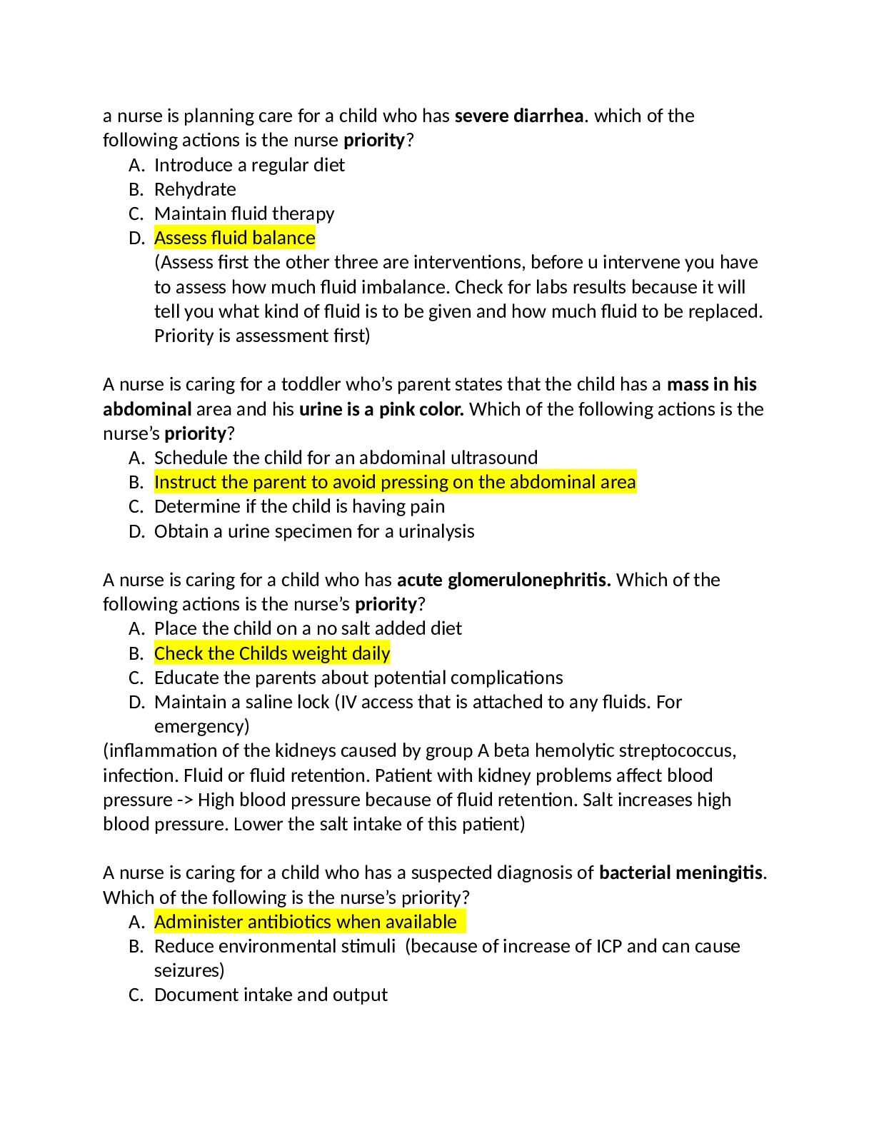
ATI proctored exam Chapter 1 & 2: • Medications: amiodarone, lidocaine, magnesium, procainamide, and vasopressin these are antiarrhythmic used in life threatening cardiac problems Alpha 1... - cause skin and mucous membranes to vasoconstrict Beta 1- stimulate the heart (you have 1 heart) Beta 2- affects heart and lungs (you have 2 lungs) bronchodilation in lungs, and uterine smooth muscle to relax. Dopamine- causes renal blood vessels to dilate Epinephrine- triggers alpha 1, and beta 1 and 2 receptors. Vasoconstriction, increased HR, and bronchodilation SE: Hypertensive crisis, dysrhythmias, angina, Dopamine- triggers dopamine receptors, beta 1 receptors, useful for shock and heart failure Dobutamine- triggers beta 1 receptors (heart rate increased, and used for heart failure) CH. 3 Neurologic diagnostic procedures Cerebral angiogram: catheter inserted in groin or neck. Contrast dye is used so be aware of pregnancies, shellfish and iodine allergies, check for patient’s renal function (BUN, creatnine). Check is patient is on anticoagulant because bleed risk. NPO 4-6 hrs before procedure. After procedure, check distal pulses and bleeding at site. EEG: Used to detect seizures, but can also check for sleep disorders and behavioral changes. Wash hair prior to hair, and be sleep deprived, don’t have to fast for this. This stress can trigger seizures or abnormal brain activities. Glasgow Coma scale: how we determine level of consciousness. Highest score is 15; anything less then 8 is severe head injury and coma. 4 are eyes opening. Intracranial pressure & monitoring: If someone has a low Glasgow scale more monitoring needs to be done. High infection risk with the machine that is taking pressure. Symptoms in increase ICP: irritability, headache, decreasing LOC, no pupil response, alterations in breathing, decorticate posturing. Normal intracranial pressure should be between 10-15. Lumbar puncture: Used to withdrawal small amt of CSF to treat for diseases. Lay in cannonball position, have patient lay flat for an hour. If clotting doesn’t occur afterwards, CSF may leak and they’ll get a really bad cranial headache. MRI: Remove jewelry, check for claustrophobia (might need sedation), check for implants containing metal. Ch. 4: Pain management Acute pain: temporary, protective and usually resolves with tissue healing Chronic pain: goes past 6 months and includes depression, fatigue Nociceptive pain: damage or inflammation of tissue. Localized. Described as throbbing or aching -Somatic: Bones, joints, muscles -Visceral: internal organs -Cutaneous: in the skin or subQ tissue Check on aggravating vs. Relieving symptoms- what makes it worse/what makes it better. Non-pharmacological and pharmacological methods of managing pain Non-pharmacological: imagery, acupuncture, relaxation, TRANS, application of heat and cold, therapeutic touch/massage Pharmacological: Percocet (be away there is Tylenol in this), Tylenol, tinnitus or vertigo is a side effect associated with aspirin (salicylates), gastric ulcers are side effects for NSAID’s. Opioids are go to for moderate to severe pain. Opioid SE: constipation, urinary retention, orthostatic hypotension, n/v, sedation, respiratory depression. Have order for narcan on hand incase respirations get too low. Ch 5: Meningitis What is it? Inflammation of the meninges. These are the membranes that surround the brain and spiral cord. Can either be viral or bacterial. Viral is more common and will self-resolve. Bacterial meningitis is very worse and requires antibiotics. Key vaccines= HIV vaccine, and then MCV4 vaccine. Symptoms of meningitis: headache, neck stiffness, photophobia, fever & chills, n/v, altered level of consciousness, tachycardia, seizures, red macular rash, and increased ICP. Positive brudinski sign (lay pt flat and pull up on their head and that will cause them to flex in their knees bc it hurts. Remember it because “Bro- that hurts my neck- brudinski sign). Kernig- knee bending sign. Diagnosing meningitis: CSF sample. Cloudy= bacterial, viral= clear. Either way you’re likely to see increased WBC, bacterial meningitis you can see a decreased glucose value. Nursing care: Droplet precautions. Viral meningitis can be put on standard precautions after 24 hrs. Maintain quiet environment and no noise. Decrease intracranial pressure; have HOB increased at 30 degrees. No coughing/deep breathing for patient. Complications: increased intracranial pressure (SE: irritability, decreased LOC, change in pupil response, SIADH). Seizures Risk factors for seizures: Genetics Fevers Head trauma Cerebral edema Infection (such as meningitis) Metabolic disorder (such as hypoglycemia/hypernatremia) Hypoxia Fluid or electrolyte imbalances Other triggers: Excessive stress Overwhelming fatigue Excess caffeine Flashing lights Generalized seizure “tonic/clonic: Some people have an aura before it happens. Tonic means stiffening of muscles and loss of consciousness. Clonic episode is 1-2 minutes of jerking movements of extremities. Absent seizure: loss of consciousness lasting a few seconds looks like spacing out. This is common in school age people. They’ll do eye flutter, staring etc. Atonic seizure: no tone, you lose all muscle tone and they end up falling down. Ch 6. Diagnostic procedures for seizures: • EEG • Cat scan • MRI • CSF sample Nursing interventions during a seizure: • Gently lower patient to the ground • Place on their side • Move anything away from them • Loosen restrictive clothing • Do NOT try to move them or put anything in their mouth • Document the onset and duration of seizure • After seizure: keep in side lying position, check vitals, do neurological checks, re-orient patient, put on seizure precautions, determine if there was a trigger for the seizure Seizure meds: • Antiepileptic drugs o Phenytoin- they will need to have periodic blood checks to check the levels of phenytoin in the blood. SE: gingival hyperplasia o Phenytoin decreased effectiveness of oral contraceptives, Coumadin, and warfarin. Surgical interventions: -Patient might have nerve stimulator implanted and when they have a seizure they can hold the magnetic over the implantable device. No MRI’s, and no use of microwaves. Patient might have a interruption of brain tissue and may require a craniotomy. Have patient wear a medical identification badge. Status epilepticus- Prolonged seizure activity that occurs over a 30-minute time frame. EKG monitoring, o2, medications, vitals, continuous infusion of phenytoin for seizure prevention. Ch 7: Parkinson’s disease Mostly affects motor function. The balance of dopamine and acetylcholine are off balance in the body (acetylcholine too high). This is because the substantia nigra is off balance. Symptoms: Slow shuffling gait Mask like expression Difficulty chewing and swallowing Drooling Difficulty with ADL’s Mood swings Cognitive impairments Nursing care: Monitor swallowing. May need to thicken foods. Encourage exercise and ROM exercises Speak slowly May need to use alternate forms of communication Meds: Levodopa often combined with carbidopa Levodopa- increases dopamine levels in the body Anticholinergics- Be aware of the side effects which are dry mouth, constipation, urinary retention. Complications of Parkinson’s: Aspiration pneumonia- keep patient upright, have suction equipment nearby Ch. 8: Alzheimer’s disease Occurs after the age of 65. It is memory loss, personality changes, and problems with judgment. Stage 1: no impairment Stage 2: mild cognitive decline Stage 3: mild cognitive decline with short term memory loss Stage 4: moderate cognitive decline. This stage has personality changes, and memory loss Stage 5: moderate severe cognitive decline Stage 6: severe cognitive decline. Likely to have episodes of fecal and urinary incontinence. Stage 7: Very severe cognitive decline. Pt loses ability to speak, move, eat, and swallow. Diagnostics: Brain tissue biopsy once patient dies for definitive diagnosis of Alzheimer’s. Therapeutic procedures: Avoid over stimulation, stick to routines, reorient patient frequently. Home safety measures: remove rugs, install door locks, have good light, use colored tape on the edge of the stairs, remove clutter, put mattress on floor to prevent fall. Meds: Donepazil- increases amount of acetylcholine available and presents breakdown and helps improve behavior and cognition. *If a patient has a brain tumor, the hypothalamus can become damaged and lead to SIADH or diabetes insipitus. Ch. 9: MS- An autoimmune disorder, plaque damages the myelin sheath. Chronic disease, no known cure, affects patients between 20-40 and normally women. Viruses, cold climates, physical injuries, stress, pregnancy, fatigue, hot baths and shower can trigger an exacerbation. MS symptoms: Diplopia Decreased visual acuity Tinnitus Hearing acuity Dysphagia Slurred/nasal speech Nystagmus Bowel dysfunction Urinary incontinence Impaired judgment Sexual dysfunction Muscle spasticity Diagnostic for MS: MRI, which will show plaques on brain Meds: cyclosporine, prednisone, muscle relaxers (dantrolene, baclofen) ALS: Degenerative neurologic disorder causes progressive paralysis. Affects respiratory muscles and causes respiratory failure and then death. Cause unknown. No known cure. ALS Symptoms: Muscle atrophy Difficulty swallowing Respiratory affects Meds: Rilusole- glutamate antagonist and can help the deterioration of causes Complications: Pneumonia because of the respiratory problems Ch. 10: Myasthenia Gravis What is it? An autoimmune disorder which causes weakness. What causes exacerbations? Fatigue, pregnancy, hot water, infection These patients have hyperplasia (enlargement) of the thymus gland Symptoms of myasthenia gravis: Muscle weakness Diplopia Difficulty chewing and swallowing Respiratory function Bowel/bladder incontinence Drooping eyelids Diagnosis of MG: These patients’ symptoms mimic symptoms of a cholinergic crisis (muscle weakness, and impaired respiratory function). So in order to determine which it is you will give patient idraphonium, which increases amount of acetylcholine at the junction, if symptoms improve it, was a MG exacerbation, if the symptoms worsen, then it was a cholinergic crisis. So you will want to have atropine, which is the antidote for cholinergic crisis. Patient care for patient with MG: Maintain patent airway Make sure o2, intubation, and suctioning is nearby Allow for rest periods Provide small frequent high calorie meals Have patient sit upright Lubricating eye drops (you may have to put tape when going to sleep for cornea) MG meds: Pyredostigme, and neostigmine- inhibits breakdown of acetylcholine at junction. Therapeutic procedure: plasmopharesis, which removes antibodies in plasma which helps to improve symptoms Surgical interventions for MG: Thymectomy- removal of thymus gland to allow patient go into remission. Ch. 11: Headaches Migraines, and cluster headaches Causes: Odors, bright lights, fatigue, noises, hormone changes, foods containing tymarime MSG or nitrates Migraine headache symptoms: Photophobia, phonophobia, nausea, and vomiting, unilateral pain. Typically last 4-72 hours. Some people get this with an aura. Nursing interventions: Have cool, dark, quiet, and environment Elevate HOB to 30 degrees NSAID’s and an antiemetic for the n/v Educate patient to reduce intake of tyramine, MSG, and preservatives Cluster headache: Brief cluster of intense pain. Lasts for 30min-4hr. Usually occur daily, at the same time, during the spring and fall. No warning signs. Less common then migraines. Found in men between 20-5o years. You will also find tearing of the eye with a runny nose and nasal congestion, facial sweating, and a drooping eyelid. Meds for cluster headache: Triptan Ergotamine NSAID’s Ch. 12: Eye disorders Macular degeneration: #1 cause of vision loss. No cure. Symptoms: Blurred vision Loss of central vision Can cause blindness Nursing care: Eat foods high in antioxidants, vitamin E Cataracts: Opacity in the lens of the eye that impairs vision. Progressive, and painless loss of vision. No red reflex. Treatment: Offer large print reading material Cataract surgery- they remove the lense of the eye. Vision will be fixed 4-6wks after. Post surgery nursing care: Instruct patient to wear sunglasses outside Report signs of infection Yellow/green discharge Avoid activity that increases IOP (no bending at waist, avoid sneezing, coughing, straining, bearing down, hyper flexing head, tilting head back, limit house chores, avoid driving/sports) Glaucoma: Function or structural issue with the optic nerve. Leading cause of blindness. 2 types= Open angle symptoms: mild eye pain, loss of peripheral vision, elevated IOP (over 21) Closed angle symptoms: Severe pain, nausea, photophobia, and rapid onset of elevated IOP Tonometry: Measures IOP should be between 10-21 Meds: Medicated eye drops Q 12 hrs. Pylocarpine- constricts pupil Beta-blockers Mannitol- diuretic that can be used to decrease IOP Pylocarpy Encourage patient to limit activities that increase IOP. Ch. 13: Middle and inner ear disorders Middle ear infection= aka otitis media. Risk factors: recurrent colds, respiratory infection, and large adenoids Symptoms: red/inflamed ear canal and tympanic membrane Inner ear issues: Meniere’s disease **Symptoms: Tinnitus, vertigo, lateral hearing loss Risk factors: viral/bacterial infection, damage due to toxicity Ch. 14: Head injuries Head injury due to an accident: Understand this patient is at risk for a cervical spine injury so stabilize this until it’s ruled out. Assess patient for increased cranial pressure Symptoms of ICP: Severe headache Deteriorating LOC Low Glasgow coma scale ** Irritability * first sign Nonreactive pupils Alterations in breathing patterns Cushing’s reflex (severe hypertension, widening pulse pressure, bradycardia) Leakage from nose/ears (if it looks like blood or yellow halo sign fluid it is injury to the head) Ch. 15: Stroke/cerebral vascular accident Hemorrhagic- patient has a ruptured artery or aneurism Embolis- a blood clot travels and ends up in the cerebral artery Risk factors of stroke: Hypertension Obese Lack of physical activity Diabetes Symptoms of CVA: Visual disturbances Dizziness Slurred speech Facial drooping Weak extremity Right-sided stroke complications: Affects visual and spatial awareness May overestimate abilities Poor impulse control and judgment Left sided stroke complication: Deficits to language, math, and analytical think Expressive dysphagia Difficulty reading or writing Hemianopia Diagnosis: MRI CT Watch out for dysphagia and monitor for a gag reflex Patient teaching: Have patient keep neck forward and flexed to swallow Risk for skin injury to reposition patient frequently Provide safe environment Help prevent falls Homonymous hemianopia- loss of visual field, if they have this then you want to teach the patient to turn their head from unaffected side to affected side to see everything Medications: Thrombolytics- retiplase, alteplase- help break up the clot. Give within 4.5 hours of initial stroke Surgical intervention: Carotid artery angioplasty with stenting Ch. 16: Spinal Cord Injury If a patient injured the cervical region this causes quadriplegia, below level of T1 causes paraplegia, is injury above level of C4 then they are at risk for respiratory issues. Neurogenic shock- sometimes happens after the surgery. This causes total loss of voluntary functioning. They’ll have hypotension, dependent edema Nursing interventions for spinal cord injuries: Daily stool softeners Catheterization Dantrolene- muscle relaxer than can be given for spastic muscle tone loss. Complications: Orthostatic hypotension Change clients position slowly Treat hypotension and bradycardia with meds (atropine) *Autonomic dysreflexia* Extreme hypotension, pallor, diaphoresis. Notify provider, check for catheter and kinking, check for fecal impaction, loosen tight clothing, check for lower extremity fracture Ch. 17: ABG’s Taking arterial blood Hold pressure to this site for 5 minutes, if patient is on blood thinners then hold for 20 min Complications: Hematoma Air embolism- if this is suspected, position patient on left side in tredenburg position. SOB, anxiety, air hunger Diagnostics: Bronchoscopy- patient should be NPO 8-12 hrs. Prior to procedure Thoracentesis- chest is perforated and you go into pleural space and most likely remove fluid Unexpected findings: Bronchospasm Complications: Mediastina shift Pneumothorax (SxS=deviated trachea, pain on the affected side) Ch. 18: Chest tubes Help to remove excess blood, fluid, or air Water chamber should have 2 cm of water in there There should not be bubbling in the water seal Nursing care for chest tube: Cough and deep breathe Q 2 hrs. Greater then 70 mL per hour of drainage you need to contact provider Monitor insertion site and position patient upright for lung expansion and drainage After procedure you want to get x-ray to make sure it is placed properly Keep 2 enclosed hemostats, sterile water, and occlusive dressing at the bedside. Monitor water seal chamber, there should not be bubbling Air leak You can immerse the tip into sterile water to maintain the seal If the chest tube falls out of patient you want to put an occlusive dressing and tape it on 3 sides to patient Tension pneumothorax- usually happens because there’s a kink in the tubing Deep breath, bare down, while chest tube is being taken out. Ch. 19: Oxygen and mechanical ventilation Nasal cannula: can deliver between 1-6 L per min Partial re-breather mask-6-11 L per min Non-re-breather mask-10-15 L per min, bag should be 2/3 full and perform hourly assessments Venturi mask- the right method if you need precise oxygen concentration Aerosol mask- can be used it patient has a facial trauma or burn Why would a patient need oxygen? Hypoxia Tachypnea Tachycardia Restlessness Signs of respiratory distress (use of accessory muscles etc.) Late signs= confusion/stupor, cyanotic skin, bradypenia, bradycardia, hypotension Oxygen toxicity- Signs include non-productive cough, sub sternal pain, nasal stuffiness, sore throat, and hypoventilation. Low pressure alarms: Can be caused due to a disconnection, cuff leak, or tube displacement. High-pressure alarms: Can indicate excess secretions, client might be biting on tube; kink in tubing, client may be coughing, or indicative of pulmonary edema bronchospasm or a pneumothorax. [Show More]
Last updated: 2 years ago
Preview 1 out of 12 pages

Buy this document to get the full access instantly
Instant Download Access after purchase
Buy NowInstant download
We Accept:

Reviews( 0 )
$10.50
Can't find what you want? Try our AI powered Search
Document information
Connected school, study & course
About the document
Uploaded On
Mar 08, 2021
Number of pages
12
Written in

Additional information
This document has been written for:
Uploaded
Mar 08, 2021
Downloads
0
Views
110


.png)
 (1).png)