AHA Exam 2 Review
● Test consists of cardiac, respiratory, skin, and Neuro (Headaches)
● Make sure that you read the questions before answering- this test will have multiple scenarios, chief complaints, and assessment
...
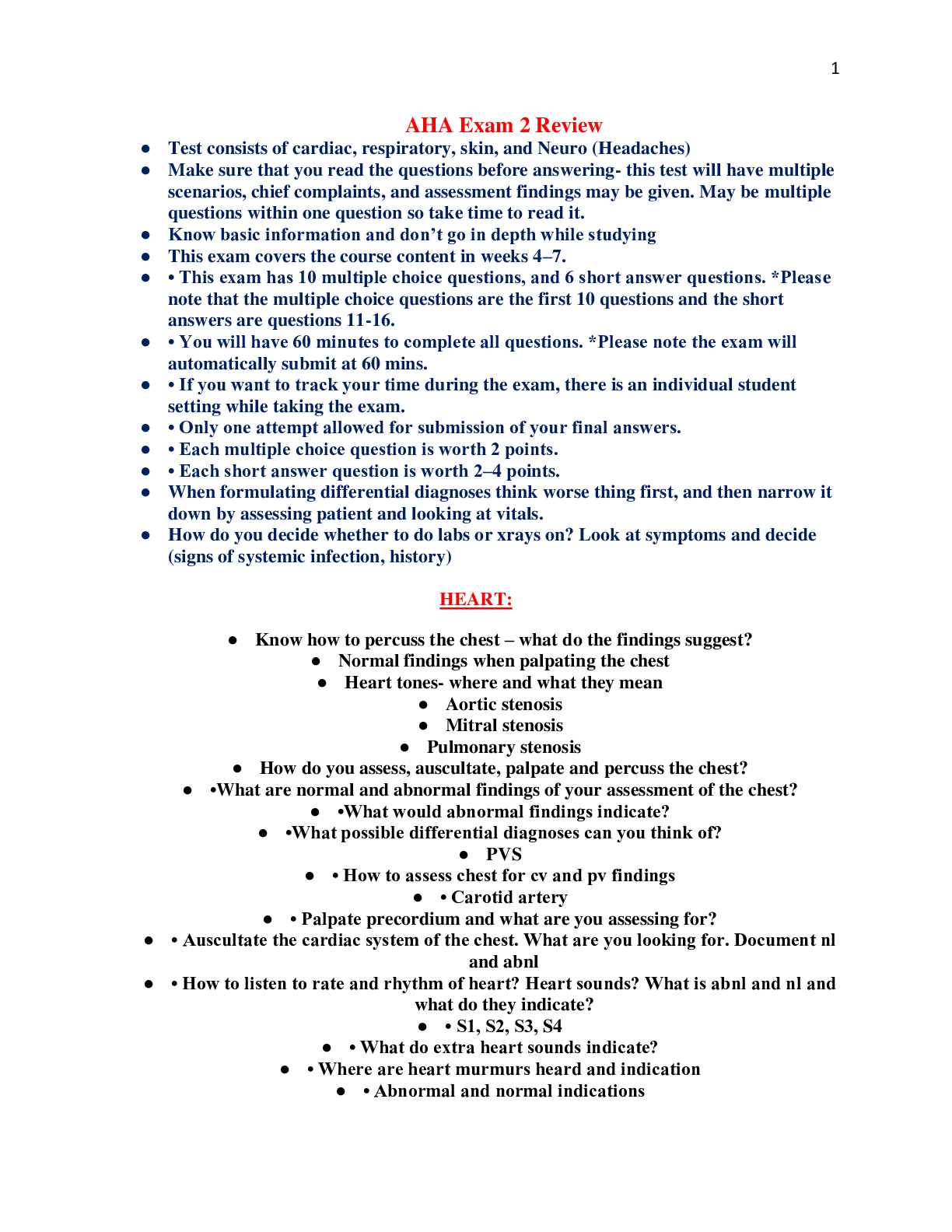
AHA Exam 2 Review
● Test consists of cardiac, respiratory, skin, and Neuro (Headaches)
● Make sure that you read the questions before answering- this test will have multiple scenarios, chief complaints, and assessment findings may be given. May be multiple questions within one question so take time to read it.
● Know basic information and don’t go in depth while studying
● This exam covers the course content in weeks 4–7.
● • This exam has 10 multiple choice questions, and 6 short answer questions. *Please note that the multiple choice questions are the first 10 questions and the short answers are questions 11-16.
● • You will have 60 minutes to complete all questions. *Please note the exam will automatically submit at 60 mins.
● • If you want to track your time during the exam, there is an individual student setting while taking the exam.
● • Only one attempt allowed for submission of your final answers.
● • Each multiple choice question is worth 2 points.
● • Each short answer question is worth 2–4 points.
● When formulating differential diagnoses think worse thing first, and then narrow it down by assessing patient and looking at vitals.
● How do you decide whether to do labs or xrays on? Look at symptoms and decide (signs of systemic infection, history)
HEART:
● Know how to percuss the chest – what do the findings suggest?
● Normal findings when palpating the chest
● Heart tones- where and what they mean
● Aortic stenosis
● Mitral stenosis
● Pulmonary stenosis
● How do you assess, auscultate, palpate and percuss the chest?
● •What are normal and abnormal findings of your assessment of the chest?
● •What would abnormal findings indicate?
● •What possible differential diagnoses can you think of?
● PVS
● • How to assess chest for cv and pv findings
● • Carotid artery
● • Palpate precordium and what are you assessing for?
● • Auscultate the cardiac system of the chest. What are you looking for. Document nl and abnl
● • How to listen to rate and rhythm of heart? Heart sounds? What is abnl and nl and what do they indicate?
● • S1, S2, S3, S4
● • What do extra heart sounds indicate?
● • Where are heart murmurs heard and indication
● • Abnormal and normal indications
● • Know ekg strips.
● • Review the sample documentation. May see short answers about it.
● Pearl of Wisdom…
● •When I learned S3 and S4 in undergraduate nursing school, I learned S3 is a gallop and I often heard this in ER and ICU with the bell of my stethoscope.
● •I heard ken tuck EE ken tuck EE ken tuck EE.
● •An S4 I learned in undergraduate nursing school too. This is often in diastolic heart failure, severe left ventricular hypertrophy, active cardiac ischemia. It goes S4, S1 S3. tenUHseetenUHseetenUHsee
● •Go go www.medzcool.com - It’s a site I like really well for heart sounds.
Questions about chest pain and what you should be asking
▪ Subjective Data:
o Substernal pain or intense pressure radiating to the neck, jaws, and arms; particularly in the left arm
o Often accompanied by shortness of breath, fatigue, diaphoresis, faintness, and syncope
▪ Objective Data:
o Are you having current chest pain?
o What medicines are you currently taking?
o Do you have any comorbidities (other medical dx)?
o Is this your 1st time having CP or is this recurrent?
o How long has the chest pain been occuring?
o Is it constant or intermittent?
o Does the pain radiate anywhere?
o How long does the chest pain last?
o What does the pain feel like (sharp, crushing, dull ache)?
o What number would you state it is from 1-10?
o Do you have any other symptoms with the chest pain (shortness of breath, nausea, vomiting, dizziness, syncope, palpitations, cough)?
o What makes the chest pain worse (activity, intercourse, eating, stress, laying flat)?
o What makes the pain better (rest, medications, sitting up)?
o Have you had an injury to your chest recently?
What are some causes of an increase or decrease in heart sounds
▪ Split S1:
o May occur in RBBB and with PVCs
▪ Fixed splitting of S2:
o Occurs with ASD and RV failure
▪ Wide splitting of S2:
o Associated with delayed closure of the pulmonic valve
o Caused by pulmonic stenosis
o RBBB
o Early closure of aortic valve in MR
▪ Paradoxical splitting of the S2:
o Occurs only on expiration
o Associated with delayed closure of the aortic valve s/t LBBB
▪ Physiological S3:
o Confined to children, young adults, and pregnant women as a result of rapid early ventricular filling
▪ Low pitched at the apex or left sternal border with the bell of the stethoscope
▪ Pathological S3 (aka ventricular gallop):
o Heard in adults with decreased myocardial contractility, HF, and volume overload
o MR or TR
▪ Same as physiological S3, heard after S2 with pt supine or left lateral recumbent position
▪ The sound is very soft and difficult to hear
▪ S4 (atrial gallop):
o Occasionally occurs in normal adults or well-trained athletes
o Usually d/t increased resistance to filling of the ventricle
o Left sided S4 caused by HTN, CVD, CM, AS
o Right sided S4 caused by pulmonic stenosis and pulmonary HTN
▪ Heard just before S1 with the pt supine or in left lateral recumbent position
▪ Sound can be as loud as S1 & S2
▪ It is not heard in pts with AF due to no atrial kick
▪ Opening snap:
o Opening of a stenotic mitral or tricuspid valve
▪ High pitched with the diaphragm of the stethoscope
▪ Friction rub:
o Occurs after MI
o With pericarditis
▪ High pitched grating, scratching
▪ Increased S3
o Detection: bell at apex; patient in left lateral recumbent position
o Timing and description: Early diastole; low pitch
▪ Increased S4
o Bell at apex; patient supine in left lateral recumbent position
o Late diastole or early systole; low pitch
▪ Gallops
o Bell at apex; patient supine or in left lateral recumbent position
o Presystole, intense, easily heard
▪ Mitral valve opening snap
o Diaphragm medial to apex, may radiate to base; any position, second left intercostal space
o Early diastole briefly, before S3: high pitch, sharp snap or click; not affected by inspiration; easily confused with S2
▪ Aortic valve ejection click
o Apex, base in second right intercostal space; patient sitting or supine\
o Early systole, intense, high pitch; radiates; not affected by respirations
▪ Pulmonary valve ejection click
o Second left intercostal space at sternal border; patient sitting or supine
o Early systole, less intense than aortic click; intensifies on expiration, decreased on inspiration
▪ Pericardial friction rub
o Widely heard, sound clearest to apex
o May occupy all of systole and diastole; intense, grating, machine-like; may have 3 components, may sound like a murmur
Mitral Stenosis: heard with bell at apex, patient in left lateral decubitus position
Description:
▪ Narrowed valve restricts forward flow; forceful ejection into ventricle
▪ Often occurs with mitral regurgitation
▪ Caused by rheumatic fever or cardiac infection
Findings of Exam:
▪ Low-frequency diastolic rumble, more intense in early and late diastole, does not radiate
▪ systole usually quiet
▪ palpable thrill at apex in late diastole common
▪ S1 increased and often palpable at left sternal border
▪ S2 split often with accented P2
▪ opening snap follows P2 closely
▪ Visible lift in right parasternal area if right ventricle hypertrophied
▪ Arterial pulse amplitude decrease
Aortic stenosis: heard over aortic area; ejection sound at second right intercostal border
Description:
▪ Calcification of valve cusps restricts forward flow; forceful ejection from ventricle into systemic circulation
▪ Caused by congenital bicuspid (usually tricuspid) valve, rheumatic heart disease, atherosclerosis
▪ May be cause of sudden death, particularly in children and adolescents, either at rest or during exercise; risk apparently related to degree of stenosis
Findings of Exam:
▪ Midsystolic (ejection) murmur, medium pitch, coarse, diamond-shaped, crescendo-decrescendo
▪ radiates along the left sternal border (sometimes to the apex) and to carotid with palpable thrill
▪ S1 often heard best at apex, disappearing when stenosis is severe, often followed by ejection click
▪ S2 soft or absent and may not be split
▪ S4 palpable
▪ Ejection sound muted in calcified valves
▪ The more severe the stenosis, the later the peak of the murmur in systole
▪ Apical thrust shifts down and left and is prolonged if left ventricular hypertrophy is also present
Subaortic stenosis: heard at apex and along left border
Description:
▪ Fibrous ring, usually 1-4 mm below aortic valve
▪ Most pronounced on ventricular septal side
▪ May become progressively severe with time
▪ Difficult to distinguish from aortic stenosis on clinical grounds alone
Findings of Exam:
▪ Murmur fills systole, diamond-shaped, medium pitch coarse
▪ Thrill often palpable during systole at apex and right sternal border
▪ Multiple waves in apical impulses
▪ S2 usually split
▪ S4 often present
▪ Arterial pulse brisk, double wave in carotid common
▪ Jugular venous pulse prominent
Pulmonic stenosis: heard over pulmonic area radiating to left and into the neck; thrill in second and third left intercostal spaces
Description:
▪ Valve restricts forward flow
▪ Forceful ejection from ventricle into pulmonary circulation
▪ Cause is almost always congenital
Findings of Exam:
▪ Systolic (ejection) murmur, diamond-shaped, medium pitch, coarse
▪ Usually with thrill
▪ S1 often followed quickly by ejection click
▪ S2 often diminished, usually wide split
▪ P2 soft or absent
▪ S4 common in right ventricular hypertrophy
▪ Murmur may be prolonged and confused with that of a ventricular septal defect
Tricuspid stenosis: heard with bell over tricuspid area
Description:
▪ Calcification of valve cusps restricts forward flow; forceful ejection into ventricles
▪ Usually seen with mitral stenosis, rarely occurs alone
▪ Caused by rheumatic heart disease, congenital defect, endocardial fibroelastosis, right atrial myxoma
Findings of Exam:
▪ Diastolic rumble accentuated early and late in diastole, resembling mitral stenosis but louder on inspiration
▪ Diastolic thrill palpable over right ventricle
▪ S2 may be split during inspiration
▪ Arterial pulse amplitude decreased
▪ Jugular venous pulse prominent, especially a wave
▪ Slow fall of V wave
Mitral regurgitation: heard best at apex; loudest there, transmitted into left axilla
Description:
▪ Valve incompetence allows backflow from ventricle to atrium
▪ Caused by rheumatic fever, myocardial infarction, myxoma, rupture of chordae
Findings of Exam:
▪ Holosystolic, plateau-shaped intensity, high pitch, harsh blowing quality, often quite loud and may obliterate S2
▪ Radiates from the apex to base or to left axilla
▪ Thrill may be palpable at apex during systole
▪ S1 intensity diminished
▪ S2 more intense with P2 often accented
▪ S3 often present
▪ S3-S4 gallop common in late disease
▪ If mild, late systolic murmur crescendos
▪ If severe, early systolic intensity crescendos
▪ Apical thrust more to left and down in ventricular hypertrophy
Mitral valve prolapse: heard at apex and left lower sternal border; easily missed in supine position; also listen with patient upright
Description:
▪ Valve is competent early in systole but prolapses into atrium later in systole
▪ May become progressively severe, resulting in a holosystolic murmur
▪ Often concurrent with pectus excavatum
Findings of Exam:
▪ Typically late systolic murmur preceded by midsystolic clicks, but both murmur and clicks highly variable in intensity and timing
Aortic regurgitation: heard with diaphragm patient sitting and leaning forward; Austin-Flint murmur heard with bell; ejection click heard in second intercostal space
Description:
▪ Valve incompetence allows backflow from aorta to ventricle
▪ Caused by rheumatic heart disease, endocarditis, aortic diseases (like marfan syndrome, medial necrosis), syphilis, ankylosing spondylitis, dissection, cardiac trauma
Findings of Exam:
▪ Early diastolic, high pitch blowing, often with diamond-shaped mid-systolic murmur, sounds often not prominent
▪ duration varies with blood pressure
▪ Low-pitched, rumbling murmur at apex common (Austin-Flint)
▪ Early ejection click is sometimes present
▪ S1 soft
▪ S2 split may have drum-like quality
▪ Mitral (M1) and A2 often intensified, S3-4 gallop common
▪ In left ventricular hypertrophy, prominent prolonged apical impulse down and to left
▪ Pulse pressure wide
▪ Water-hammer or Corrigan pulse common in carotid, brachial, and femoral arteries
Pulmonic regurgitation
Description:
▪ Valve incompetence allows backflow pulmonary artery to ventricle
▪ Secondary to pulmonary hypertension or bacterial endocarditis
***************************************************CONTINUES..................................................
[Show More]