NUR 2063 Pathophysiology Final Exam Study Guide {REVISED} Spring 2020
Final Exam Concept Guide
Know the Etiology, Signs/Symptoms, Diagnosis/Diagnostics, Clinical Manifestation, Risks, Treatment and Complications fo
...
NUR 2063 Pathophysiology Final Exam Study Guide {REVISED} Spring 2020
Final Exam Concept Guide
Know the Etiology, Signs/Symptoms, Diagnosis/Diagnostics, Clinical Manifestation, Risks, Treatment and Complications for the following:
Gastritis
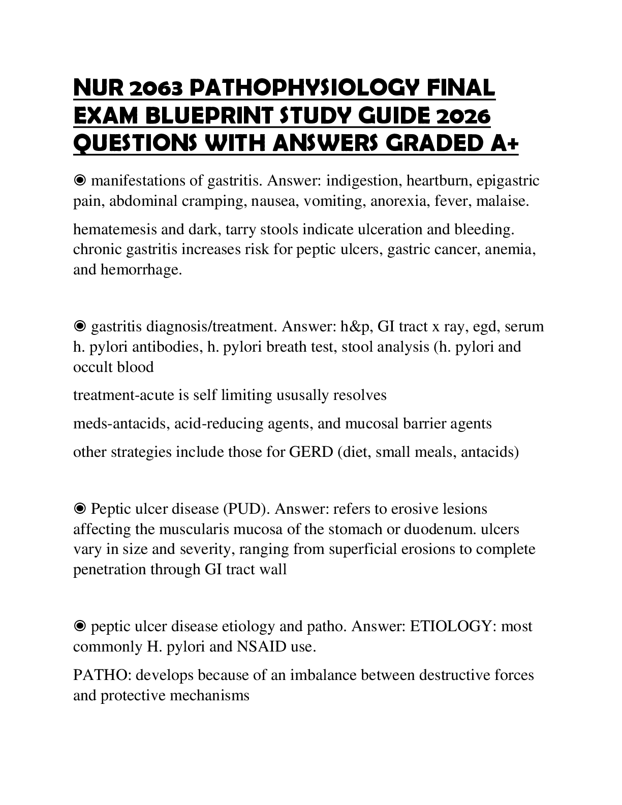
Gastritis – inflammation of the stomach lining
Acute Gastritis – (just acquired) ingestion of toxins, alcohol, aspirin or other irritating substances
Chronic- 2 months to become chronic
Triggers of Gastritis: Alcohol, caffeine, autoimmune disease, viral or bacteria
Chronic Gastritis: H Pylori is always a factor
H Pylori goes very deep in the lining of the stomach and It causes persistent inflammation
S/S: N/V – Anorexia- postcranial discomfort
Post Cranial Discomfort- after eating- goes away and come back 1-2 hrs
Gastritis- hematemesis- blood in the vomit- coffee brown color
Treatment: Treat H pylori treat GERD, change lifestyle, PPI
Peptic Ulcer Disease
Inflammation and ulceration in the stomach (acid and pepsin)
Gastric: stomach location
Duodenal: duodenal location
PUD is a complication of Gastritis
PUD is caused by aspirin, H pylori, Nsaids, Stress, Smoking
S/S Gastric N/V Anorexia Chest discomfort, asymptotic, Dyspepsia
Duodenal – normal weight
Biggest complication of PUD- GI bleeding due to Ulcer perforation- hole in the lining and bleed
It is life-threatening if it keep bleeding (Anemic, electrolytes imbalance (losing volume)
Duodenal – Blood in the stool – black and tarry
Bleeding profusely-frank with cloth
Hematemesis- Bleeding in vomiting
Treatment: Cortery of perforation, treatment of H. pylori, PPI, Cessation of smoking
Ulcerative Colitis and Crohn’s the difference in the complications
Complication in UC Malnutrition – dehydration, increased risk factor of colon cancer
7-10 yrs, rarely in megacolon
Complication of Chron- Fistulas, perianal fissures, abscesses. The risk of colorectal cancer
Bowel Obstruction Manifestations
Obstructions in the jejunal area: Vomiting, dehydration, electrolyte depletion
Obstructions of the distal portion of the small bowl or ileum, dehydration to hypovolemic schock
Obstructions of the colon: Massive gas distention
Blockage of the colon by a tumor is the most common cause of colonic obstruction and perforation of the bowel wall adjacent to the tumor.
What percentage of the pancreas is dedicated to endocrine functions?
Only 5%
Pancreatic Cancer
Pancreatic Cancer – 2% of all cancers
Ranked 4th among death in all malignancies
Risk Factors; cigarette smoking, obesity
S/S; head: Jaundice, malabsorption, weight loss tail: Abd pain, nausea’
Hepatic Encephalopathy is due to?
Hepatic encephalopathy is a decline in brain function due to severe liver disease
Hepatic encephalopathy is usually precipitated by certain well-defined clinical developments, including hypokalemia, hyponatremia, alkalosis, hypoxia, hypercarbia, infection, use of sedatives, GI hemorrhage, protein meal gorging, renal failure, and constipation. In some patients, progressive liver failure leads to chronic encephalopathy without other exacerbating factors.
Hepatic encephalopathy is graded 1 to 4:
• Grade 1: Confusion, subtle behavioral changes, no flap
• Grade 2: Drowsy, clear behavioral changes, flap present
• Grade 3: Stuporous but can follow commands, marked confusion, slurred speech, flap present
• Grade 4: Coma, no flap
Gastroesophageal Varices Management
- Initial treatment: Fluid resuscitation to stop bleeding
- Large bore intravenous lines are placed
- Admin of parenteral vitamin K and plasma, platelet infusion if thrombocytopenia is present
- Octreotide acetate (synthetic analog) no more vasopressin 3-5 days
- Metoclopramide and B blockers
- Esophagogastroduodenoscopy EGD to determine site of bleeding
Difference between Diverticulosis and Diverticulitis
Diverticulosis (diverticular disease) presence of diverticula in the colon.
Diverticula are acquired herniations of the mucosa and submucosa through the muscular coat of the colon
Diverticulosis The presence of one or more diverticula vs diverticulitis inflammation of one or more diverticula
Kidney Disease- Assessment-CVA
Pain associated with intrarenal disorders are assessed by palpating or light percussion over the costovertebral angle (CVA) posteriorly and is recorded as CVA tenderness. Pain is transmitted to the spinal cord between T10 and L1
Kidney Cancer signs and symptoms
Benign renal neoplasm: S/S Hematuria and flank pain
Some may be asymptomatic until large
Renal cell carcinoma: Metastatic disease
Risk factors: smoking, obesity and hypertension
S/S CVA tenderness, hematuria, palpable mass
Dialysis- Benefits and Risks
Filter blood and rid the waist
Dialysis is the only therapeutic option for those with ESRD unable to obtain transplant
Each treatment of dialysis remove about 2/3 of the total body urea content
Dialysis maintain volume status
Prevent and treat acid-base and electrolyte disturbances
Prevent and treat uremia
Support nutritional needs.
Prevent and treat infection
Orevent and treat anemia
Improve quality of life
Lower mortalty and morbidity rates
Control pains
VS
RISKS
Electrolytes imbalance (potassium and sodium) -need to check hyper or hypo
Low blood pressure (have to check vitals before and after)
Risk for infection
Long-term mobidity
Life threatening
Cardiovascular disease remained the most common cause of death in ESRD patient
Acute Kidney Injury- Three phases- Prodromal- Oliguric- Postoliguric
Prodomal Phase
- Normal or declining urine output
- Serum blood urea nitrogen (BUN) and creatine levels rise
Oliguric phase (10-14 days) range from 1 day to 8 weeks
- Oliguria – Small abnormal amount of urine
- Edema, Hypertension, Hypervolemia
- Distended neck veins, weight gain
- Crackles, possibly heart faily
- WBC and RBC in urine
- Protein in urine
- Hyperkalemka
- Volume overload
- Azotemia and uremia
- Metabolic acidosis
Postoliguric phase (5% recover)
- Fluid volume deficit
- Labs begin to normalize
- Polyuria – sodium waste
Acute Tubular Necrosis and the causes of Acute Tubular Necrosis
ATN is the result of tubular cell injury
- Ischemia: Exposure to nephrotoxic substances
- ½ of all cases of AKI
Nephrotoxins leading cause of ATN (aminoaglycosides, NSAIDS, amphotericin B, cisplatin, tetracycline.
CI- AKI or Contrast-induced nephropathy
Cystitis- signs and symptoms
(inflammation of the bladder) no fever or flank pain If fever is present – Might be infection (UTI)
Confusion
Cloudy urine
Frequency
Urgency
Dysuria
Intrarenal Kidney Injury- Toxic causes of
Box 28.1
Types of Acute Kidney Injury
Prerenal
• • Absolute decrease in circulating volume
o • Hemorrhage
o • Dehydration
o • Burns
• • Relative decrease in circulating volume
o • Distributive shock (neurogenic, anaphylactic, septic)
o • Third-spacing and edema
o • Decreased cardiac output
• Cardiogenic shock
• Dysrhythmias
• Cardiac tamponade
• Heart failure
• Myocardial infarction
• • Primary renal hemodynamic abnormalities
o • Occlusion or stenosis of renal artery*
o • Drug-induced impairment of renal autoregulation in susceptible persons†
Postrenal
• • Benign prostatic hyperplasia
• • Kinked or obstructed catheters
• • Intraabdominal tumors
• • Strictures
• • Calculi
Intrarenal/Intrinsic
• • Tubular (acute tubular necrosis)
o • Ischemic
• Prolonged prerenal failure
• Transfusion reactions
• Rhabdomyolysis
o • Nephrotoxic
• Prolonged postrenal failure
• Certain antimicrobials (antibiotics; antifungal and antiviral drugs)
• Radiographic contrast media
• Certain cytotoxic chemotherapy agents
• Recreational drugs (amphetamines, heroin)
• Environmental agents (heavy metals, carbon tetrachloride, insecticides)
• Snake and insect venom
• • Glomerular
o • Acute glomerulonephritis
• • Interstitial
o • Acute allergic interstitial nephritis
o • Acute pyelonephritis
• • Vascular
o • Vasculitis
o • Emboli
o • Nephrosclerosis (due to primary hypertension, hypertensive emergencies, and urgency)
Two electrolytes that are affected by the kidney’s inability to regulate.
Potassium and Sodium
Hypokalemia and Hyponatremia
Renin-Angiotensin-Aldosterone System and the relationship between the autoregulation of the kidneys
Renin-Angiotensin-Aldosterone System (RAAS) act to increase the total circulating volume in the kidney attempt to autoregulate perfusion and maintain GFR.
What is the Glomerelus?
It is a segment of nephron that filters fluid from blood into Bowman capsule, prevents passage of blood cells and proteins.
What is Glomerulonephritis?
An assortment of immune-mediated conditions that produce inflammation of the glomeruli.
Diabetes Type I and Type II
Type 1: Destruction of the B cells of the pancreas results in absolute insulin deficiency
5-20 yrs
Chromosone 6
Type 2: Most common form of DM – Insulin resistance and B cells dysfunction lead to a resistance lack of insulin
Risk factor: Female sex, obesity, and sedentary lifestyle
Risk factor: non Caucasian, elderly
Poly’s of Diabetes
3 P : Polyuria, Polyphagia, Polydipsia
Cushing’s Syndrome characterized by
Cushing Syndrome Adrenocortical Insufficiency
Truncal obesity
Moon face
Dorsocervical fat pad
Hirsutism
Muscle wasting
Striae
Petechiae
Glucose intolerance
Hypertension
Hypokalemia Weakness
Hypotension
Hypoglycemia
Hyperpigmentation (Addison disease)
Hyperkalemia
Weight loss
Traumatic Brain Injury
Traumatic brain injury (TBI) refers to injuries of brain tissues sustained as a consequence of trauma
Traumatic brain injury == head injury
Injuries to the cranials ≠ not always brain injury
TBI is the leading cause of death and disability in the U.S. 50,000 + yr
Causes TBI (falls, sports injuries, firearms, and transportation-related trauma)
Fall majority for 0-4 or 65+, motor vehicle TBI – 14-24 yr. Men are more likely to die than women
Types of TBI:
Severity
Location
Mechanisms of injury
Severity of TBI is classified by Glasgow coma scale (CGS) (standardized tool developed for the purpose assessing the LOC in acutely brain-injured patients. To evaluate patient with an altered LO
Glasgow coma scale: Mild score (13-15), Moderate (9-12), severe (8 – below)
Primary injury- initial trauma injury on brain cells
- Focal injuries (coup)
- Polar injuries (coup contrecoup)
- Diffuse injury
In addition, as location, Primary Injury can also differentiated by Mechanism of injury
- Concussion
- Contusion
- Intracranial hematoma.
Concussion also known as Mild traumatic brain injury (MTBI) most common for military and athletes. Alterations or LOC≤30 mn but no evidence of brain damage on CT.
Symptoms: Headache, n/v, dizziness, fatigue, blurred vision, cognitive, emotional disturbances
Contusion CT or MRI reveals area of Brain Tissue damage (necrosis, laceration, bruising)
Intracranial hematoma: Localized collection of blood within the cranium.
Three types; Epidural, subdural, subachnoid.
Secondary Injury : Body’s response to initial injury may cause more harm than the initial injury.
Can cause: Ischemia, hypoxic events, vasogenic/neurogenic edema, and other processes that leads to brain swelling and increased ICP.
Treatment: Cardiopulmonary stabilization. Radiologic screening, maintenance of normal body temperature or mild hypothermia, normal PaCO2, normal glucose level and noema intravascular volume recommended. Acutely elevated ICP, mannitol(osmotic diuretic) sedation, hypothermia and mild hyperventilation.
Reperfusion Injury- Definition
Reperfusion injury is the secondary injury that occurs after reestablishing blood flow
Increased Cranial Pressure- ICP
ICP is the pressure exerted by the contents of the cranium and normal ranges from 0-15 mmhg
The volume of the cranium nerve is made up 3 parts: brain tissue, cerebrospinal, blood
Monroe-kellie hypothesis – the relationship between the 3 components of cranium
Compliance – the ability to accommodate changes in volume w/o sign. Increase in pressure
The most common causes of ICP: stroke, trauma and tumors
Most sensitive indicator of altered brain function?
A change in Level of consciousness LOC is one of the most sensitive indicators of altered brain function
Assessment of the brainstem function- How do you assess it?
Pupil light reflex
Oculovestibular reflex
Corneal reflex
CVA- Types and causes
Transient ischemic attack (TIA) – Episodes of stroke s/s with a duration of less than 24 hr and less
than1hr (30% pt with stroke have prior TIA)
Ischemic stroke- Sudden occlusion of cerebral artery secondary to thrombus formation/embolization
- Thrombotic strokes: atherosclerosis and hypercoagulable states
- Embolic strokes: cardiac sources
Hemorrhagic stroke = Hemorrage within the brain parenchyma and after a severe and long-standing
Hypertension. (30 % mortality rate)
Test used to diagnosed CVA
Non-contrast CT scan /diffusion weighted brain MRI
Meningitis what is it?
Meningitis is the pyogenic infection that invades the leptomeninges and the subarachnoid space.
the most comment sequela to microbial invasion of the CNS. (most bacterial but can be viral/fungal)
Bacteria: Streptoccocus pneumoniae
Encephalitis What is it?
Encephalitis is the inflammation of the brain caused by viruses, bacteria, fungi and parasites
Seizures- how they are classified?
1. Generalized seizures: Entire brain Surface
• Absence (petit mal)
• Atypical absence
• Myoclonic
• Atonic (drop attack)
• Clonic
• Tonic
• Generalized tonic-clonic (grand mal)
2. Partial Seizure: Part of the brain surface
• Simple partial: There is no impairment of consciousness during the seizure.
• Complex partial: There is impairment of consciousness during the seizure.
• With secondary generalization: Onset begins as simple partial and then progresses to impairment of consciousness.
Status Epilepticus what is it?
Status Epilepticus is a continuing series of seizures without a period of recovery between episodes
Dementia
Progressice deterioration and continuing decline of memory and other cognitive changes
60 % to 80 % of Alzheimer are dementia- Vascular dementia is the most common cause
No cure
Cause is unknown
Dementia-causing illness: - Alcoholism
- Alcoholism
- Intracranial tumor
- Normal-pressure hydrocephalus
- Parkinson disease
- Lewy body disease
- Huntington disease
- Multiple sclerosis MS
- Bocine Spongiform encephalopathy (Mad Cow)
Delerium and depression in elederly different than dementia
Delirium in global mental dysfunction – Acute Confusional state
Primary risk factor: age and family
Others: Head trauma, diabetes, depression, stroke, hypertension diabetes
Signs and Symptoms memory lost, anxiety and agitation, diffulty in judgement, problem solving and communication. Assistance needed for ADL . Difficulty eating and swallowing. Weight loss
Parkinson disease
Disorder of mobility - 1 million americans affected. 60,000 cases each year
4 % are younger than 50 Y/O
idiopatic or acquired
Common causes: Infection, intoxication, and trauma
Drug Toxicity: Phenotiazine class: (Chlorpromazine, prochlorperazine, Thioridazine) and Butyrophenone(haloperidol)
Parkinson disease results from degeneration of the pigmented dopaminergic neurons found in the subtantia nigra and to a lesser extent neurons elsewhere in the brain.
Lewy bodies are cytoplasmic inclusions are found in the surviving neurons.
GERD signs and symptoms
Heartburn
Chest pain
Regurgitation
Epigastric pain
Dry cough
Hoarseness in the morning
Esapheogeal strictures with Gerd
Multiple Sclerosis- Structures effected by the demyelination?
Chronic demyelinating disease of the CNS – sign disability in young adults
-
-
-
-
-
-
-
Continued
[Show More]











 – Chamberlain College of Nursing.png)
 – Chamberlain College of Nursing.png)