Anatomy and Physiology - A&P 1 > QUESTIONS & ANSWERS > Portage Learning BIOD 151| Module 5: Problem Set: Essential Human Anatomy & Physiology I w/Lab - Bur (All)
Portage Learning BIOD 151| Module 5: Problem Set: Essential Human Anatomy & Physiology I w/Lab - Burtt - 2025E | Answered Correctly 75/75.
Document Content and Description Below
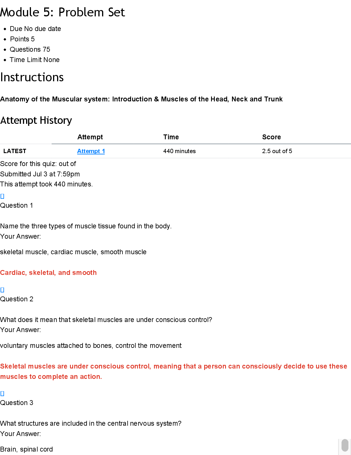
Module 5: Problem Set Instructions Anatomy of the Muscular system: Introduction & Muscles of the Head, Neck and Trunk Question 1 Name the three types of muscle tissue found in the body.... skeletal muscle, cardiac muscle, smooth muscle Cardiac, skeletal, and smooth Question 2 What does it mean that skeletal muscles are under conscious control? voluntary muscles attached to bones, control the movement Skeletal muscles are under conscious control, meaning that a person can consciously decide to use these muscles to complete an action. Question 3 What structures are included in the central nervous system? Brain, spinal cord Brain and spinal cord Question 4 Describe a motor action vs. sensory input in terms of the nervous system. Motor action sends signals from the brain to muscles to cause movement, sensory input brings information from the body to the brain. Messages from the central nervous system to a muscle are called motor actions. Nerves also carry information from the external environment and the muscles to the central nervous system, called sensation or sensory input. Question 5 True or false: The brachial plexus supplies nerves to the lower extremities. True Correct! False Question 6 Describe the difference between tendons and ligaments. Tendons connect muscle to bone and help with movement. Ligaments connect bone to bone and help stabilize joints. Tendons are connective tissues that connect skeletal muscle to bone at each end. Ligaments are connective tissue that connects bone to bone, helping to stabilize joints where bones meet. Question 7 Describe the meaning of muscle origin and insertion. Muscle origin is the fixed attachment point on a bone that doesn’t move during contraction. Muscle insertion is the point where the muscle attaches to the bone that moves during contraction. The origin is the bony site of attachment that is stationary during the movement. The insertion of a muscle is the bony site of attachment that is moved by the muscle contraction. Question 8 Describe the meaning of muscle action and innervation. Muscle action is the specific movement a muscle performs when it contracts, like bending a joint. Innervation is the nerve supply that controls the muscle's movement and response The action of the muscle is what effect is produced by the muscle’s contraction. The innervation is the peripheral nerve that supplies a muscle with the message from the brain. Practice: Muscles of Facial Expression *NOTE* This is not an exhaustive list. All the muscles in the module need to be learned for the exam. Question 9 Identify the muscle indicated below. Also name its action and innervation. orbicularis oculi. Orbicularis oculi Action: eye closure Innervation: facial nerve (CN VII) Question 10 Identify the muscle indicated below. Also name its action and innervation. orbicularis oris Orbicularis oris Action: mouth closure: closes lips, protrudes lips forward, presses lips against teeth Innervation: facial nerve (CN VII) Question 11 Identify the muscle indicated below. Also name its action and innervation. risorius Risorius Action: pulls the corners of the mouth posteriorly (grin or grimace) Innervation: facial nerve (CN VII) Question 12 Identify the muscle indicated below. Also name its action and innervation. occipitofrontalis Frontalis (occipitofrontailis) Action: raise eyebrows Innervation: facial nerve (CN VII) Question 13 Identify the muscle indicated below. Also name its action and innervation. temporalis Temporalis Action: Elevates mandible, closes jaw Innervation: trigeminal nerve (CN V, mandibular branch) Question 14 Identify the muscle below. Also name its action and innervation. masseter Zygomaticus major Action: pull corners of lips upward Innervation: facial nerve (CN VII) Question 15 Identify the muscle below. Also name its action and innervation. buccinator. Action: Compresses the cheek (as in blowing, sucking, or keeping food between teeth while chewing). Innervation: Facial nerve (cranial nerve VII) Buccinator Action: compress cheeks Innervation: facial nerve (CN VII) Practice: Muscles of the Head and Neck, Vertebral Column, Abdomen, and Breathing *NOTE* This is not an exhaustive list. All the muscles in the module need to be learned for the exam. Question 16 Identify the muscle indicated below. Also name its origin, insertion, action, and innervation. splenius capitis. Action: Extends, rotates, and laterally flexes the head and neck. Innervation: Cervical spinal nerves (posterior rami) Splenius Capitis Origin: spinous process/ligaments of inferior cervical vertebrae Insertion: mastoid process, occipital bone of skull Action: Bilateral extend head Unilateral laterally flexes neck to same side Innervation: cervical spinal nerves Question 17 Identify the muscle indicated below. Also name its origin, insertion, action, and innervation. sternocleidomastoid. Action: Rotates the head to the opposite side and flexes the neck. Innervation: Accessory nerve (cranial nerve XI) Sternocleidomastoid Origin: sternal end of clavicle and manubrium Insertion: mastoid region of skull Action: Bilateral: neck flexion Unilateral: turns face to opposite side Innervation: accessory nerve (CN XI) Question 18 Identify the muscle indicated below (seen in both figures). Also name its origin, insertion, action, and innervation. levator scapulae. Action: Elevates the scapula and helps rotate the neck to the same side. Innervation: Dorsal scapular nerve (C5) and cervical nerves (C3–C4) Longissimus cervicis Origin: transverse processes of superior thoracic vertebrae Insertion: transverse process of middle and superior cervical vertebrae Action: Bilateral extend head Unilateral laterally flexes neck to same side Innervation: cervical and thoracic spinal nerves Question 19 Identify the muscle indicated below. Also name its origin, insertion, action, and innervation. erector spinae. Action: Extends and laterally flexes the vertebral column (helps with posture and standing upright) Innervation: Dorsal rami of spinal nerves Longissimus thoracis Origin: transverse process of all thoracic and lumbar vertebrae Insertion: transverse processes of all thoracic vertebrae Action: Bilateral extension of spine Unilaterally: lateral flexion of spine Innervation: thoracic and lumbar spinal nerves Question 20 Identify the muscle indicated below. Also name its origin, insertion, action, and innervation. semispinalis capitis. Action: Extends the head and neck; rotates the head to the opposite side. Innervation: Dorsal rami of cervical spinal nerves Semispinalis Capitis Origin: articular processes of inferior cervical & transverse process of superior thoracic vertebrae Insertion: occipital bone Action: Bilateral extend head Unilateral laterally flexes neck to same side Innervation: spinal nerves Question 21 Identify the muscle indicated below. Also name its origin, insertion, action, and innervation. anterior scalene. Action: Elevates the first rib during inhalation and flexes/laterally bends the neck.Innervation: Cervical spinal nerves (C4–C6) Thyrohyoid Origin: thyroid cartilage of larynx Insertion: hyoid bone Action: elevates thyroid, depresses hyoid bone Innervation: hypoglossal nerve Question 22 Identify the muscle indicated below. Also name its origin, insertion, action, and innervation. anterior scalene. Action: Elevates the first rib during inhalation and flexes/laterally bends the neck.Innervation: Cervical spinal nerves (C4–C6) Middle Scalenes Origin: transverse processes of C2-C7 Insertion: first and second ribs Action: elevates ribs 1 & 2 Innervation: cervical spinal nerves Question 23 Identify the muscle shown below of the Erector Spinae group and name its three divisions. spinalis Action: Extends and laterally flexes the vertebral column. Innervation: Dorsal rami of spinal nerves Spinalis: spinalis thoracis, spinalis cervicis, and spinalis capitis Question 24 Identify the muscle shown below of the Erector Spinae group and name its three divisions. longissimus. Action: Extends and laterally flexes the spine; also helps extend and rotate the head. Innervation: Dorsal rami of spinal nerves Longissimus: Made up of three divisions (longissimus thoracis, longissimus cervicis, longissimus capitis) Question 25 Identify the muscle shown below of the Erector Spinae group and name its three divisions. iliocostalis. Action: Extends and laterally flexes the vertebral column. Innervation: Dorsal rami of spinal nerves Iliocostalis: Made up of three divisions (iliocostalis lumborum, iliocostalis thoracis, iliocostalis cervicis) Question 26 Identify the muscle indicated below. Also name its origin, insertion, action, and innervation. rectus abdominis. Action: Flexes the vertebral column (as in doing a sit-up) and compresses abdominal contents. Innervation: Thoracic spinal nerves (T7–T12) Rectus abdominis Origin: pubic crest, pubic symphysis Insertion: cartilages of ribs 5-7, xiphoid process Action: flexion of spine, compression of abdominal viscera Innervation: spinal nerves (T 7-T 12) Question 27 Identify the muscle indicated below. Also name its origin, insertion, action, and innervation. external oblique. Action: Compresses the abdomen, flexes and rotates the trunk. Innervation: Thoracic spinal nerves (T7–T12) Transverse abdominis Origin: lateral inguinal ligament, inner iliac crest Insertion: linea alba, pubis Action: compression of abdomen Innervation: first lumbar nerve (T 7- L1), iliohypogastric (T12-L1), ilioinguinal (T12-L1) Question 28 Identify the muscle indicated below. Also name its origin, insertion, action, and innervation. diaphragm. Action: Primary muscle of respiration; it contracts to enlarge the thoracic cavity and draw air into the lungs. Innervation: Phrenic nerve (C3–C5) Diaphragm Origin: cartilage of ribs 7-12, xiphoid process, lumbar vertebrae Insertion: anterior longitudinal ligament (vertebral column) Action: expands thoracic cavity, compresses abdominal cavity Innervation: phrenic nerve (C3-5) Question 29 Identify the muscle indicated below. Also name its origin, insertion, action, and innervation. internal intercostals. Action: Depress the ribs during forced exhalation. Innervation: Intercostal nerves (T1–T11) Internal Intercostals Origin: superior border of ribs 2-12 Insertion: inferior of ribs above (1-11) Action: depresses ribs (forced expiration) Innervation: intercostal nerves Question 30 Identify the muscle indicated below. Also name its origin, insertion, action, and innervation. external intercostals. Action: Elevate the ribs during inhalation to expand the thoracic cavity. Innervation: Intercostal nerves (T1–T11) External Intercostals Origin: lower border of ribs 1-11 Insertion: upper border of ribs below (2-12) Action: elevates ribs (normal inspiration) Innervation: intercostal nerves Question 31 Your patient has bilateral damage to the facial nerves (CN VII). Name at least three muscles that would be impaired. Orbicularis oculi – difficulty closing the eyes Orbicularis oris – trouble puckering or closing the lips Buccinator – problems with chewing and keeping food in the mouth Could include any three: Orbicularis oculi, Orbicularis oris, Zygomaticus major/minor, Risorius, Frontalis, Buccinator Question 32 Your patient injured her ankle while playing soccer. She sustained injuries to the tendons of peroneus longus and peroneus brevis. What actions would be impaired due to this injury? Eversion of the foot – difficulty turning the sole of the foot outward Plantarflexion – weakness when pointing the foot downward (especially during push-off while walking or running) Plantarflexion and eversion of foot Question 33 Your patient is having difficulty with scapular retraction. Name two muscles that are most likely involved in this limitation. Rhomboid major (and rhomboid minor), Trapezius (especially the middle fibers) Trapezius, rhomboids (minor/major) Practice: Muscles of the Shoulder and Upper Extremity *Note* This is not an exhaustive list of the muscles for the exam. All the muscles in the module must be learned for the exam. Question 34 Identify the muscle below. Also name its origin, insertion, action, and innervation. trapezius. Action: Upper fibers elevate the scapula, middle fibers retract the scapula, and lower fibers depress the scapula. Innervation: Accessory nerve (cranial nerve XI) Trapezius Origin: occipital bone, spinous process of T1-12 Insertion: lateral clavicle, acromion, and scapular spine of scapula Action: rotation, retraction, elevation, depression of scapula; extends neck and stabilizes shoulder Innervation: Accessory nerve (Cranial Nerve 11) Question 35 Identify the muscle below (in blue). Also name its origin, insertion, action, and innervation. rhomboid minor (top arrow) and rhomboid major (bottom arrow). Action: Retract (adduct) the scapula and rotate it to depress the glenoid cavity Innervation: Dorsal scapular nerve (C5) Levator scapulae Origin: transverse process of C1-4 Insertion: medial border of scapula Action: elevates scapula Innervation: dorsal scapular nerve Question 36 Identify the muscle below (in blue). Also name its origin, insertion, action, and innervation. rhomboid major.Action: Retracts and elevates the scapula; rotates it to depress the glenoid cavity Innervation: Dorsal scapular nerve (C5) Rhomboids (major) Origin: spinous process T2-5 Insertion: medial border of scapula Action: retraction of scapula Innervation: dorsal scapular nerve Question 37 Identify the muscles below (A and B). Also name their origins, insertions, actions, and innervations. Muscle A is the serratus anterior. Action: Protracts and stabilizes the scapula; assists in upward rotation. Innervation: Long thoracic nerve. Muscle B is the external oblique. Action: Compresses the abdomen, flexes and rotates the trunk. Innervation: Thoracic spinal nerves (T7–T12) A= Pectoralis minor Origin: ribs 3-5 Insertion: coracoid process of scapula Action: elevates ribs, draws scapula down and medially Innervation: medial pectoral nerve B= Serratus Anterior Origin: upper 8-9 ribs Insertion: medial border of scapula Action: protraction of scapula Innervation: long thoracic nerve Question 38 Identify the muscles below (A, B, and C). Also name their origins, insertions, actions, and innervations. Muscle A: Deltoid Origin: Lateral third of clavicle Acromion Spine of scapula Insertion: Deltoid tuberosity of the humerus Action: Anterior fibers: flex and medially rotate arm Middle fibers: abduct arm Posterior fibers: extend and laterally rotate arm Innervation: Axillary nerve (C5–C6) Muscle B: Pectoralis Major Origin: Clavicular head: anterior surface of medial clavicle Sternocostal head: sternum, upper 6 costal cartilages, and aponeurosis of external oblique Insertion: Lateral lip of the intertubercular sulcus (bicipital groove) of the humerus Action: Adducts and medially rotates the arm Clavicular head: flexes the arm Sternocostal head: extends the flexed arm Innervation: Lateral and medial pectoral nerves (C5–T1) Muscle C (in blue): Latissimus Dorsi Origin: Spinous processes of T7–T12 Thoracolumbar fascia Iliac crest Inferior 3 or 4 ribs Insertion: Floor of intertubercular sulcus (bicipital groove) of humerus Action: Extends, adducts, and medially rotates the arm Assists in movements like climbing or swimming Innervation: Thoracodorsal nerve (C6–C8) A= Deltoid Origin: clavicle and scapula Insertion: deltoid tuberosity of humerus Action: abduction at shoulder (whole muscle) Innervation: axillary nerve B= Pectoralis major Origin: ribs 2-6, body of sternum Insertion: greater tubercle of humerus Action: flexion, adduction and medial rotation at shoulder Innervation: pectoral nerves C= Latissimus Dorsi Origin: spinous process of inferior thoracic and lumbar vertebrae, ribs 8-12 Insertion: intertubercular groove of humerus Action: extension, adduction, and medial rotation at shoulder Innervation: thoracodorsal nerve Question 39 Is this a view of the right or left shoulder? Identify the muscles below (A, B, C, and D) . Also name their origins, insertions, actions, and innervations. Which muscle is not part of the rotator cuff? Muscle A is the supraspinatus, which originates from the supraspinous fossa of the scapula and inserts on the superior facet of the greater tubercle of the humerus. Its primary action is to initiate the first 15 degrees of arm abduction, and it is innervated by the suprascapular nerve (C5–C6). Muscle B is the infraspinatus, located just below the supraspinatus. It originates from the infraspinous fossa of the scapula and inserts onto the middle facet of the greater tubercle of the humerus. The infraspinatus laterally rotates the arm and is also innervated by the suprascapular nerve (C5–C6). Muscle C, shown in blue, is the teres minor, a small rotator cuff muscle that originates from the lateral border of the scapula and inserts on the inferior facet of the greater tubercle of the humerus. It functions to laterally rotate the arm and stabilize the shoulder joint. It is innervated by the axillary nerve (C5–C6). Muscle D is the teres major, which is not part of the rotator cuff group. It originates from the inferior angle of the scapula and inserts on the medial lip of the intertubercular sulcus of the humerus. Its actions include adduction, medial rotation, and extension of the arm. The teres major is innervated by the lower subscapular nerve (C5–C6). This is a (posterior) view of the right shoulder. (D) is not part of the rotator cuff. A= Supraspinatus Origin: supraspinatus fossa of scapula Insertion: greater tubercle of humerus Action: abduction at the shoulder Innervation: suprascapular nerve B= Infraspinatus Origin: infraspinatus fossa of scapula Insertion: greater tubercle of humerus Action: lateral rotation at shoulder Innervation: suprascapular nerve C= Teres Minor Origin: lateral border of scapula Insertion: greater tubercle of humerus Action: lateral rotation at shoulder Innervation: axillary nerve D= Teres Major – not part of the rotator cuff Origin: inferior angle of scapula Insertion: intertubercular groove of humerus Action: extension, adduction, and medial rotation at shoulder Innervation: lower subscapular nerve Question 40 Is this a view of the left or right shoulder? Identify the muscles below (A and B). Then, name their origins, insertions, actions, and innervations. Muscle A is the subscapularis, which originates from the subscapular fossa of the scapula and inserts on the lesser tubercle of the humerus. It medially rotates the arm and helps stabilize the shoulder joint. The subscapularis is innervated by the upper and lower subscapular nerves (C5–C7). It is also part of the rotator cuff muscle group. Muscle B is the teres major, which originates from the inferior angle of the scapula and inserts on the medial lip of the intertubercular sulcus of the humerus. Its actions include adduction, extension, and medial rotation of the arm. It is innervated by the lower subscapular nerve (C5–C6). Though located near the rotator cuff muscles, it is not considered one of them. This is an anterior view of the left shoulder. (Note, the sternum and clavicle are both visible.) A= Subscapularis Origin: subscapular fossa of scapula Insertion: lesser tubercle of humerus Action: medial rotation at the shoulder Innervation: subscapular nerves B= Coracobrachialis Origin: coracoid process of scapula Insertion: medial shaft of humerus Action: adduction and flexion at shoulder Innervation: musculocutaneous nerve Practice: Muscles of the Forearm and Elbow *Note* This is not an exhaustive list of the muscles for the exam. All the muscles in the module must be learned for the exam. Question 41 A. Looking at the figure below, is this an anterior or posterior view of the body? Explain your reasoning. B. Identify the muscle as the short head or the long head and explain how you came to this response. Also include if it is the left or right. C. Identify the origin, insertion, action, and innervation of the muscle. A. This is a posterior view of the body. You can tell because the scapulae (shoulder blades) are visible, and the orientation of the ribs and humeri show the back of the arms and torso. B. The highlighted muscle is the long head of the triceps brachii on the left side of the body (from the subject's perspective). This is determined because the long head is located medially and originates from the scapula, whereas the lateral and medial heads originate from the humerus and lie more lateral or deep. Since it's a posterior view and the muscle is on the subject’s left, it is the left long head. C. Origin: Infraglenoid tubercle of the scapula Insertion: Olecranon process of the ulna Action: Extends the forearm at the elbow; assists in extension and adduction of the arm at the shoulder Innervation: Radial nerve (C6–C8) This is an anterior view. Multiple responses are correct. Here are a few acceptable responses: The body of the sternum can be seen on the anterior side of the body. The view of the vertebral column. The anterior sides are smooth, while the posterior sides have a pointed spinous process. In addition, the biceps brachii is on the anterior surface of the arm. Therefore, this is an anterior view of the body. Since this is an ANTERIOR view of the body (see reasoning above in part A), this is, therefore, the RIGHT biceps brachii (short head). Origin: short head- coracoid process Insertion: tuberosity of radius Action: flexion at elbow and shoulder; supination Innervation: musculocutaneous nerve Question 42 A. Looking at the figure below, is this an anterior or posterior view of the body? Explain your reasoning. B. Identify the muscle as the short head or the long head of the triceps and explain how you know. Also include if it is the left or right. C. Identify the origin, insertion, action, and innervation of the muscle. A. This is a posterior view of the body. You can tell because the scapula is clearly visible from behind, and the ribs and vertebrae are shown from the back side. Also, the muscle highlighted is on the posterior side of the upper arm. B. The highlighted muscle is the long head of the triceps brachii on the right side of the body (subject's right). The long head is identified because it runs from the scapula across the shoulder joint, making it the only head of the triceps that crosses both the shoulder and elbow joints. It lies more medially compared to the lateral head in posterior views. C. Origin: Infraglenoid tubercle of the scapula Insertion: Olecranon process of the ulna Action: Extends the forearm at the elbow; assists with extension and adduction of the arm at the shoulder Innervation: Radial nerve (C6–C8) A. This is a posterior view. Multiple responses are correct. Here are a few acceptable responses: The sternum is visible on the opposite aspect of the body, through the rib cage. The posterior surface of the scapula is visible, as the spine of the scapula is on the posterior aspect of the body. The orientation of the vertebral column. The anterior sides are smooth, while the posterior sides have a pointed spinous process. In addition, the triceps muscle is located on the posterior surface of the arm. Therefore, this is a POSTERIOR view of the body. B. Since this is a POSTERIOR view of the body (See reasoning above in part A), this is, therefore, the LEFT long head of the triceps. (**Note** The long head is medial to the lateral head. The medial head of the triceps is deep to the long and the lateral heads.) C. Origin: Long head – Infraglenoid tubercle of scapula Insertion: olecranon of ulna Action: extension at elbow Innervation: radial nerve Question 43 Identify muscles A and B in the figure below. Then name their origins, insertions, actions, and innervations. Muscle A: Brachioradialis Origin: Lateral supracondylar ridge of the humerus Insertion: Styloid process of the radius Action: Flexes the forearm at the elbow (especially when the forearm is in mid-pronation/supination) Innervation: Radial nerve (C5–C6) Muscle B: Pronator teres Origin: Humeral head: medial epicondyle of the humerus Ulnar head: coronoid process of the ulna Insertion: Middle of the lateral surface of the radius Action: Pronates the forearm and assists in elbow flexion Innervation: Median nerve (C6–C7) A: Supinator Origin: lateral epicondyle of humerus Insertion: anterolateral surface of radius Action: supination Innervation: deep radial nerve B: Pronator Teres Origin: medial epicondyle of humerus Insertion: mid-lateral surface of radius Action: pronation Innervation: median nerve Question 44 Name the muscle below (including left or right). Identify the origin, insertion, action, and innervation. The highlighted muscle is the biceps brachii on the right arm. Origin: Short head: Coracoid process of the scapula Long head: Supraglenoid tubercle of the scapula Insertion: Radial tuberosity of the radius Bicipital aponeurosis into the fascia of the forearm Action: Flexes the forearm at the elbow Supinates the forearm Assists with shoulder flexion Innervation: Musculocutaneous nerve (C5–C7) Right Brachialis Origin: anterior/distal surface of humerus Insertion: tuberosity of ulna Action: flexion at elbow Innervation: musculocutaneous nerve and radial nerve Practice: Muscles of the Hand and Wrist *Note* This is not an exhaustive list of the muscles for the exam. All the muscles in the module must be learned for the exam. Question 45 Identify the muscles below (A, B, and C), including left or right. Name the action and innervation for each muscle. Muscle A: Brachioradialis Action: Flexes the forearm at the elbow (most active when the forearm is in a mid-pronated position) Innervation: Radial nerve (C5–C6) Muscle B: Flexor carpi radialis Action: Flexes and abducts the wrist (radial deviation) Innervation: Median nerve (C6–C7) Muscle C: Palmaris longus Action: Flexes the wrist and tenses the palmar aponeurosis Innervation: Median nerve (C7–C8) A: Flexor carpi radialis (right) Action: wrist flexion, radial deviation of the hand Innervation: median nerve B: Palmaris longus (right) Action: wrist flexion Innervation: median nerve C: Flexor carpi ulnaris (right) Action: wrist flexion, ulnar deviation of the hand Innervation: ulnar nerve Question 46 Identify the muscles below (A, and B), including left or right. Name the action and innervation for each muscle. Muscle A: Flexor carpi ulnaris Action: Flexes and adducts the wrist (ulnar deviation) Innervation: Ulnar nerve (C7–T1) Muscle B: Flexor digitorum superficialis Action: Flexes the proximal interphalangeal (PIP) joints of digits 2–5; assists in flexion at the metacarpophalangeal (MCP) joints and wrist Innervation: Median nerve (C7–T1) A= Right Flexor pollicis longus Action: flexion of thumb Innervation: median nerve B= Right Flexor digitorum profundus (FDP) Action: wrist flexion, flexion of digits 2-5 (distal phalanx) Innervation: palmar interosseous nerve Question 47 Identify the muscles below (A, and B), including left or right. Name the action and innervation for each muscle. Muscle A: Flexor digitorum superficialis Action: Flexes the proximal interphalangeal (PIP) joints of digits 2–5; also flexes the metacarpophalangeal (MCP) joints and wrist Innervation: Median nerve (C7–T1) Muscle B: Flexor digitorum profundus Action: Flexes the distal interphalangeal (DIP) joints of digits 2–5; assists in wrist and MCP joint flexion Innervation: Lateral half (digits 2–3): Median nerve (anterior interosseous branch) Medial half (digits 4–5): Ulnar nerve (C8–T1) A= Right Extensor digitorum Action: wrist extension, extension of digits 2-5 Innervation: deep radial nerve B= Right Extensor carpi ulnaris Action: extension, adduction of the wrist Innervation: deep radial nerve Question 48 Identify the muscles below (A, B, and C). Name the action and innervation for each muscle. Muscle A: Flexor carpi radialis Action: Flexes and abducts the wrist (radial deviation) Innervation: Median nerve (C6–C7) Muscle B: Palmaris longus Action: Flexes the wrist and tenses the palmar aponeurosis Innervation: Median nerve (C7–C8) (Note: This muscle is absent in about 10–15% of people) Muscle C: Flexor digitorum superficialis Action: Flexes the proximal interphalangeal (PIP) joints of digits 2–5; also helps flex MCP joints and wrist Innervation: Median nerve (C7–T1) A= Extensor carpi radialis longus Action: extension, abduction of the wrist Innervation: radial nerve B= Extensor carpi radialis brevis Action: extension, abduction of the wrist Innervation: radial nerve C=Extensor digiti minimi Action: wrist extension, extension of digit 5 Innervation: deep radial nerve Question 49 Identify the muscle below, including left or right. Explain how the orientation or location of this muscle helps you identify the actions. the flexor carpi ulnaris on the left forearm. Its position on the medial (ulnar) side helps identify it, as it flexes and adducts the wrist (ulnar deviation). This orientation gives it leverage to pull the wrist toward the pinky side. It’s innervated by the ulnar nerve (C7–T1). Extensor carpi radialis longus (left)- this is a posterior view of the LEFT arm. The wrist extensors are on the posterior side of the forearm, helping with wrist extension. Since this muscle inserts on the thumb side of the wrist (follows the radius), this muscle would also help with wrist ABduction. Question 50 Identify the muscles below (A and B). Name the action and innervation for each muscle. Explain how you know these are flexor or extensor muscles. Muscle A is the extensor carpi radialis brevis, and Muscle B is the extensor digitorum. Both are on the posterior right forearm, confirming they are extensors. Extensor carpi radialis brevis extends and abducts the wrist; extensor digitorum extends the fingers and wrist. Both are innervated by branches of the radial nerve (C7–C8). Their location on the back of the forearm and origin from the lateral epicondyle indicate their extensor function. These are extensor muscles, as they are on the posterior aspect of the forearm. Therefore, they will contribute to wrist and/or digit extension actions. Note the orientation of the ulna. The olecranon process can be seen, which forms a projection on the posterior aspect of the elbow joint. (See Figure 4.26 for a posterior view of the bones and bone landmarks of the ulna.) A= Extensor pollicis brevis (EPB) Action: thumb extension, wrist abduction Innervation: deep radial nerve B= Extensor indicis (EI) Action: extension of wrist and digit 2 Innervation: deep radial nerve Practice: Muscles of the Lower Extremity *Note* This is not an exhaustive list of the muscles for the exam. All the muscles in the module must be learned for the exam Question 51 Identify the muscle below. Name the origin, insertion, action, and innervation. Explain how you know these are flexor or extensor muscles. the iliacus, part of the iliopsoas group. It originates from the iliac fossa and inserts on the lesser trochanter of the femur. Its action is to flex the thigh at the hip, and it’s innervated by the femoral nerve (L2–L4). Its anterior position on the pelvis indicates it’s a hip flexor, not an extensor, as extensors lie posteriorly (e.g., gluteus maximus). Iliacus Origin: iliac fossa of ilium Insertion: lesser trochanter of femur Action: hip flexion Innervation: femoral nerve This muscle is on the anterior portion of the pelvis. Contraction of this muscle (bringing the insertion closer to the origin) moves the femur closer to the pelvis, closing the angle of this joint. Therefore, this is a flexor muscle. Question 52 Identify the muscles below (A, B, and C). Name the origin, insertion, action, and innervation for each muscle. Muscle A is the gluteus medius, which originates on the outer surface of the ilium and inserts on the greater trochanter. It abducts and medially rotates the thigh, innervated by the superior gluteal nerve (L4–S1). Muscle B is the gluteus maximus, originating from the ilium, sacrum, and coccyx, and inserting on the gluteal tuberosity and iliotibial tract. It extends and laterally rotates the thigh, innervated by the inferior gluteal nerve (L5– S2). Muscle C is the tensor fasciae latae, originating from the anterior iliac crest and inserting into the iliotibial tract. It flexes, abducts, and medially rotates the thigh, innervated by the superior gluteal nerve (L4–S1). This is a posterior view of the thigh. A= TFL – tensor fascia latae Origin: ASIS (anterior superior iliac spine) Insertion: iliotibial tract Action: abducts and internally rotates thigh, stabilizes hip and knee joints Innervation: superior gluteal nerve B= Gluteus medius Origin: gluteal surface of the ilium Insertion: greater trochanter of the femur Action: abducts thigh, stabilizes the hip joint Innervation: superior gluteal nerve C= Gluteus maximus Origin: posterior gluteal line of the ilium, lower sacrum, side of the coccyx Insertion: gluteal tuberosity Action: abducts, extends, laterally rotates hip joint Innervation: inferior gluteal nerve Question 53 Identify the muscles below (A, B, C, and D). Name the action and innervation for each muscle. Muscle A is the gluteus medius, which abducts and medially rotates the thigh; it’s innervated by the superior gluteal nerve (L4–S1). Muscle B is the piriformis, which laterally rotates and abducts the thigh; innervated by nerve to piriformis (S1–S2). Muscle C is the superior gemellus, which laterally rotates the extended thigh and is innervated by nerve to obturator internus (L5–S2). Muscle D is the inferior gemellus, with the same action as C and innervated by nerve to quadratus femoris (L4–S1). A= Gluteus minimus (*also need to know the origin and insertion of this muscle for the exam) *Origin: Gluteal surface of the ilium *Insertion: Greater trochanter of the femur Action: Abducts thigh, stabilizes hip joint Innervation: Superior gluteal nerve B= Piriformis Action: lateral rotation, adduction, extension of hip joint Innervation: spinal nerves L5-S2 C= Obturator internus Action: lateral rotation of thigh Innervation: spinal nerves S1-3 D= Quadratus Femoris Action: lateral rotation, adduction of hip joint Innervation: spinal nerves L5, S1 Question 54 Is this a view of the right or left leg? Explain your reasoning. Identify the muscles below (A, B, and C). Name the origin, insertion, action, and innervation for each muscle. Muscle A is the sartorius, which flexes, abducts, and laterally rotates the hip; it also flexes the knee. It originates at the ASIS, inserts on the medial tibia, and is innervated by the femoral nerve. Muscle B is the vastus medialis, which extends the knee. It originates from the linea aspera and inserts on the tibial tuberosity via the patellar ligament; innervated by the femoral nerve. Muscle C is the gracilis, which adducts the thigh and flexes the leg. It originates from the pubic ramus, inserts on the medial tibia, and is innervated by the obturator nerve. This is the right leg. The fibula in the lower leg is visible and is always on the lateral side of the tibia. The large condyles of the femur are visible on the posterior aspect of the leg, so therefore this is a posterior view of the right leg. A= Semimembranosus Origin: ischial tuberosity Insertion: medial condyle of tibia Action: knee flexion Innervation: tibial nerve B= Semitendinosus Origin: ischial tuberosity Insertion: medial surface of tibia Action: knee flexion Innervation: tibial nerve C= Biceps Femoris Origin: Long head, ischial tuberosity; Insertion: head of the fibula, lateral surface Action: knee flexion, hip extension Innervation: Long head - tibial nerve Question 55 Identify the muscle below. Also name its origin and innervation. the vastus intermedius, part of the quadriceps group. Origin: Anterior and lateral surfaces of the shaft of the femur Innervation: Femoral nerve (L2–L4) Biceps Femoris, short head. Origin: linea aspera of femur, lateral surface Innervation: Common fibular nerve Question 56 Identify the muscles in the diagram below (A, B, and C). Also name their origins, insertions, actions, and innervations. Which muscle needs to be removed in order to see vastus intermedius? Muscle A: Rectus femoris Origin: Anterior inferior iliac spine (AIIS) Insertion: Tibial tuberosity via patellar ligament Action: Extends the knee and flexes the hip Innervation: Femoral nerve (L2–L4) Muscle B: Vastus lateralis Origin: Greater trochanter and lateral lip of the linea aspera Insertion: Tibial tuberosity via patellar ligament Action: Extends the knee Innervation: Femoral nerve (L2–L4) Muscle C: Vastus medialis Origin: Medial lip of the linea aspera Insertion: Tibial tuberosity via patellar ligament Action: Extends the knee Innervation: Femoral nerve (L2–L4) A= Sartorius Origin: ASIS (anterior superior iliac spine) Insertion: superior shaft of tibia Action: Hip: flexion, abduction, external rotation; Knee: flexion, internal rotation Innervation: femoral nerve B= Vastus Lateralis Origin: greater trochanter of femur Insertion: patella via quadriceps tendon; tibial tuberosity Action: knee extension Innervation: femoral nerve C= Rectus Femoris Origin: AIIS (anterior inferior iliac spine) Insertion: common tendon of the quadriceps to tibial tuberosity Action: knee extension, hip flexion Innervation: femoral nerve *This muscle must be removed to view vastus intermedius, which is deep to rectus femoris. Question 57 Identify the muscles in the diagram below (A, B, C, and D). Also name their origins, insertions, actions, and innervations. Muscle A: Tensor fasciae latae Origin: Anterior iliac crest and ASIS Insertion: Iliotibial tract Action: Flexes, abducts, and medially rotates the thigh Innervation: Superior gluteal nerve (L4–S1) Muscle B: Sartorius Origin: Anterior superior iliac spine (ASIS) Insertion: Medial surface of proximal tibia (pes anserinus) Action: Flexes, abducts, laterally rotates thigh; flexes knee Innervation: Femoral nerve (L2–L3) Muscle C: Vastus intermedius Origin: Anterior and lateral surfaces of the femoral shaft Insertion: Tibial tuberosity via patellar ligament Action: Extends the knee Innervation: Femoral nerve (L2–L4) Muscle D: Vastus medialis Origin: Medial lip of linea aspera Insertion: Tibial tuberosity via patellar ligament Action: Extends the knee Innervation: Femoral nerve (L2–L4) A= Pectineus Origin: pectineal line Insertion: posterior surface of the femur Action: adducts thigh Innervation: femoral nerve B= Adductor Brevis Origin: superior and inferior rami of the pubis Insertion: medial shaft femur, into the upper linea aspera Action: adducts, medially rotates thigh Innervation: obturator nerve C= Adductor Longus Origin: front of the pubis Insertion: linea aspera Action: adducts, medially rotates thigh Innervation: obturator nerve D= Adductor Magnus Origin: inferior ramus of pubis Insertion: adductor tubercle, medial condyle of the femur, linea aspera Action: adducts, medially rotates thigh Innervation: obturator nerve Question 58 If the obturator nerve was injured, what muscles would be impaired? Name at least two. Adductor longus Gracilis Adductor longus, adductor brevis, adductor magnus, and gracilis are all acceptable responses. Question 59 Identify the muscles in the diagram below (A, B, C, and D). Also name their origins, insertions, actions, and innervations. Muscle A: Biceps femoris (tendon visible proximally) Origin: Long head – ischial tuberosity; Short head – linea aspera Insertion: Head of fibula Action: Flexes knee, extends hip (long head), laterally rotates leg Innervation: Long head – tibial nerve; Short head – common fibular nerve (L5–S2) Muscle B: Tibialis anterior Origin: Lateral condyle and upper two-thirds of lateral tibia Insertion: Medial cuneiform and base of 1st metatarsal Action: Dorsiflexes and inverts foot Innervation: Deep fibular nerve (L4–L5) Muscle C: Extensor digitorum longus Origin: Lateral condyle of tibia and anterior surface of fibula Insertion: Middle and distal phalanges of digits 2–5 Action: Extends toes 2–5 and dorsiflexes foot Innervation: Deep fibular nerve (L5–S1) Muscle D: Extensor hallucis longus Origin: Anterior surface of fibula and interosseous membrane Insertion: Distal phalanx of great toe (hallux) Action: Extends big toe and dorsiflexes foot Innervation: Deep fibular nerve (L5–S1) A= Gastrocnemius Origin: medial and lateral heads: posterior surfaces of medial and lateral femoral condyles Insertion: posterior surface of calcaneus via Achilles tendon Action: plantarflexion of foot, knee flexion Innervation: tibial nerve B= Soleus Origin: posterior surface of fibula, medial region of posterior tibia Insertion: posterior surface of calcaneus via Achilles tendon Action: plantarflexion of foot Innervation: tibial nerve C= Tibialis Anterior Origin: lateral condyle of tibia Insertion: base of metatarsal 1, medial cuneiform Action: dorsiflexion, inversion of foot Innervation: deep peroneal nerve D= Peroneus Longus Origin: posterior surface of tibia, superior fibula Insertion: base of metatarsal 1 Action: plantarflex and evert foot (pronate) Innervation: superficial peroneal nerve Question 60 Identify the muscle in the diagram below. Also name the origin, insertion, action, and innervation. tibialis anterior. Origin: Lateral condyle and superior two-thirds of the lateral surface of the tibia Insertion: Medial cuneiform and base of the 1st metatarsal Action: Dorsiflexes and inverts the foot Innervation: Deep fibular (peroneal) nerve (L4–L5) Extensor Digitorum longus Origin: lateral condyle of tibia Insertion: distal phalanges of digits 2-5 Action: extends digits 2-5, dorsiflexion of foot Innervation: deep peroneal nerve Question 61 In order to view the muscles shown in the diagram below, what muscles must be removed? Identify the muscles in the diagram below (A, B, and C). Also name their origins, insertions, actions, and innervations. Muscle A: Popliteus Origin: Lateral condyle of femur Insertion: Posterior surface of the tibia, above the soleal line Action: Unlocks the knee by medially rotating the tibia on the femur (or laterally rotating the femur on the tibia in a fixed foot) Innervation: Tibial nerve (L4–S1) Muscle B: Flexor digitorum longus Origin: Posterior surface of the tibia (inferior to the soleal line) Insertion: Distal phalanges of digits 2–5 Action: Flexes toes 2–5; plantarflexes the ankle; supports the longitudinal arch Innervation: Tibial nerve (S2–S3) Muscle C: Tibialis posterior Origin: Posterior surfaces of the tibia, fibula, and interosseous membrane Insertion: Navicular, cuneiforms, cuboid, and bases of metatarsals 2–4 Action: Plantarflexes and inverts the foot Innervation: Tibial nerve (L4–L5) Gastrocnemius and soleus must be removed in order to view these deeper muscles on the posterior surface of the leg: A= Tibialis Posterior Origin: posterior surface of tibia, interosseous membrane, medial surface of fibula Insertion: navicular bone, cuneiforms (3), cuboid, and metatarsals 2-4 Action: plantarflexion, inversion and adduction of foot Innervation: tibial nerve B= Flexor digitorum longus Origin: posterior surface of tibia Insertion: inferior surface of distal phalanges 2-5 Action: flexion of digits 2-5; inversion and plantarflexion of foot Innervation: deep peroneal nerve C= Flexor hallucis longus Origin: posterior fibula Insertion: inferior surface of distal phalanx of digit 1 (great toe) Action: flexion of digit 1, inversion and plantarflexion of foot Innervation: tibial nerve (deep peroneal branch) Question 62 What types of muscle tissue are under involuntary control? Cardiac muscle – found in the heart; beats automatically without conscious effort Smooth muscle – found in the walls of internal organs like the stomach, intestines, and blood vessels Smooth muscle and cardiac muscle are under involuntary control. Question 63 Under the microscope, what types of muscle appear to be striated or striped in appearance? Under the microscope, skeletal muscle and cardiac muscle appear striated or striped in appearance. This striated pattern comes from the organized arrangement of actin and myosin filaments within the sarcomeres. Smooth muscle, on the other hand, does not appear striated. Under the microscope, skeletal and cardiac muscle appear to be striated or striped in appearance. Question 64 True or false: Muscles can only push, not pull. True Correct! False muscles can only pull, not push. Question 65 What muscle would be in an antagonistic pair with the tibialis anterior? Muscles need to work in pairs because they can only pull, not push. For movement to occur in both directions at a joint, one muscle contracts to create the movement, while the opposing muscle relaxes. To reverse the movement, the roles switch. Gastrocnemius Question 66 Why do muscles need to work in pairs? Muscles need to work in pairs because they can only pull, not push. When one muscle contracts to create movement (the agonist), the opposite muscle (the antagonist) must relax. To reverse the motion, the roles switch. This pairing allows smooth, controlled movement in both directions at a joint. Muscles can only pull, so one muscle must act to contract in one direction, while the opposite muscle in the antagonistic pair moves the bone in the opposite direction. Question 67 Define the following terms: Flexion, Extension, Dorsiflexion, Plantarflexion, Radial Deviation, and Ulnar Deviation Flexion: Bending a joint to decrease the angle between two bones (e.g., bending the elbow or knee). Extension: Straightening a joint to increase the angle between two bones (e.g., straightening the arm or leg). Dorsiflexion: Lifting the foot upward toward the shin (e.g., standing on your heels). Plantarflexion: Pointing the foot downward (e.g., standing on your toes). Radial Deviation: Moving the wrist toward the thumb (radius) side. Ulnar Deviation: Moving the wrist toward the pinky (ulna) side. Flexion - closing of a joint, “bending” Extension - opening of a joint, “straightening” Dorsiflexion - flexion superiorly occurring at the subtalar (ankle) joint Plantarflexion - flexion inferiorly occurring at the subtalar (ankle) joint Radial Deviation - lateral movement of the wrist towards the radius Ulnar Deviation - medial movement of the wrist towards the ulna Question 68 True or false: Myosin is known as the thin filament. True Correct! False Question 69 True or false: One sarcomere is from one Z line to one Z line. Correct! True False Question 70 When a muscle contraction occurs, what lines move closer together towards the center of the sarcomere (M line)? When a muscle contraction occurs, the Z lines (or Z discs) move closer together toward the center of the sarcomere, the M line. This shortens the sarcomere and the muscle fiber, producing contraction. Z lines Question 71 What is the name of the junction where a motor neuron meets with a muscle? The junction where a motor neuron meets a muscle is called the neuromuscular junction (NMJ). This is the site where the nerve communicates with the muscle fiber to initiate contraction by releasing acetylcholine into the synaptic cleft. Neuromuscular junction Question 72 The release of acetylcholine from the motor neuron triggers the influx of which ion? The release of acetylcholine (ACh) from the motor neuron triggers the influx of sodium ions (Na⁺) into the muscle cell. This sodium influx depolarizes the sarcolemma, initiating an action potential that spreads across the muscle fiber and leads to contraction. Sodium Question 73 In response to the action potential in the sarcolemma, the sarcoplasmic reticulum releases which ion? In response to the action potential in the sarcolemma, the sarcoplasmic reticulum releases calcium ions (Ca²⁺). These calcium ions bind to troponin, which leads to the exposure of binding sites on actin so that muscle contraction can occur. Calcium Question 74 What needs to happen for muscle relaxation to occur? Nerve signal stops – The motor neuron stops sending action potentials to the muscle. Acetylcholine is broken down – The neurotransmitter acetylcholine (ACh) is removed from the synaptic cleft by the enzyme acetylcholinesterase. Calcium is pumped back – Calcium ions are actively transported from the cytoplasm back into the sarcoplasmic reticulum. Troponin-tropomyosin complex resets – With calcium gone, tropomyosin moves back into position, blocking myosin from binding to actin. Cross-bridges release – Myosin heads detach from actin, and the muscle fiber returns to its resting length. Once the calcium ions return to the sarcoplasmic reticulum, relaxation of the muscle occurs. Review all figures in Module 5. All muscles listed in the module must be memorized for the exam (location, action, innervation, origin, and insertion- if listed) * It is highly recommended to use the Visible Body Courseware to review the muscles for this exam. If you have questions about how to access this included courseware, please reach out to your instructor. Question 75 2.5 / 5 pts As a reminder, the questions in this review quiz are a requirement of the course and the best way to prepare for the module exam. Did you complete all questions in their entirety and in your own words? yes [Show More]
Last updated: 2 weeks ago
Preview 1 out of 63 pages
Buy this document to get the full access instantly
Instant Download Access after purchase
Buy NowInstant download
We Accept:

Reviews( 0 )
$14.50
Can't find what you want? Try our AI powered Search
Document information
Connected school, study & course
About the document
Uploaded On
Jul 15, 2025
Number of pages
63
Written in
Additional information
This document has been written for:
Uploaded
Jul 15, 2025
Downloads
0
Views
7


