Emergency Medicine EOR Exam Questions with Answers 2025
Document Content and Description Below
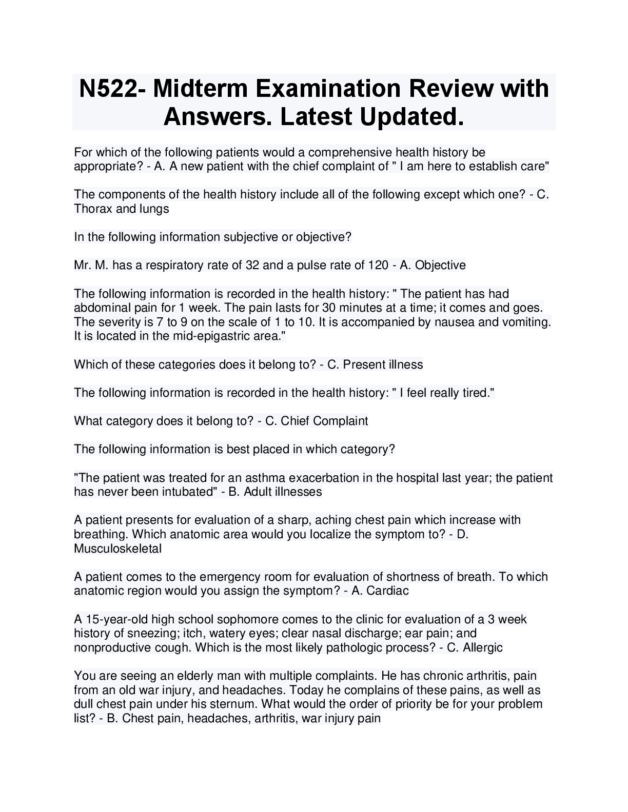
Dilated Cardiomyopathy: most common cause is . Others? - alcohol; may also be idiopathic, myocarditis, or drugs (doxorubicin) -- 1 in 3 cases of heart failure are caused by dilated cardiomyopathy
What PE and EKG changes
...
[Show More]
Last updated: 1 year ago
Preview 5 out of 329 pages
Instant download

Loading document previews ...
Buy this Document to get the Full Access Instantly
Provided by Students Who Aced it
We Verify Document Content to Gurantee Accuracy
Reviews( 0 )
Document information
Connected school, study & course
About the document
Uploaded On
Aug 01, 2025
Number of pages
329
Written in
All

Additional information
This document has been written for:
Uploaded
Aug 01, 2025
Downloads
0
Views
71
Topic Tags
Recommended For You
Get more on EXAM »
$15.5
95 Pages
FNP PRETEST EXAM NEWEST 2025/2026 ACTUAL EXAM TEST BANK COMPL...


$23
202 Pages
Solutions Manual for Fluid Mechanics Fundamentals and Applicat...
.png)
$19
185 Pages
Test Bank for Drugs, Behavior and Society 3rd Canadian Edition...
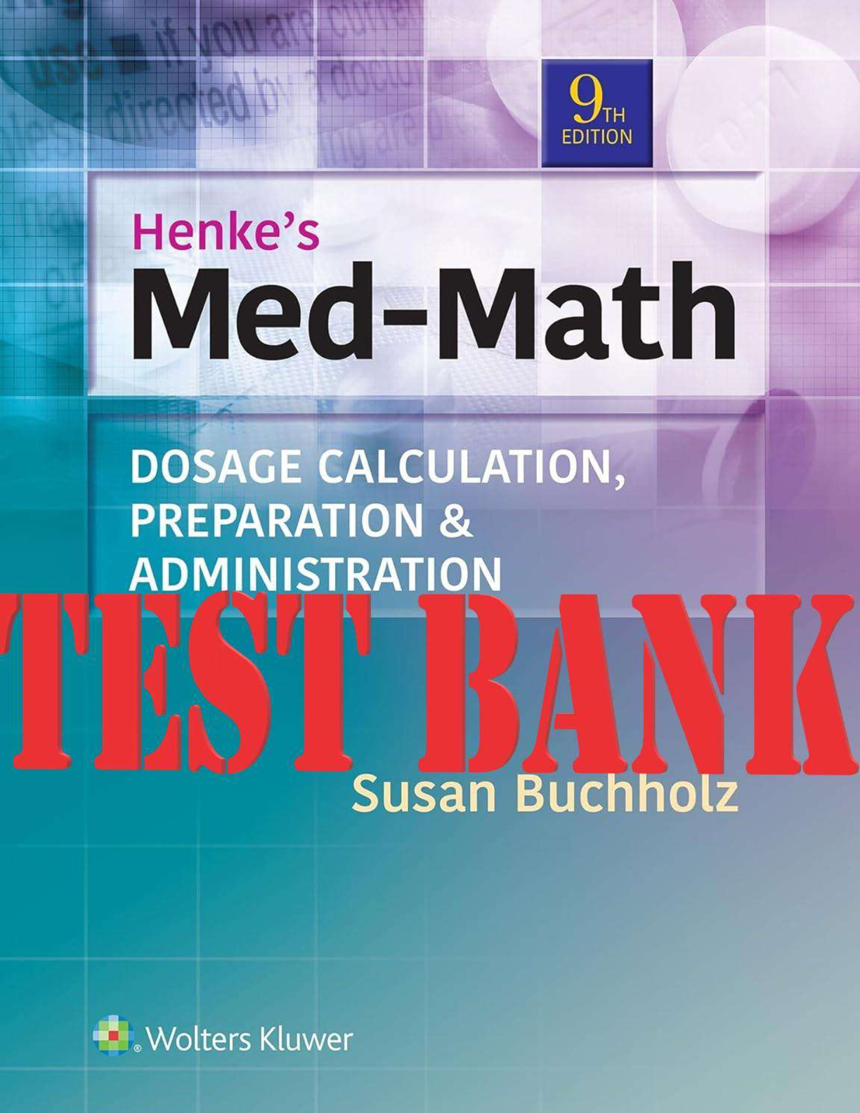
$24
235 Pages
Henke's Med-Math Dosage Calculation, Preparation, & Administra...
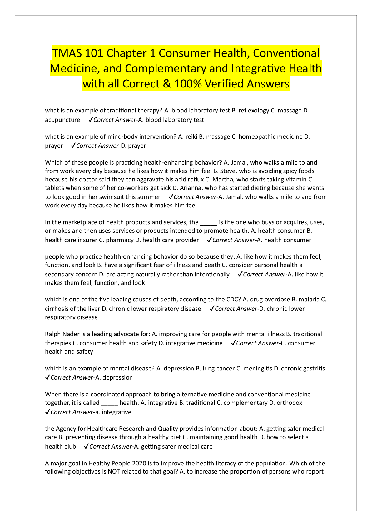
$8.5
3 Pages
TMAS 101 Chapter 1 Consumer Health, Conventional Medicine, and...
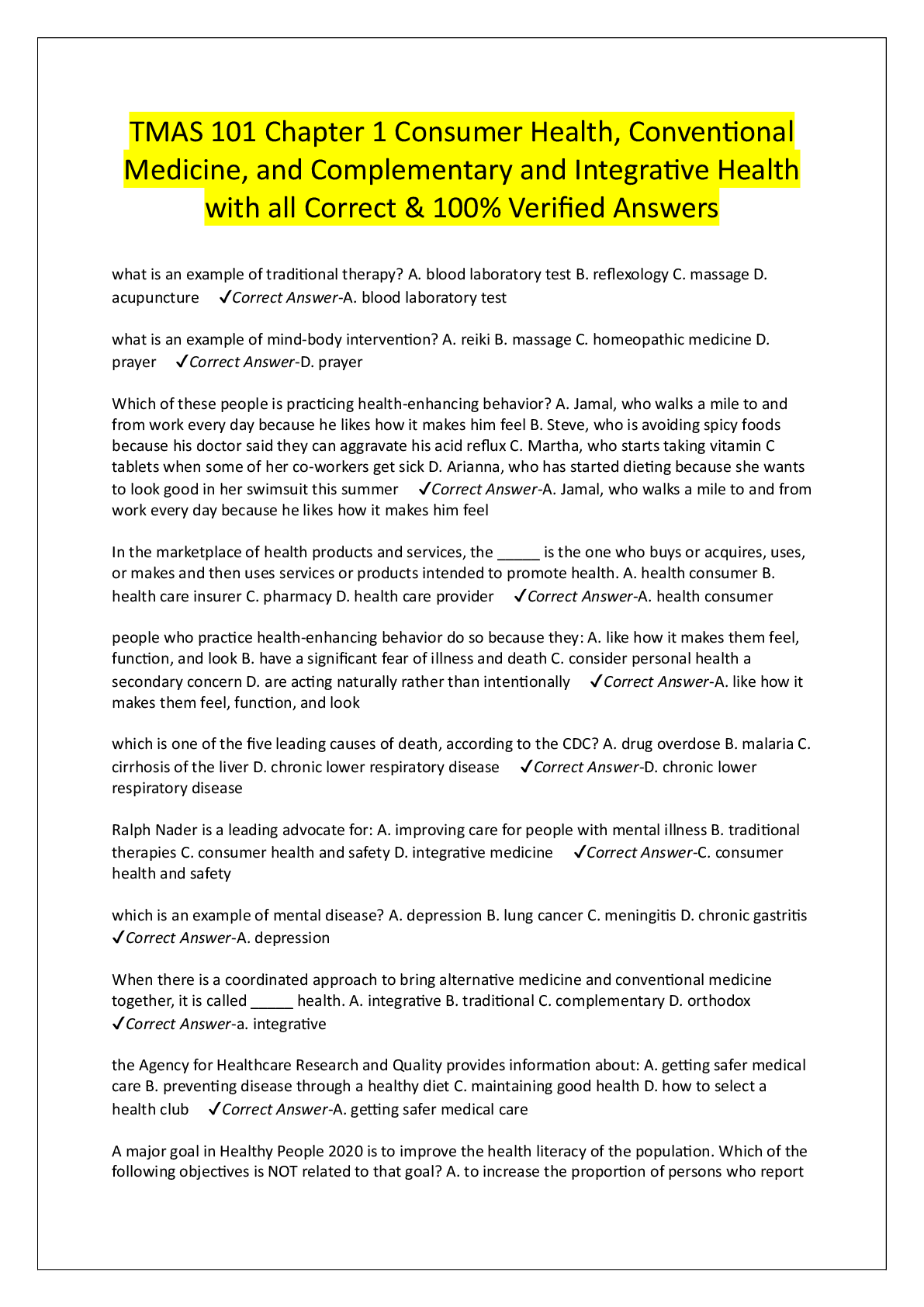
$8.5
3 Pages
TMAS 101 Chapter 1 Consumer Health, Conventional Medicine, and...
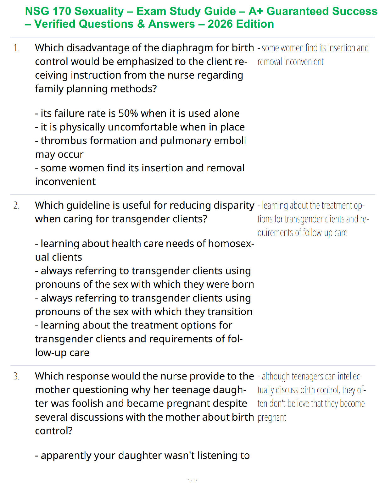
More related documents below