SCI 1001 – Lab #1 Body Orientation, Histology, Integumentary System
The Appendix, located at the end of the lab, lists the structures for which you are responsible.
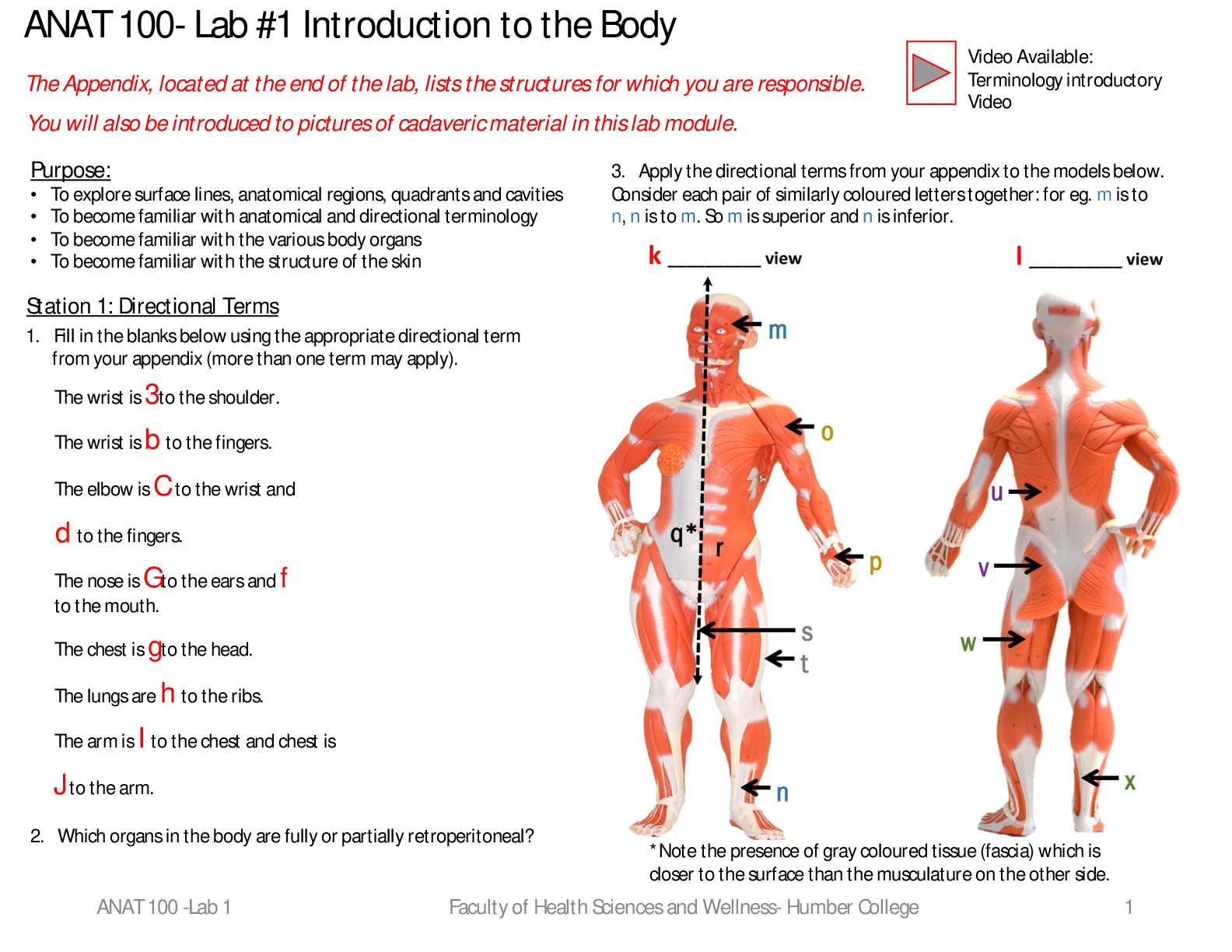
3. Apply the directional terms from your appendix to
...
SCI 1001 – Lab #1 Body Orientation, Histology, Integumentary System
The Appendix, located at the end of the lab, lists the structures for which you are responsible.
3. Apply the directional terms from your appendix to the models below.
Consider each pair of similarly coloured letters together: for eg. m is
superior to n, n is inferior to m. So m is superior
and n is inferior.
1. Fill in the blanks below using the appropriate directional term
from your appendix (more than one term may apply).
The wrist is a distal to the shoulder.
The wrist is proximal to the fingers.
The elbow is c proximal to the wrist and
d inferior to the fingers.
The nose is medial to the ears and medial
to the mouth.
The chest is g inferior to the head.
The lungs are h Deep to the ribs.
The arm is I superior to the chest and the
chest is
j inferior to the arm.
Station 1: Directional Terms K anterior view l posterior view
SCI 1001 – Lab 1 1
You will also be introduced to pictures of cadaveric material in this lab module.
Purpose:
• To become familiar with anatomical and directional terminology
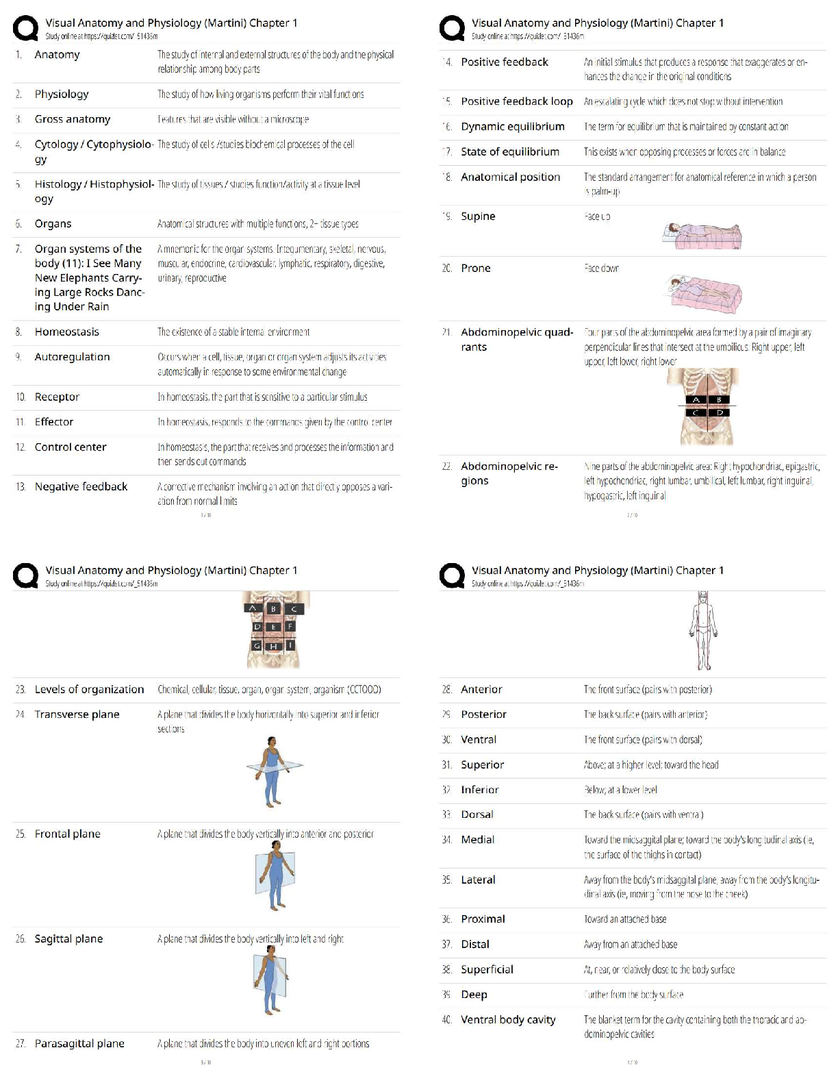
• To explore surface lines, planes, quadrants and cavities
• To become familiar with the various body organs
• To become familiar with the structure of the skin
• To be able to identify the various tissue types
Video Available:
Terminology introductory
video
O to P proximal
P to O distal
Q is surface
R is deep
S to T medial
T to s lateral
U and V Ipsilateral
w & x: Contralateral
SCI 1001 – Lab 1 Faculty of Health Sciences and Wellness - Humber College 2
a) frontal plane
b) sagittal
plane
C Transverse
a b
c
Station 2: Lines, Planes and Sections
1. Identify the planes/sections on the
model below.
Video Available:
Planes and sections
2. Identify the planes/sections of each of the following:
Stomach
d Transverse
E frontal
f Sagittal
G Coronal
trachea
esophagus
Female pelvic cavity
j Sagittal
h Transverse
I Transverse
3
Station 3: Body Cavities
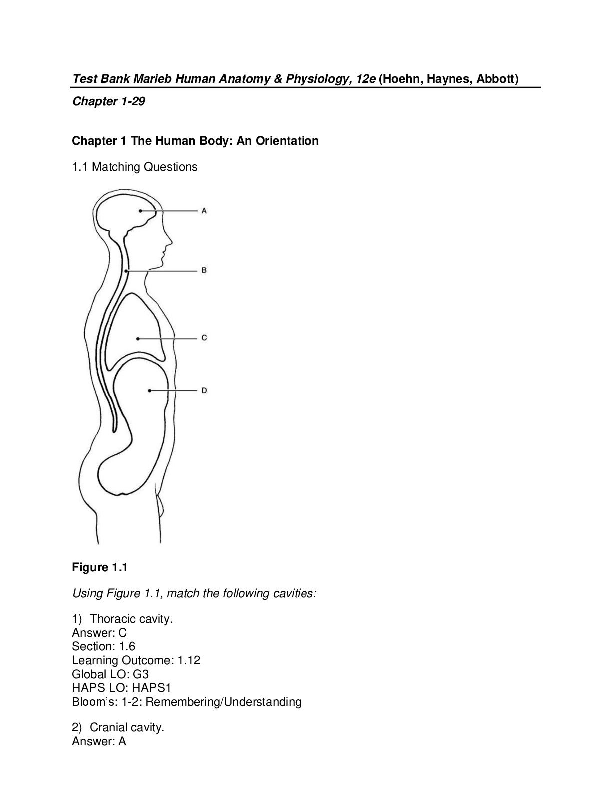
1. Label the different body cavities visible on the torso model.
Faculty of Health Sciences and Wellness - Humber College
A Cranial
B Spinal
C Dorsal
D Thoracic
E Abdominal
F pelvic
G abdominopelvic
H Ventral
SCI 1001 – Lab 1
Video Available:
Organs and Body
Cavities
4
Station 4: Identification of Body Organs
1. Identify the organs visible on the different views of the torso models below.
SCI 1001 – Lab 1 Faculty of Health Sciences and Wellness - Humber College
A larynx
B Lungs left
D Transverse Colon
Ileum e
g F
H
Liver
I
Heart
C Stomach
Superficial
J
Lungs right
Video Available:
Superficial Structures of the Thoracic
and Abdominal Cavity - Cadaveric in situ
Jejunum
Gallbladder10/8/202
[Show More]