NURSING > EXAM > PATHOPHISIOLOGY II – SUMMER 2016 TEST 1 CHAPTER 31- Structure and Function of the Cardiovascular a (All)
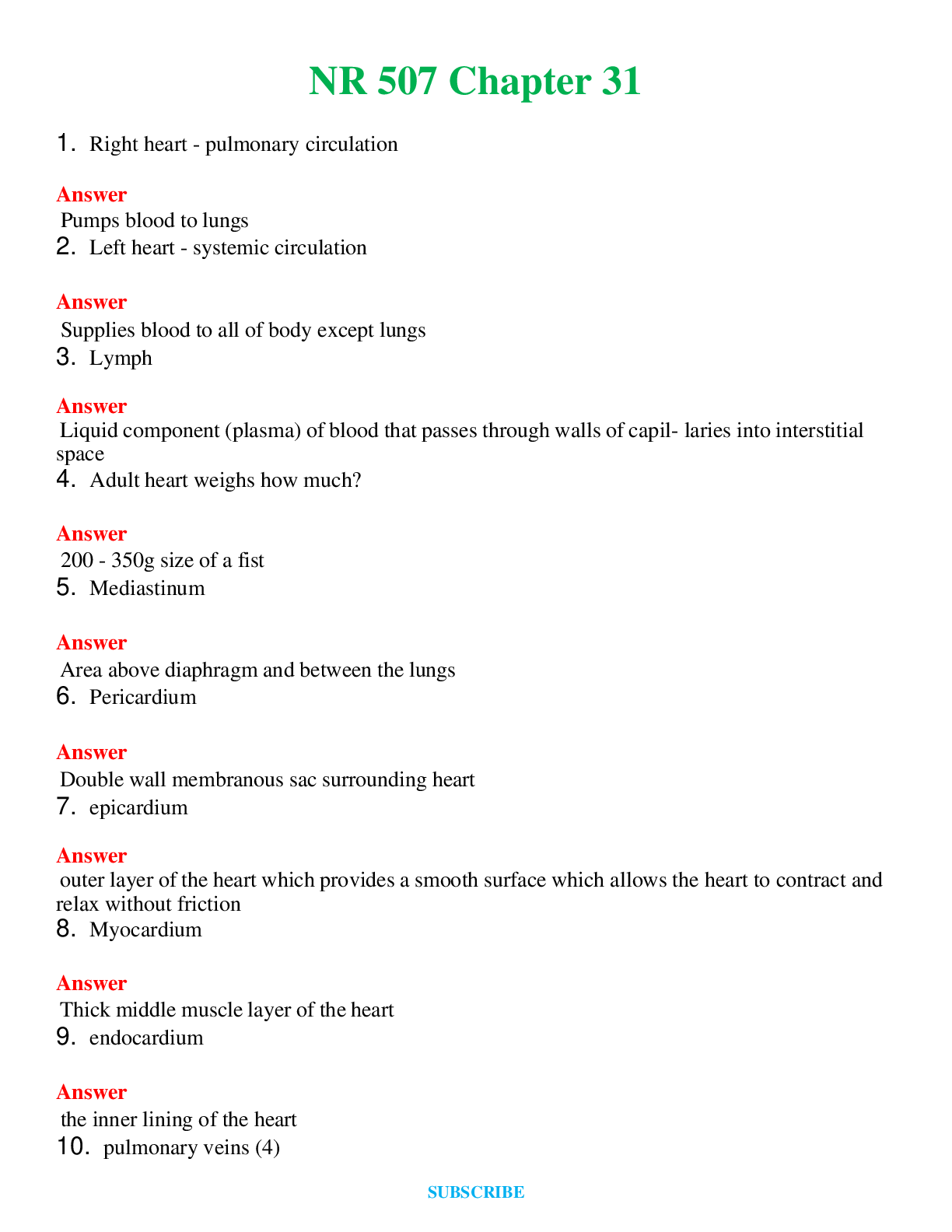
PATHOPHISIOLOGY II – SUMMER 2016 TEST 1 CHAPTER 31- Structure and Function of the Cardiovascular and Lymphatic System
Document Content and Description Below
Last updated: 3 years ago
Preview 1 out of 22 pages
Instant download
.png)
Buy this Document to get the Full Access Instantly
Provided by Students Who Aced it
We Verify Document Content to Gurantee Accuracy
Reviews( 0 )
Document information
Connected school, study & course
About the document
Uploaded On
May 27, 2021
Number of pages
22
Written in
All

Additional information
This document has been written for:
Uploaded
May 27, 2021
Downloads
0
Views
137
Topic Tags
Recommended For You
Get more on EXAM »
NURS 501 ADVANCED PHYSIOLOGY AND PATHOPHYSIOLOGY : Ultimate Te...
Test Bank For Leading and Managing in Nursing, 7th Edition By...

NR 566 Exam Preparation: Week 6 Chapter 31 Contraception (TOP...

NURS 501 ADVANCED PHYSIOLOGY AND PATHOPHYSIOLOGY : Ultimate Te...
Primary Care Art and Science of Advanced Practice Nursing - Te...

Test Bank-Chapter 31. Nursing Care of Patients With Lower Resp...
Foundations for Population Health in Community Public Health N...
.png)
Chapter 31: Older Adults Halter: Varcarolis’ Foundations of Ps...
NURS 4403 Chapter 31: The Infant and Family,100% CORRECT

University of Mary Hardin-Baylor - NURS 3320 Chapter 31- Disor...
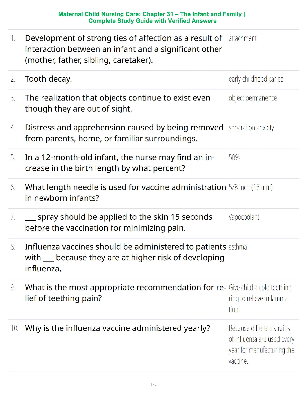
Maternal Child Nursing Care: Chapter 31 – The Infant and Famil...

NORRIS PORTHS PATHOPHYSIOLOGY 10TH EDITION TEST BANKS CHAPTE...
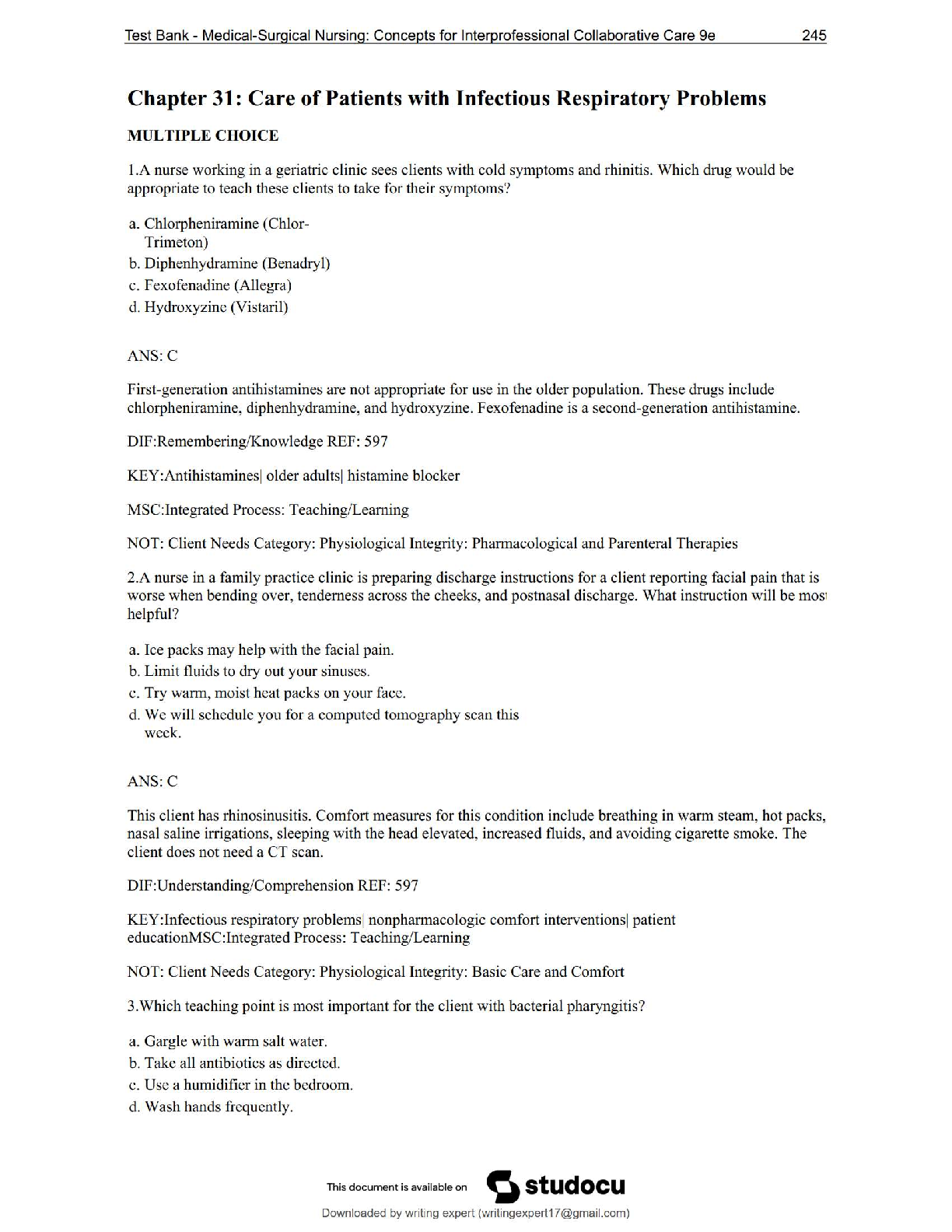
 3RD EDITION.png)
PEDIATRIC TRAUMA CHAPTER 31 (Questions with Complete Solutions...