Biology > QUESTIONS & ANSWERS > Mammalian+Eye+Dissection_PreLab+Q.docx (All)
Mammalian+Eye+Dissection_PreLab+Q.docx
Document Content and Description Below
Last updated: 3 years ago
Preview 1 out of 1 pages
Instant download


Buy this Document to get the Full Access Instantly
Provided by Students Who Aced it
We Verify Document Content to Gurantee Accuracy
Reviews( 0 )
Document information
Connected school, study & course
About the document
Uploaded On
Jun 15, 2021
Number of pages
1
Written in
All

Additional information
This document has been written for:
Uploaded
Jun 15, 2021
Downloads
0
Views
70
Document Keyword Tags
Recommended For You
Get more on QUESTIONS & ANSWERS »
![Preview of eBook [PDF] General Biology 1st Edition By Mariëlle Hoefnagels](https://browseimages.nyc3.digitaloceanspaces.com/paper-images/2024/Aug/29/7E1h3v7v2024-08-29-02-0766d0568027c90.png)
eBook [PDF] General Biology 1st Edition By Mariëlle Hoefnagels
![Preview of Essentials Of Biology 7th Edition By Michael Windelspecht, Sylvia Mader [eBook] [PDF]](https://browseimages.nyc3.digitaloceanspaces.com/paper-images/2024/Aug/29/2WloBjnG2024-08-29-10-3866d0257cc3214.png)
Essentials Of Biology 7th Edition By Michael Windelspecht, Syl...
![Preview of eBook [PDF] AP Biology 2024 1st Edition By Mark Anestis, Kelcey Burris](https://browseimages.nyc3.digitaloceanspaces.com/paper-images/2024/Aug/29/HQDEEwr22024-08-29-11-1666d02e461f2df.png)
eBook [PDF] AP Biology 2024 1st Edition By Mark Anestis, Kelce...
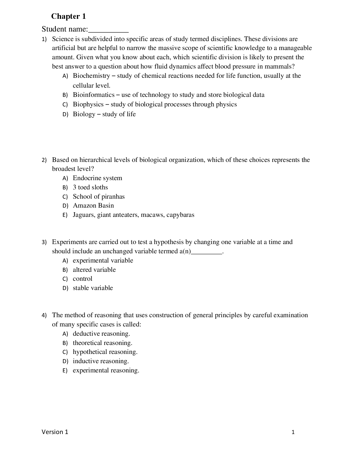
Campbell Biology 12th Edition Urry Test Bank Complete All 56...

Introduction to Spectroscopy 5th Edition By Donald Pavia, Gary...

Introduction to Marine Biology 4th Edition By George Karleskin...


 (1).png)