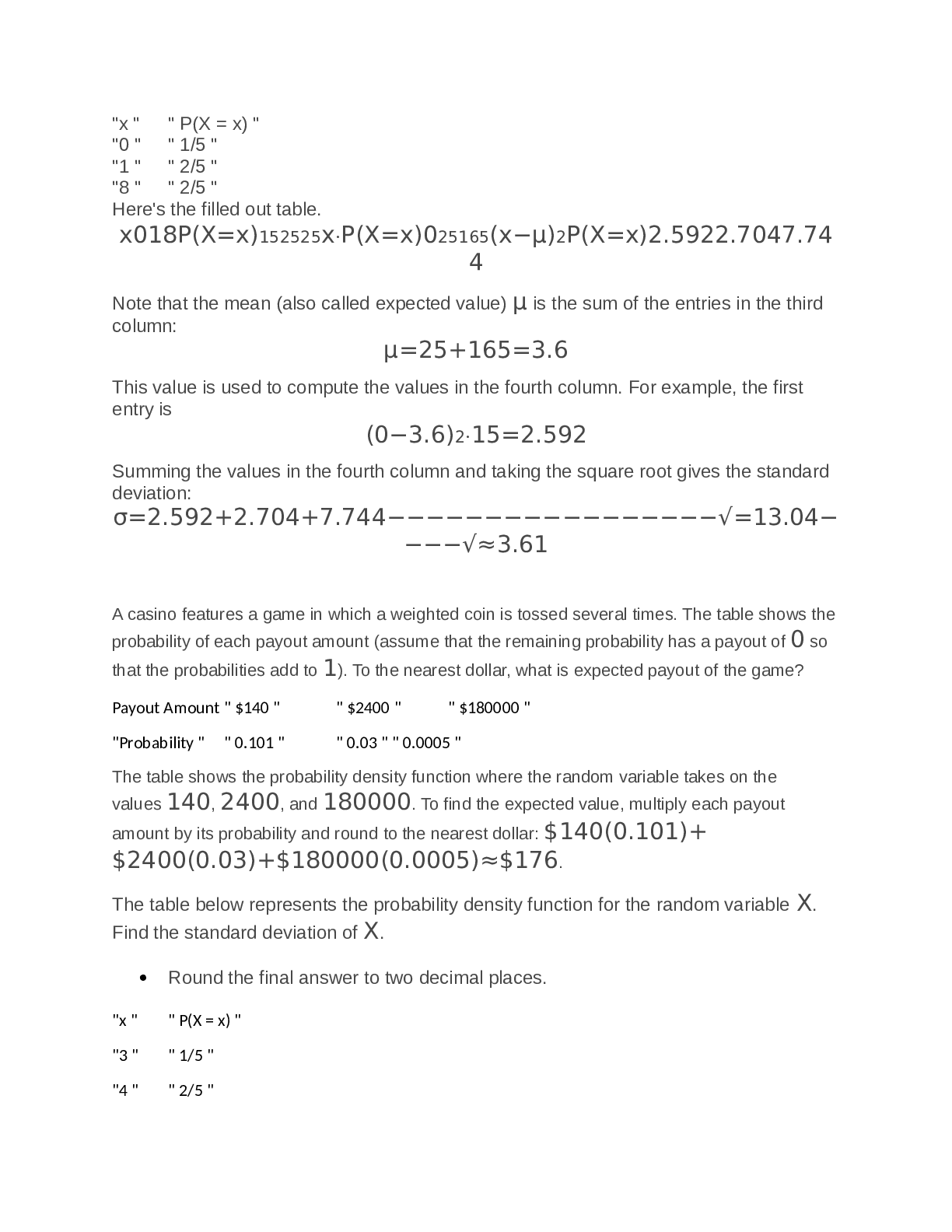
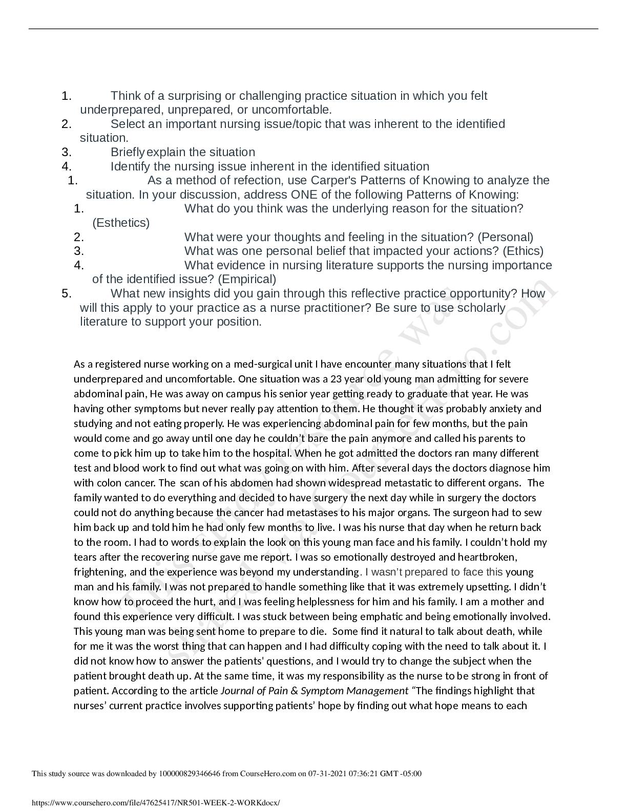
NURSING > QUESTIONS & ANSWERS > NURSING MS NR509week 4 quiz. (All)
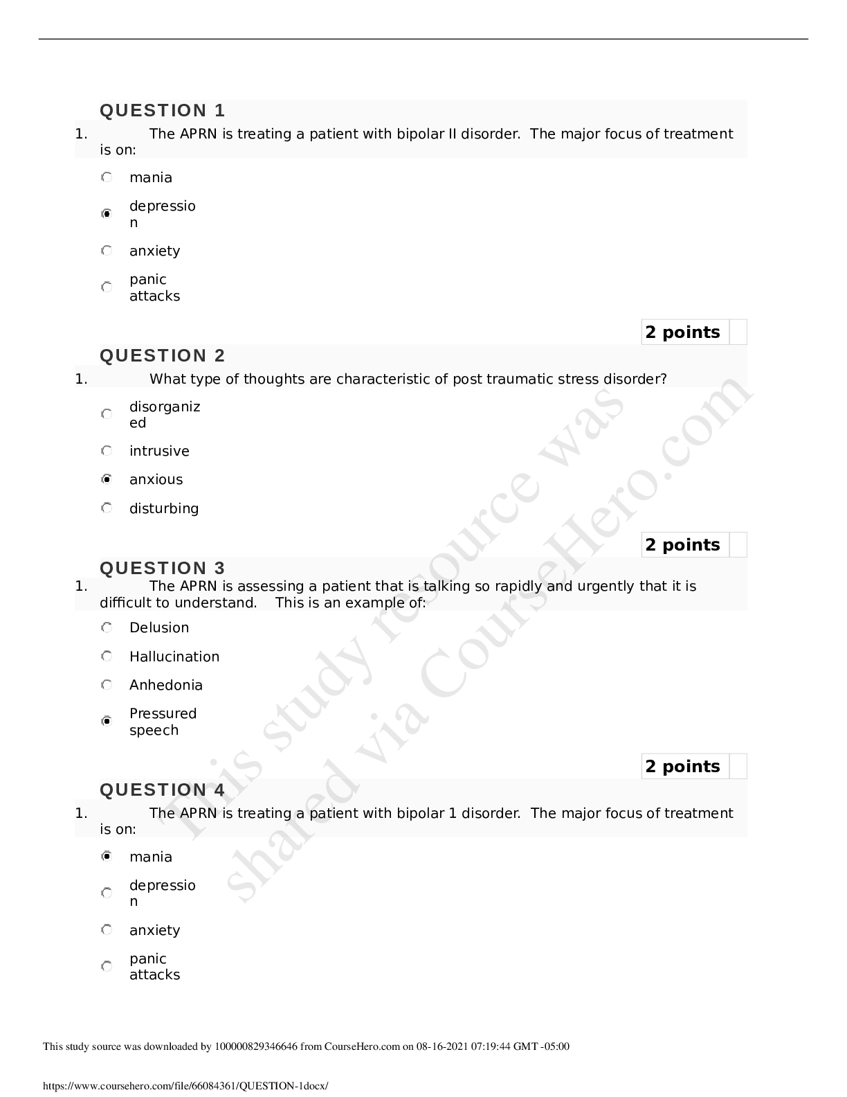
NURSING MS NR509week 4 quiz.
Document Content and Description Below
Last updated: 3 years ago
Preview 1 out of 14 pages
Instant download

Buy this Document to get the Full Access Instantly
Provided by Students Who Aced it
We Verify Document Content to Gurantee Accuracy
Reviews( 0 )
Document information
Connected school, study & course
About the document
Uploaded On
Jul 07, 2021
Number of pages
14
Written in
All

Additional information
This document has been written for:
Uploaded
Jul 07, 2021
Downloads
0
Views
162
Topic Tags
Related Subjects & Courses
Explore similar study resources and related concepts.
Recommended For You
Get more on QUESTIONS & ANSWERS »
TEST BANK for Davis Advantage for Psychiatric Mental Health Nu...
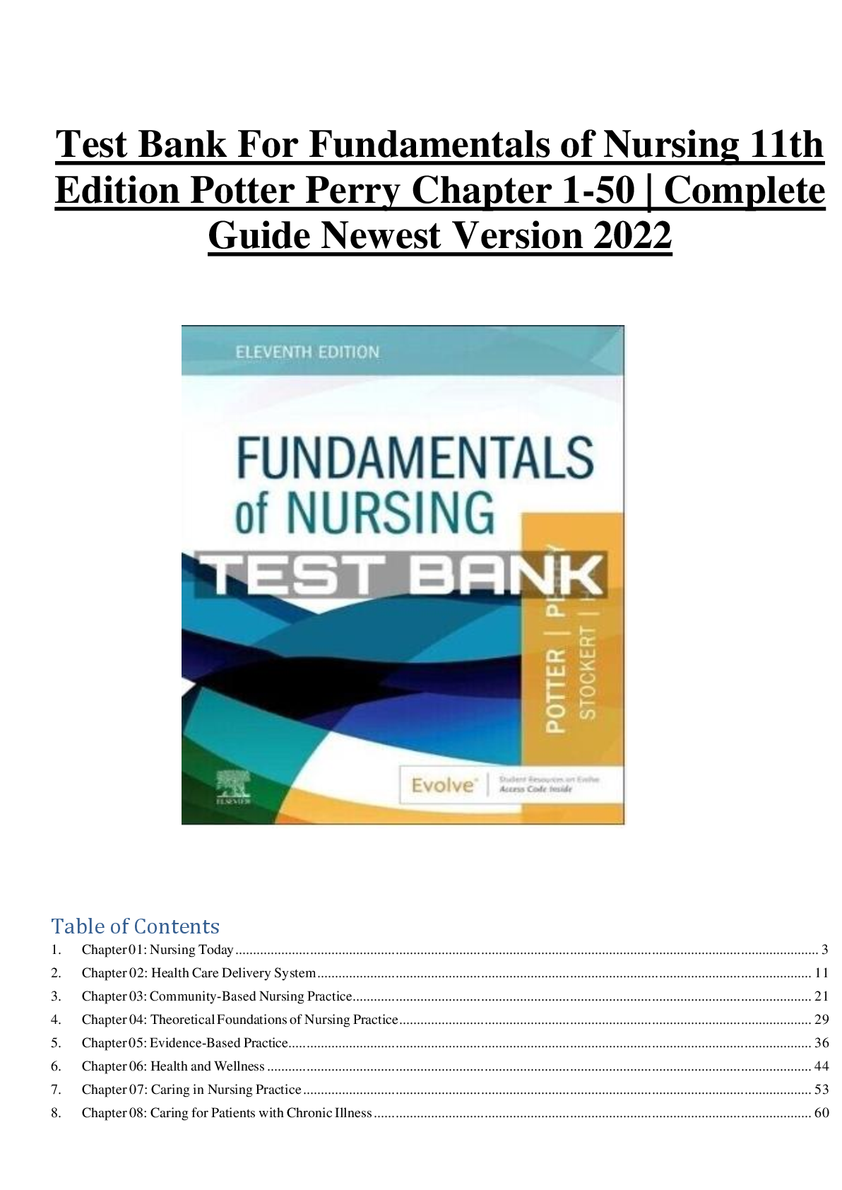
Test Bank For Fundamentals of Nursing 11th Edition Potter Perr...
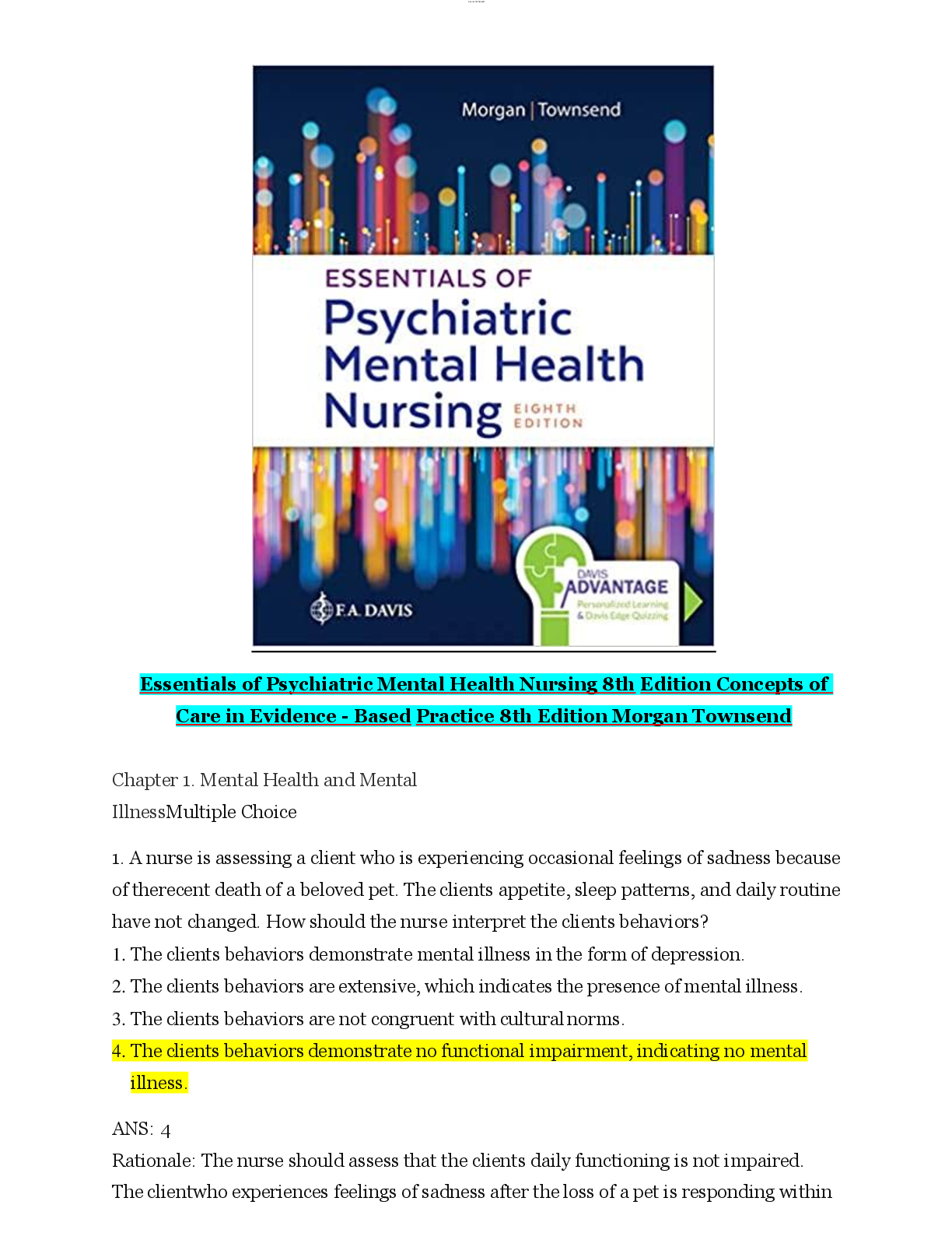
TEST BANK for Essentials of Psychiatric Mental Health Nursing:...
Essentials of Psychiatric Nursing 2nd Edition Boyd TEST BANK
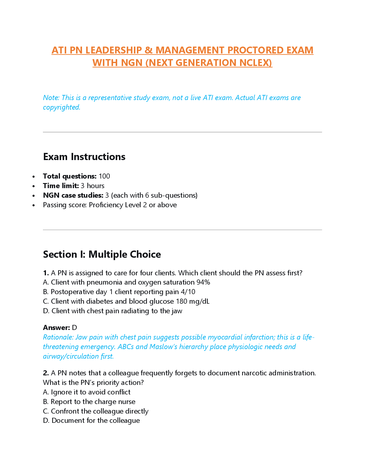
.png)



.png)