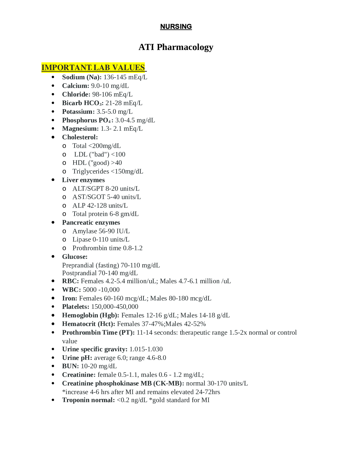
NURSING > EXAM REVIEW > Critical care exam 2 personal notes (All)
Critical care exam 2 personal notes
Document Content and Description Below
Last updated: 3 years ago
Preview 1 out of 12 pages
Instant download
.png)
Buy this Document to get the Full Access Instantly
Provided by Students Who Aced it
We Verify Document Content to Gurantee Accuracy
Reviews( 0 )
Document information
Connected school, study & course
About the document
Uploaded On
Jul 09, 2021
Number of pages
12
Written in
All

Additional information
This document has been written for:
Uploaded
Jul 09, 2021
Downloads
0
Views
96
Topic Tags
Related Subjects & Courses
Explore similar study resources and related concepts.
Recommended For You
Get more on EXAM REVIEW »
NR508 Midterm Exam Possible Questions-Answers: Chamberlain Col...

NUR2474 / NUR 2474: Pharmacology for Professional Nursing Exam...

NUR 2502 / NUR2502 Exam 2 Review Updated: Multidimensional Car...

NUR2474 / NUR 2474 Final Exam 3 Review Updated: Pharmacology f...
BIOS 255 Practical Exam Questions and Answers (Verified Answ...

![Preview of eBook [PDF] Barash, Cullen, and Stoelting's Clinical Anesthesia 9th Edition By Bruce F. Cu](https://browseimages.nyc3.digitaloceanspaces.com/paper-images/2024/Aug/29/8DF2QRTq2024-08-29-10-3966d0259b7c4b5.png)
eBook [PDF] Barash, Cullen, and Stoelting's Clinical Anesthesi...
![Preview of eBook [PDF] Fundamentals of Nursing 3rd Edition By Joanne Tollefson, Sue C. DeLaune, Patri](https://browseimages.nyc3.digitaloceanspaces.com/paper-images/2024/Aug/26/U6SQ8mmq2024-08-26-01-4766cc5d40d7385.png)
eBook [PDF] Fundamentals of Nursing 3rd Edition By Joanne Toll...
![Preview of eBook [PDF] Burns' Pediatric Primary Care 7th Edition BY Dawn Lee Garzon Maaks,, Nancy Bar](https://browseimages.nyc3.digitaloceanspaces.com/paper-images/2024/Aug/26/WR2rowXX2024-08-26-02-0966cc625bea839.png)
eBook [PDF] Burns' Pediatric Primary Care 7th Edition BY Dawn...

Test Bank for Burns' Pediatric Primary Care 7th Edition by Daw...
Burns Pediatric Primary Care 7th Edition All Chapters Complete...
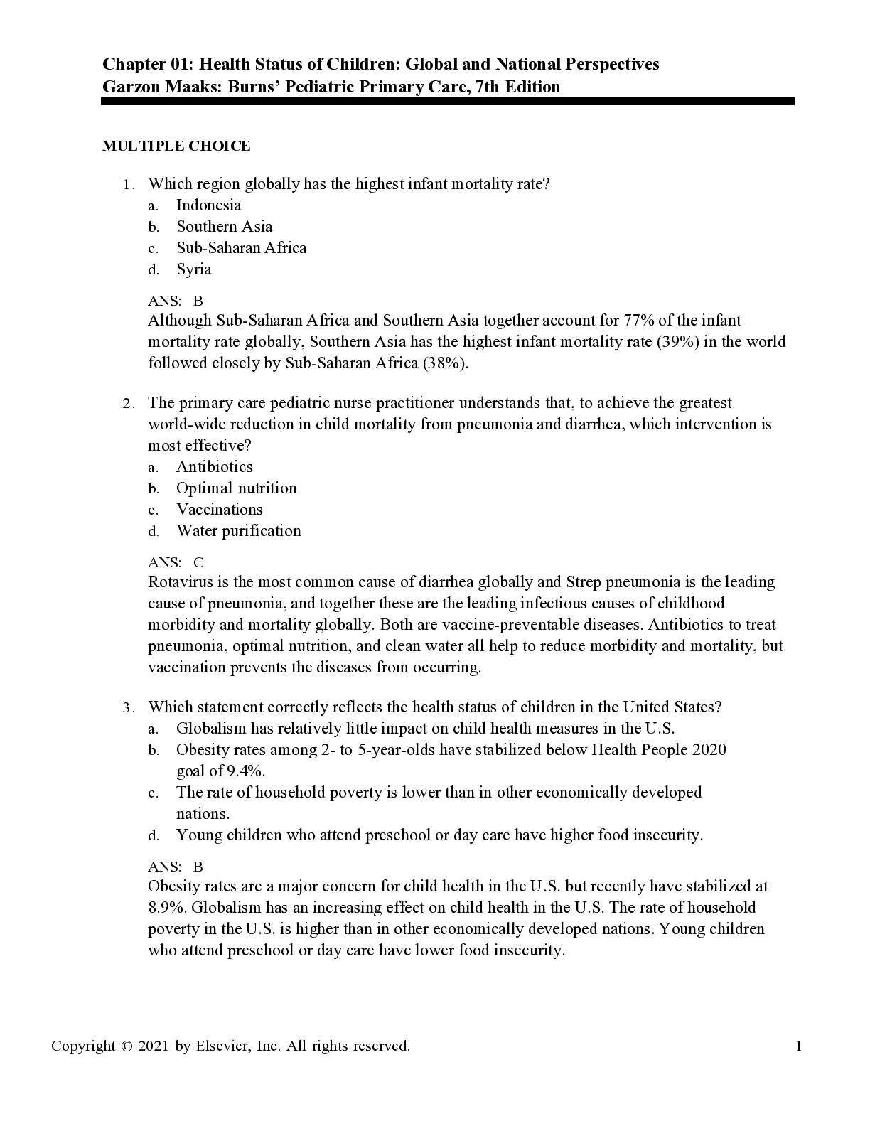
Burns' Pediatric Primary Care, 7th Edition by Garzon, Barber S...