*NURSING > EXAM > NURSING 100COTAC Final Exam Outline (AF)[5617] (1).docx/ COMPLETE SOLUTION/; RATED A (All)
NURSING 100COTAC Final Exam Outline (AF)[5617] (1).docx/ COMPLETE SOLUTION/; RATED A
Document Content and Description Below
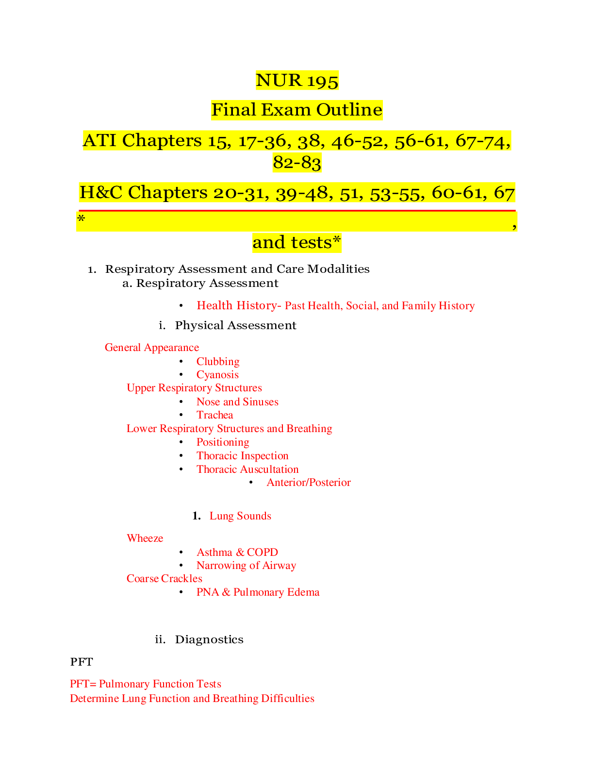
NURSING 100 COTAC Final Exam Outline (AF)[5617] (1).docx 1. Respiratory Assessment and Care Modalities a. Respiratory Assessment • Health History- Past Health, Social, and Family History i. Phys... ical Assessment General Appearance • Clubbing • Cyanosis Upper Respiratory Structures • Nose and Sinuses • Trachea Lower Respiratory Structures and Breathing • Positioning • Thoracic Inspection • Thoracic Auscultation • Anterior/Posterior Wheeze 1. Lung Sounds • Asthma & COPD • Narrowing of Airway Coarse Crackles • PNA & Pulmonary Edema ii. Diagnostics PFT PFT= Pulmonary Function Tests Determine Lung Function and Breathing Difficulties ABG’ Arterial Blood Gases (ABG) Reports the Status of Oxygenation and Acid-Base Balance in the Blood pH PaO2 • Amount of H+ • Partial pressure of oxygen PaCO2 • Partial pressure of carbon dioxide HCO - • Concentration of bicarbonate SaO2 • Percentage of oxygen bound to HgB as compared to the total amount that can be carried Bronchoscopy Directly Examine Larynx, Trachea, Bronchi Indications • Visualize and Biopsy Tissue • Visualize Inflammation or Strictures • Aspirate Secretions, Sputum or Lung Abscesses • Removal of Foreign Bodies Considerations • Pt is sedated and given anesthetic • NPO 4-8 hours before exam • ADAT after procedure when gag/cough reflex is present Thoracentesis Aspiration of Fluid from the Pleural Space • Pleural Fluid Lubricates the Movement of the Lungs in the Thorax Indications Sputum Cultures • Empyema • Pneumonia • Best collected in the morning before the patient has had anything to eat or drink Use a sterile container X- Ray Patient Education=Contraindicated in pregnancy • Patient needs to take a deep breath and HOLD it Patient Preparation= Gown and take off jewelry/metal • May use lead shield over thyroid, ovaries, and testicles • Nurse should observe the three principles of time, distance, and shielding CT Patient Education= Contraindicated in allergy, pregnancy, morbid obesity Patient Preparation= Must remain supine, sit still; NPO for 4 hours prior if contrast needed MRI Patient Education=Uses magnet and radiofrequency instead of radiation • May also use contrast • More detailed images • Contraindicated in morbid obesity, claustrophobia, confusion/agitation, implanted metal a. Some medical devices are okay Patient Preparation=Remove all metal, must lay flat and still • OFFER EAR PLUGS Pulse Ox Noninvasive • Measures oxygen saturation of hemoglobin • Sensor can be placed on finger, forehead, earlobe, nare, lip, bridge of nose Chest Tubes 2. Fluoroscopy a. Patient Education b. Patient Preparation Water Seal=Allow excess air/fluid to exit pleural space with exhalation and stops air from entering with inhalation c. Sterile Water at Bedside 3. Tidaling= expected Bubbling=Air Leak in System Indications=Pneumothorax • Hemothorax • Pulmonary Empyema Complications=Air Leaks • Do not d/c with air leak Accidental Disconnection • Keep Sterile Water at Bedside • Keep Occlusive Dressing at Bedside Tension Pneumothorax • Clamped/Kinked Tubing Removal=Pain Medication 30 Min Before • Remove Sutures • Patient will Valsalva • Apply Airtight, Sterile Petroleum Jelly Gauze Dressing • Obtain Chest X-Ray • Monitor Wound Drainage b. Oxygen Modalities i. Nasal Cannula _1-6_ L ii. Simple Mask 5-8_ L iii. Venturi Mask _4-10_ L iv. Nonrebreather 10-15_ L CPAP- Continuous Positive Airway Pressure (CPAP) • Sleep Apnea Requires Leak-Proof Mask BiPap Bi-level Positive Airway Pressure (BiPAP) • COPD or Ventilatory Assistance • Requires Leak-Proof Mask v. Mechanical Ventilation Breathes for patient Hypoxemia Hypoventilation Respiratory failure Compromised airway Respiratory distress with confusion Increased work of breathing not relieved by other interventions Controlled hyperventilation vi. High Flow Nasal Cannula c. Treatments i. Incentive Spirometer 1. Patient Education Sit upright in a chair or in bed. 2. Put the mouthpiece in your mouth and close your lips tightly around it. 3. Breathe in (inhale) slowly through your mouth as deeply as you can. 4. Try to get the piston as high as you can, while keeping the indicator between the arrows. Tracheostomy Surgical procedure • Complications • Can occur early or late (years after removal) Small Volume Nebulizer Indications • Asthma attack • SOB Nursing Management • Smoking when it is working Albuterol is most common drug used in SVN Breathing Exercises • Breathe slowly and rhythmically to exhale completely and empty lungs completely • Inhale through the nose to filter, humidify, and warm the air before it enters the lungs • If you feel out of breath, breathe more slowly by prolonging the exhalation time • Keep the air moist with a humidifier • Pursed lip exhalation Tripod breathing 2. Upper and Lower Respiratory Disorders a. Acute Respiratory Disorders i. Influenza- Flu Nursing Interventions • Monitor respiratory status • Administer antivirals if within 24 hours of symptoms (Can shorten infection) • Administer flu vaccines • Patient – wash hands, cough into shirt, avoid getting others sick, etc. Medications • Flu vaccine • Anti-viral: amantadine, rimantadine, and ribavirin Patient Education • Begin antivirals within 24-48 hours of symptom onset. • Annual flu vaccination • Hand hygiene, cough etiquette, and avoid places where people gather. • When flu manifests: Increase fluids, rest, stay home ii. Pneumonia- Inflammatory process producing excess fluid that fill the alveoli. a. Types- Viral and bacterial b. Chronic Respiratory Disorders i. Asthma- Chronic hyper-response of the airway producing edema and mucus 1. Medications Long lasting medications Short Acting Medications • Inhale short-acting Beta2-agonist (SABA): Albuterol HFA’s (increase Heart Rate) a. Patient Education • Smoking cessation • Recognize and avoid triggers • Self-administer inhaler • Teach about importance of adhering to medication regimen ii. COPD- Having both Emphysema & chronic Bronchitis 1. Emphysema- Loss of lung elasticity, hyperinflation of lungs, destruction of alveoli 2. Chronic Bronchitis- inflammation of bronchi/bronchioles due to chronic exposure to irritants 3. Medications Bronchodilators • Short-acting beta2-agonist: Albuterol • Cholinergic Antagonist: Ipratropium Anti-inflammatory agents Mucolytic agents • guaifenesin a. Patient Education • ICOUGH • Pneumonia vaccines • Pulmonary rehab • 2-3 liters of liquids/day 4. Complications • Respiratory infection • Right-sided heart failure (cor pulmonale) c. Tuberculosis- Infectious disease transmitted through aerosolization (airdrop precautions) i. Medications • Regimen for 6 to 12 months • Monitor for hepatotoxicity: LFT’s often and assess for jaundice • Rifampin (RIF): All body secretion will be orange • Recommended that patient should be taking a combination therapy of two or more of these medications at a time. Patient Education • Importance of adhering to the medication regiment • Rifampin turns secretions orange • Wear a mask in public areas • Report jaundice • RIPES d. Pulmonary Embolism- Pulmonary vasculature obstruction (Life threatening emergency) i. Therapies Medications • Anticoagulants: Heparin (drip), enoxaparin (SubQ), Warfarin (Oral) • Thrombolytic therapy (Alteplase (TPA) Procedures • Embolectomy, vena cava filter ii. Prevention • Promote smoking cessation • Encourage a healthy diet and physical activity • Prevent DVT to encouraging client to do leg exercises, wear compression stockings, and avoid sitting for long period of time. e. Pneumothoraxes- Presence of air or fluid in pleural space that collapses lung i. Types • Tension pneumothorax: air enters pleural space that cannot exists -Increases intrathoracic pressure, decreasing cardiac output. • Open pneumothorax: Open to environment (COVER THE HOLE) • Hemothorax: Accumulation of blood in the pleural space • Spontaneous pneumothorax: Lung spontaneously collapses f. Respiratory Failure i. ARF- Failure to adequately ventilate and/or oxygenate ii. ARDS- Alveoli collapse away from capillary bed • Need PEEP (positive end expiratory pressure) to keep their alveoli open. g. Obstructive Sleep Apnea- Apnea during sleep caused by obstruction h. Atelectasis- Collapse of alveoli i. Aspiration- Inhalation of foreign material into lung j. Pleural Effusion- Excess fluid the pleural space k. Pulmonary Edema 3. CV Assessment a. Trace a Drop of Blood Through the Heart i. Forwards and Backwards. 1. (Inferior & Superior) Vena cava, 2. Right atrium, 3. Tricuspid valve, 4. Right ventricle, 5. Pulmonary semilunar valve, 6. Pulmonary artery, 7. Lungs, 8. Pulmonary vein, 9. Left atrium, 10. Mitral valve, 11. Left Ventricle, 12. Aortic semilunar valve, 13. Ascending aorta, 14. Aorta, 15. Systemic circulation. b. Trace Electricity of the Heart. 1. SA node, 2. AV node, 3. Bundle of HIS, 4. Left & Right bundle branches, 5. Purkinje fibers. c. Cardiovascular Assessment i. Chest Pain: 1. Hiatal hernia/GERD/Spasm; 2. Pneumonia/Pulmonary Embolism; 3. Unstable Angina/MI; 4. Pericarditis; 5. Anxiety/Pain/Costochondritis. ii. Heart Sounds: 1. Normal: a. S1-Systole; b. S2-Diastole iii. Lung Sounds: 2. Cough; 3. Crackles; 4. Wheezes. iv. Abdomen: 1. Distension; 2. Hepatojugular reflux; 3. Bladder distension. v. Diagnostics: 1. Labs a. Troponin <0.03 ng/mL. Higher level indicates MI. Lasts for 7-10 days. b. BNP <100 pg/mL. Higher level indicates heart failure. Lasts 24 hours. 2. Rhythm Monitor a. Continuous Monitoring (Hardwired) – Continuous observation EKG that allows for continuous observation with the machine at bedside. Commonly used in ED and ICU. Signal sent to a main console. Telemetry – 5- lead EKG that also monitors heart rate and rhythm. It allows for ambulation (like a Holter monitor). b. EKG (See Section 5) – Rhythm strip or 12-lead EKG. Electrodes placed in standard positions on chest and extremities. Hooked up for a one-time print-out reading, which traces 12 different electrical flows between different positions around the heart, providing different viewpoints of the heart. 3. Echocardiograms a. TEE – small transducer put through mouth into esophagus to provide images from the esophageal position behind the heart. i. Patient Preparation: NPO from 2400 hrs; Sedation; Consent form; no alcohol for a few days before procedure. Cardiac meds may be withheld. b. TTE – ultrasound performed on the exterior of the chest wall. It measures ejection fraction and examines the size, shape, motion of cardiac structures. i. Patient Preparation: No special preparations (non-invasive ultrasound)Cardiac meds may be withheld. 4. Stress Test a. When to Stop – 1. Reaching target heart rate; 2. Any chest pain; 3. EKG changes. 5. Right Heart Catheterization a. Location – inserted into vein. b. Indication – diagnoses pulmonary hypertension. c. Results – Measure pressure and O2 on right side (from vena cava to right ventricle). d. CVP Monitoring – See hemodynamic monitoring below. Normal is 2-6 mm Hg. <2 is hypovolemia and >6 is hypervolemia. e. Complications – myocardial ischemia (LHC), bleeding, dysrhythmias, venous spasm (RHC), infection, perforation. 6. Left Heart Catheterization a. Location – inserted into artery. b. Indication - evaluate aortic arch & branches, coronary artery patency, left ventricle, mitral/aortic valves. c. Results - Remove when finished. d. Complications – myocardial ischemia (LHC), bleeding, dysrhythmias, venous spasm (RHC), infection, perforation. 7. Hemodynamic Monitoring – Indwelling catheters that provide info about blood volume and perfusion, fluid status and how well heart is pumping. a. Types: i. CVP: Normal is 2-6 mm Hg. <2 is hypovolemia and >6 is hypervolemia. ii. Art line. b. Dressing changes: i. Transparent. ii. Every 7 days or when soiled/coming off. c. Heart failure leads to increased CVP (>6 mm Hg) decreased CO. d. Elevated: Crackles in lungs, JVD, hepatomegaly, peripheral edema, taut skin turgor. Decreased: Poor skin turgor, dry mucous membranes. d. Coronary Disorders i. Angina – feelings of chest pain, mild indigestion to choking sensation. 2. Stable - predictable/consistent pain that occurs on exertion or emotional stress and is relieved by rest or nitroglycerin. 3. Acute Coronary Syndrome a. Unstable Angina – Occurs with exercise or at rest but increases in occurrence, severity, and duration. b. MI –Death of heart muscle tissue because it doesn’t get the O2 that it needs. STEMI or Non-STEMI. i. Symptoms include sudden chest pain. ii. SOB, indigestion, nausea, anxiety, diaphoresis, cool/pale/moist skin, increased HR & RR. 4. Nitroglycerin c. Patient Education - 1. Take while sitting and stopping all activity; 2. PRN for chest pain or in preparation for exertion (only every 12 hours); 3. 1 tablet every 5 minutes for up to 3 doses; 4. If pain not unrelieved after 1 pill, call 911; 5. Contraindicated with erectile dysfunction medication; 6. Store in dark, dry place; 7. Replace every 6 months; 8. Side effects are headache and hypotension ii. Percutaneous Coronary Interventions (PCI) are performed through a skin puncture rather than a surgical incision. 1. Percutaneous Transluminal Coronary Angioplasty (PTCA) - a balloon is inflated within a coronary artery to dilate the arterial lumen improving coronary artery blood flow. 2. Stent - a metal mesh that provides structural support to a coronary vessel, preventing its closure. 3. Postprocedural Nursing Indications – Monitor VS, insertion site for bleeding/hematomas, for thrombosis. Maintain bed rest, straight leg (if femoral approach), apply pressure. Continuous cardiac monitoring. Administer antiplatelets or thrombolytics, analgesics, IV fluids. Remove catheter sheath and apply pressure 1 inch proximal from insertion, then apply a pressure dressing. iii. Coronary Artery Bypass Graft (CABG) is a surgical procedure in which a blood vessel is grafted to an occluded coronary artery so that blood can flow around the occlusion. 1. Traditional a. Cardiopulmonary Bypass is a technique in which a machine temporarily takes over the function of the heart and lungs during surgery, maintaining the circulation of blood and the oxygen content of the patient's body. 2. Alternative (Off-Pump) uses beta blockers to slow heart with myocardial stabilization device. iv. Coronary Artery Disease (CAD) 1. Coronary Atherosclerosis – Injury causes inflammation. Inflammatory cells ingest lipids and deposit on the arterial wall resulting in atheromas (plaques), which cause lumen stenosis. Over years, the artery stops producing normal antithrombotic and vasodilating agents. a. Leads to hypercholesterolemia, chest pain, angina, MI, cardiac arrest. e. Central Line (CVC) Management i. Blood Draws – 1. Clean port (15 secs); 2. Flush with 10 mL NS; 3. With same syringe, pull back/waste 5 mL of blood; 4. Attach a sterile syringe; 5. Draw blood for labs; 6. Flush with 10 mL NS; 7. Withdraw syringe; 8. Apply alcohol cap. ii. Medication Administration - 1. Clean port (15 secs); 2. Flush with 10 mL NS; 3. Inject medication; 4. Flush with 10 mL NS; 5. Withdraw syringe; 6. Apply alcohol cap. iii. Dressing Changes – Transparent dressing for visualization. Change per protocol – usually every 7 days or when indicated (wet, loose, soiled). iv. Complications – Infection (Central Line Bundle: 1. Hand hygiene, 2. Sterile barrier precautions during line insertion, 3. Chlorhexidine skin antisepsis, 4. Optimal catheter site selection, 5. Daily review of necessity). 4. CV Structural Disorders and Complications a. Valves Mitral and Aortic- left side Tricuspid and Pulmonic- right side i. Diseases 1. Insufficiency a. Prolapse- Valve is bulging into the upper chamber. Can cause valve leakage which is regurgitation. b. Regurgitation 2. Stenosis- Narrowing, impeding blood flow 3. Symptoms Decreased Cardiac Output A FIB, chest pain, hypotension, narrowed pulse pressure, nocturnal angina with diaphoresis, palpitations. Increased Pulmonary Congestion Anxiety. Cough, crackles, diminished lung sounds, dyspnea and exertional dyspnea, hemoptysis, pulmonary edema, repeated respiratory infections, SOB, Wheezing. ii. Treatments 1. Mechanical Replacement 2. Biologic Replacement b. Inflammatory Diseases of the Heart Pericarditis manifestation: Pericardial Friction Rub heard at left lower sternal boarder, SOB, relief when sitting and leaning forward. Chest pressure and pain that is aggravated by breathing, coughing and swallowing. Myocarditis (inflammatory process of the heart) viral, bacterial, fungal, systemic inflammatory disease (like crohns). Prevent by immunizations Manifestations: cardiomegaly Rheumatic Endocarditis Due to Group A Beta-Hemolytic Strep. (strep throat leads to rheumatic fever, leads to rheumatic endocarditis that leads to rheumatic heart disease). Prevent by treating the strep infection promptly. Infective Endocarditis (microbial infection of the endothelial surface of the heart) Those at risk are: IV substance abusers, prosthetic heart valves, cardiac devices, structural cardiac malformations. Manifestations: petechiae on trunk and mucus membranes, splinter hemorrhages. Nursing Management: monitoring for indications of bleeding infections, and alteration in cardiac output. c. Heart Failure i. Left-Sided • Dyspnea • Fatigue • Displaced apical pulse • Pulmonary congestion (dyspnea, cough, bibasilar crackles) ii. Right-Sided • Jugular vein distention • Ascending dependent edema • Abdominal distension • Fatigue, weakness • Liver enlargement • Weight gain BNP iii. Physical Assessment iv. Diagnostics BNP is what is released from the heart when there is stretch of the heart. 100-300pg/ml suggest heart failure is present. Echo used to measure the systolic and diastolic functioning of the heart Ejection Fraction Amount of blood that leaves the heart with each contration 5. EKG’s i. Treatment 1. Medications 2. Ventricular Assist Device (VAD) -Mechanical pump that assists a heart that’s too weak to pump through the body. 3. Heart Transplant - For patients with end-stage heart failure 4. (Immunosuppressant therapy is required to avoid rejection) - Eligibility will look at life expectancy, age, psychosocial status, and abstinence of drugs and alcohol. 5. Lifestyle Changes- a. Diet- sodium and fluid restriction. b. Exercise- Know limitations, consult exercise with doctor. Encourage bed rest and energy conservation. ii. Complications- Watch for transplant rejection, risk for injection 1. Pericardial Tamponade- Fluid in the pericardial sac. Hypotension, jugular venous distension, muffled heart sounds. 2. Pulmonary Edema- The increased pulmonary vascular pressure (secondary to severe cardiac dysfunction) a. Acute (Flash)- Medical emergency, put patient in high fowlers. Administer oxygen, morphine, diuretics, and PEEP, watch output. b. Nonacute- Follow medication regimen, lower sodium, fluid restrictions, diet and exercise. a. Cardiac Monitoring Setup i. Continuous 1. 5 Lead ii. Telemetry 1. 5 Lead. iii. EKG 1. 12 Lead When doing a 12 lead you always start with V2 and place it at the 4th intercostal space b. Rhythm Interpretation i. NSR Sinus Brady -If it is symptomatic give 1. Atropine 3. Pacemaker (atropine usually works) Sinus Tach (A fast NSR)- This is caused from something- treat the cause to solve the problem. SVT- Very rapid and narrow complex Treat with vagal maneuver, adenosine push, synchronized cardioversion, Beta-Blocker or Calcium Channel Blocker A Fib If you know when it started, and it was within 48 hours treat it with cardioversion, also, if they are unstable, treat with cardioversion, if stable give warfarin and come back in 6 weeks for cardioversion. If you have A-fib for more than 48 hours you could form clots If they are on coumadin check their levels You can live with A-fib but if people don’t feel good and they have tried to reverse it multiple times they can go to the lab for EPS surgery and have the nerves ablated A Flutter If it’s stable use Vagal maneuver, Beta Blocker or Calcium Channel Blocker if it’s unstable cardioversion PVC’s & PAC’s PVC (premature ventricular complex) abnormally wide QRS that appears earlier than expected & QRS – T go in opposite directions – one up one down PAC (premature atrial complex) abnormal P wave followed by a narrow QRS that appears earlier than expected ii. Heart Blocks 1. 1° asymptomatic - R wave is far away from P wave – could be caused by cardiac drugs 2. 2° x 1 can be asymptomatic or symptomatic – PR intervals progressively increase until a QRS is completely dropped 3. 2° x 2. P waves don’t always appear . Treat with atropine push dopamine or epinephrine while waiting to get a pacemaker -this can rapidly progress to a complete heart block, it must be treated 4. 3° There are more P waves than QRS complexes and the PR interval is nonexistant. Complete AV block Symptomatic – teat with Atropine, Dopamine or epinephrine infusion waiting for Transcutaneous Pacemaker (Emergency) waiting for permanent pacemaker V Tach Pulse- Vagal Maneuver, Procainamide or Amiodarone drip, Cardioversion Pulseless- Defibrillate CPR, Defibrillate, Epinephrine and CPR, Defibrillate, Amiodarone and CPR V Fib Treat with CPR, defibrillation, epi and CPR, defibrillation, amiodarone and CPR Asystole- Give CPR and epinephrine iii. Pulseless Electrical Activity (PEA)- CPR and epinephrine iv. Cardiopulmonary Resuscitation- Indicated with no pulse v. MI EKG Changes 1. STEMI a. Hypertrophic T Waves 2. NSTEMI 3. T-Wave Inversion -indication of ischemia or change indicating a STEMI c. ACLS Medications i. Atropine- Speeds up your heart- only used in symptomatic bradycardia like heart blocks ii. Adenosine- Treats SVT iii. Amiodarone- Treats tachy rhythm (V-tach, V-fibs, SVT, and possibly A-Fib *Not first choice*) used prophylactically to prevent arrhythmias iv. Epinephrine d. Cardioversion (They have a pulse, synchronize it) i. Indications- Tachycardia with a pulse A-fib, V-tach with pulse SVT e. Defibrillation i. Indications- ONLY use with VFib and Pulseless VT f. Pacemaker i. Indications- Temporary ones are used for emergencies, permanent ones are installed for patients with chronic dysrhythmia. Pacemakers are programmed to pace the atrium, ventricles or both. g. Implanted Cardioverter Defibrillator (ICD) an electronic device that detects and terminates life-threatening episodes of tachycardia or fibrillation i. Indications • those who have survived sudden cardiac death • have experienced spontaneous, symptomatic VT 6. Peripheral Disorders a. Peripheral Vascular Disease (PVD) i. Peripheral Artery Disease (PAD) 1. Rubor/Pallor 2. Intermittent Claudication 3. Diagnostics a. Exercise Tolerance Testing b. Magnetic Resonance Angiography (MRA)- MRI scanner that can isolate blood vessels c. Ankle-Brachial Index (ABI)- Compares ankle to brachial pressure 4. Nursing Interventions a. Medications Antiplatelets- Reduces blood viscosity which increases blood flow to extremities (monitor for bleeding) Statin- Keeps plaques in place while lowering LDL’s (ex: Simvastatin, Atorvastatin) 5. Treatments a. Percutaneous Transluminal Angioplasty • Balloon and stent placement • Watch for bleeding at insertion site • Anticoagulants during and antiplatelets after 6. Reynaud’s Phenomenon Causes- stress, changes in temperature Manifestations- Cold, pain, pallor, white discolored then blue then red, numbness, tingling, burning pain Diagnostic- Cold-stimulation test Medications- Calcium channel blockers Patient education- Avoid triggers like cold, caffeine, tobacco, emotional distress 7. Buerger’s Disease Factors- genetics, tobacco, men ages 20-35 Manifestations- cold sensitivity of hands, sore on fingers and toes STOP SMOKING, avoid cold, avoid constrictive clothing ii. Venous Disease 1. Venous Thromboembolism (VTE)- Venous stasis + Endothelial injury+ Hypercoagulability Causes- immobility, pregnancy, surgery b. 6Ps Findings- limb pain, edema, warmth, redness 1. Venous Insufficiency Risk factors- sitting, standing too long, obesity, pregnancy Findings- swelling, stasis dermatitis (brown discoloration on lower limbs), venous stasis, cellulitis 2. Varicose Veins Risk factors- female, >30 years old, prolonged standing, pregnancy, obesity, systemic disease Expected findings- Distended superficial veins, pruritis 3. Complications- Ulcer formation, pulmonary embolism i. Pain ii. Pallor iii. Pulselessness iv. Paralysis v. Poikilothermia vi. Paresthesia 7. HTN a. Diagnosing SBP > 140 mm Hg and/or DBP > 90 mm Hg • 2 or More Readings • Essential or Secondary • Gerontologic Changes (≤ 60 y/o) Structure & function change to increase BP so HTN is 150/90 b. Medications • Diuretics- Monitor K+ -Thiazide- Encourage K+ Rich Foods -Loop- Encourage K+ Rich Foods -Potassium-Sparing • Calcium-Channel Blockers- Avoid grapefruit juice • ACE Inhibitors- Monitor for cough • Angiotensin-II Receptor Antagonists- ↓ K+ foods, Angioedema • Aldosterone-Receptor Antagonists- Monitor K+, ↓K+ foods, avoid grapefruit juice and St. John’s Wort • Beta Blockers- Don’t stop suddenly, Assess for hypoglycemia • Central-Alpha2 Agonists- Sedative, Causes Impotence • Alpha-Adrenergic Antagonists- Caution with driving • Hypertensive Crisis above 180/120 -Urgency treat with po vasodilators goal to decrease below 140 in 1-2 days • Hypertensive Urgency -Must lower BP urgently, but is not yet to the point of organ damage -Causing HA, epistaxis, anxiety -Normalize BP within 24°-48° -Oral Vasodilators- labetalol, captopril, clonidine • Hypertensive Emergency -Must lower BP emergently to prevent organ damage -Gestational hypertension, acute MI, dissecting aortic aneurysm, intercranial hemorrhage -Decrease by 20%-25% in the first hour -IV Vasodilators- Nitroprusside, nicardipine, labetalol, nitroglycerin, metoprolol Emergency with iv meds goal is to decrease in 1 hour by 20 % Patient Education- Symptoms of Electrolyte Imbalance, Importance of Adhering to Medication Regimen, Schedule Regular Provider Appointments, Report Abnormal Findings, Teach Patient and Family to Take BP Readings, Ranges, Verbal and Written Information c. Hypertensive Crisis i. Hypertensive Urgency 1. Diagnosing 2. Treatment ii. Hypertensive Emergency 1. Diagnosing 2. Treatment d. Lifestyle Changes • Nutrition -DASH- Dietary Approaches to Stop Hypertension i. ↑ Fruits & Veggies, ↓ Fat -↓Na+ -↓ETOH -↑ Ca++ and Mg+ • Weight Reduction and Maintenance -Exercise 3x Week • Smoking Cessation • Stress Reduction -Yoga, Massage, Hypnosis, etc. 8. Aneurysms Risk Factors- atherosclerosis, uncontrolled hypertension, tobacco use, family history, history of syphilis, blunt force trauma. Aneurysms are a medical emergency. Abdominal Aortic Aneurysm (AAA) Most common with atherosclerosis, flank or back pain is an indication of impending rupture Aortic Dissections i. Associated with Marfan syndrome. Sudden onset of tearing, ripping, stabbing abdominal or back pain. Hypovolemic shock, decreased peripheral pulses, neurological deficits X-ray, computed tomography (CT), Transesophageal echocardiography (TEE) used to detect. Nursing Care: take vitals q15 min until stable, monitor peripheral pulses, keep blood pressure between 100-120 systolic, Medications consist of beta blockers, antihypertension, calcium blockers Cerebrovascular Disorders b. Strokes i. TIA Sudden loss of motor, sensory or visual function. TIA is stroke like symptoms that usually resolve in 1-2 hours, can last up to 24 hrs. Causes temporary ischemia to the brain. 1. Treatment for TIA is a Carotid Endartectomy ii. Ischemic TPA- Sudden loss of function resulting from disruption to the blood supply of the brain. a. Indications- older then 18 yrs, clinical diagnosis of ischemic strokes, time of onset less then 3 hrs, Contraindications- Seizures, major surgeries, head trauma, GI bleeds, pregnancy, prothrombin time greater then 15 seconds, heparin in the last 48 hrs, Hemorrhagic- Bleeding into the brain Symptoms 2. BEFAST B- balance, E- Eyes, F-face, A- arms, S- Speech, T-time c. Nursing Interventions i. NIHSS Stroke Assessment https://www.mdcalc.com/nih-stroke-scale-score-nihss Prevent Secondary Injury- Vitals q1hrs, maintain 92 O2, EKG, LOC, HOB 30 degrees, ROM exercises, Aid in ADLS, Support emotionally, assist with safe feedings. d. Complications i. Unilateral Neglect- loss of awareness of the side affected by the stroke. Need to teach client to look over at the affected side periodically. ii. Hemianopsia- visual changes. Can only see half of field of view. Patient Education- Begin ASAP with family involvement, patient will most likely not return to baseline, need to adjust the home environment. Musculoskeletal System e. Bone Maintenance i. Calcium • Foods: milk products, green leafy veggies, fortified OJ and cereals, red and white beans, and figs. • Take calcium supplements with food. Calcium and VitD supplements can slow or prevent osteoporosis. • Prolonged med use (loop diuretics, corticosteroids, thyroid meds, anticonvulsants) affects calcium absorption and bone metabolism. • Calcium Supplements: Calcium Carbonate and Calcium Citrate (can cause GI upset) ii. Vitamin D • Foods: Fish, egg yolks, fortified mils and cereals. • VitD supplements increases absorption of calcium from the intestinal tract and availability of calcium in the blood for remineralization of bone • Nursing Care: Reinforce the need for exposure to VitD (moderate sun exposure, fortified milk) f. Diagnostics i. Arthroscopy • Procedure that allows for visualization of the internal structure of a joint. • Joint infection and lack of joint mobility are contraindicated. • Indicated when the joint is injured, an arthroscopy can ascertain the extent of damage. The provider can use the arthroscope to repair the damage (a torn tendon or meniscus) or perform a synovial biopsy ii. Nuclear Scans • Consist of Bone Scans (uncommon d/t MRI), Gallium and Thallium Scans (more sensitive than Bone Scans) • Evaluate the entire skeletal system • Radioactive isotope (nephrotoxic and used in only bone scans not used in gallium and thallium scans) is injected 2-3 hours before the scan and areas of abnormal bone will appear brighter. iii. Dual-Energy X-Ray Absorptiometry (DEXA) • Estimate the density of bone mass (most common sites are hip and spine) and the presence or extent of osteoporosis 9. Indicated to r/o osteoporosis in women >40 or menopausal a. Therapeutic Procedures • Orthotic Devices: • Vertebroplasty or Kyphoplasty: Mild sedation is used, and bone cement is injected into the fractured space of the vertebral column with or without balloon inflation. Patient must lay supine 1-2 hrs following the procedure b. Arthroplasty- Surgical removal of a diseased joint d/t osteoarthritis, osteonecrosis, rheumatoid arthritis, trauma, or congenital abnormalities and replacing the joint with prosthetic or artificial components made of metal or plastic i. Hip • Total hip: involves replacing the acetabular cup, femoral head, and femoral stem • Hemiarthroplasty: half a joint replacement. Fractures of the femoral neck can be treated by only replacing the femoral component • Early ambulation • Assistive (walker, long handled shoehorn, dressing sticks) and adaptive (raised toilet seat, grab bars, shower chair) devices • Complications: Venous thromboembolism. Monitor for acute dyspnea, tachycardia and pleuritic chest pain. Promote early ambulation, prophylactic med management, antiembolic stockings, SCD’s while in bed ii. Knee • Total Knee: replacement of the distal femoral component, the tibial plate and the patellar button • Unicondylar knee replacement: when a joint is diseased in one compartment of the joint • Float heels = pillow under knee • Meds: analgesics, peripheral nerve block, antibiotics, anticoags (enoxaparin=low molecular weight heparin commonly used to prevent DVT) • Apply ice to incision site to decrease swelling and inflammation iii. Patient Education • Position restriction for hip replacement: elevated toilet seat, straight chairs with arms, abduction pillow between legs in bed, externally rotate toes, no flexion greater than 90◦, avoid low chairs, do not cross legs, do not internally rotate toes, avoid turning to operative site • Knee dislocation: less common than hip dislocation. No deep squats, avoid continuous flexion to avoid contracture c. Amputations- removal of a body part, most common an extremity, d/t complication of peripheral vascular or accidental trauma • Amputations are described in regard to whether the amputation is above or below a joint. • Monitor cap refill, edema, necrosis, lack of hair distribution (inadequate peripheral circulation) • If cannot stop bleed apply tourniquet immediately Phantom limb pain: deep burning, cramping, shooting, aching. • Calcitonin is administered 1 week after amputation to decrease phantom pain • Beta Blockers (propranolol) can relieve continual dull, burning sensation • Antiepileptics (gabapentin) can relieve sharp, stabbing, burning pain • Antispasmodics (baclofen) Buergers Disease: rare disease in the arms and legs. The vessels become occluded and tissue death occurs in finger and/or toes. d. Osteoporosis- common chronic metabolic bone disorder resulting on low bone density. Chief sign is repeated fx. Occurs when the rate of reabsorption exceeds the rate of bone formation. i. Physical Assessment 1. FRAX- fracture risk assess tool 2. Increases risk: BMI, female, age, chronic corticosteroid use, excessive ETOH use, DM1, hyperthyroid, premature menopause, malnutrition, liver disease, lactose intolerance Posture- Kyphosis (hunchback), Lordosis (pregnancy) and Scoliosis ii. DEXA • Estimate the density of bone mass (most common sites are hip and spine) and the presence or extent of osteoporosis iii. Nursing Interventions 1. Patient Education • Diet change = increase calcium and VitD intake. Limit caffeine, ETOH and carbonation. Eat sufficient amounts of protein, magnesium and VitK (bone formation) • Sun exposure = 5-30 min 2x week • Weight bearing exercises • Home safety – equipment, avoid slippery surfaces and clean walkways 2. Medications Decreases Bone Reabsorption • Thyroid hormone – Calcitonin • Selective Estrogen Receptor Modulator – Raloxifene • Bisphosphonates – Alendronate • Monoclonal Antibodies – Denosumab Stimulates Bone Formation • Teriparatide Estrogen Replacement • Estrogen Hormone Replacement – Medroxyprogesterone Promotes Healthy Bones • Calcium Supplement – Calcium Carbonate • VitD Supplement e. Fractures i. Classification 1. Open or Closed? Open (always displaced): bone broke through skin Closed: bone did not break through the skin 2. Displaced or Nondisplaced? • Displaced: Bone fragments are not aligned • Non-Displaced: Bone fragments remain in alignment 3. Type a. Comminuted- Fragmented b. Oblique- Angled across the bone c. Spiral & Avulsion- Twisting motion d. Impacted- Fractures bone wedged in opposite fractured fragment e. Greenstick- Occurs on one side but not completely through the bone ii. Nursing Interventions 1. 6Ps a. Pain b. Pallor c. Pulse d. Paresthesia e. Paralysis f. Poikilothermia iii. Immobilizers 1. Cast (ASSESS 6 P’s) Nursing Interventions: monitor neurovascular status, maintain skin integrity, ice 24-48 hrs, elevate above heart 24-48 hrs, document drainage, cover with plastic when bathing, report pain/hotspots, report SOB 2. Traction Goal: prevent soft tissue damage, realign bone fragments, decrease muscle spasms and pain, correct deformity Types of traction: • Manual – closed reduction • Straight – Skin = less effective and short term. Use of weights, Bryants traction (congenital hip dislocation in children) and Buck’s traction (preoperative for a hip fx for immobilization) • Balanced – Skeletal = significant amount of time, pin or wire put through bone. Uses slings or splints to support the fractured extremity off the bed while pulling with ropes and weights. Pin protocol: Chlorhexidine, one swab for each pin, completed once a shift (depending on facility protocol) 3. Splints a. Protect b. Rest c. Ice d. Compression e. Elevation 4. Fixation a. External Immobilization of a fx using percutaneous pins and wires attached to a rigid external frame. Used to treat comminuted fx with extensive soft tissue damage. b. Internal Open Reduction and Internal Fixation (ORIF): surgical fixation of a fx using plates, screws, pins, rods and prosthetics. 5. Reduction Open- Fracture fragments are exposed surgically by dissecting the tissue Closed- Manipulation of the bone fragments without surgical exposure iv. Complications Compartment Syndrome- Increased pressure within a confined space compromises blood flow and tissue. Most serious complication of casting and can cause tissue damage within 4-6 hrs if untreated. Disproportionate pain with passive movement. Requires an emergency fasciotomy. ASSESS 6 P’s! Fat Emboli- Rapid onset of s/s: (1)dyspnea, (2)neuro compromise, (3)petechial rash. Can be prevented by immediate mobilization of fx, early surgical intervention, minimal fx manipulation, adequate support for the fx during turning/positioning Osteomyelitis- Infection of the bone. Septic manifestations, local pain and swelling, chronic causes nonhealing ulcers, and very abnormal x-ray. Leukocytosis, increased ESR, WCX, BCX. Treated with surgical debridement, hyperbaric, antibiotic (3 months via PICC) or amputation. Can lead to avascular necrosis, VTE and failure of the fx to heal (malunion or nonunion). Avascular Necrosis-Bone death due to compromised blood flow at fx Carpal Tunnel Syndrome- Compression of the medial nerve at the wrist. Dx with Phalen’s maneuver: push wrists together, palms apart, with fingers pointed down. Positive = numbness and tingling within 30 seconds. Treated with splint/brace, steroid injection, and/or surgery • Osteoarthritis- Degenerative joint disease. Articular cartilage is worn away = bone to bone • Risk Factors: Age, genetics, joint injury, obesity, rheumatoid arthritis, Crohns • S/S: joint pain and stiffness, crepitus, joint enlargement, nodes, joint effusion, limping, back pain Low Back Pain • Nursing Intervention: Health Promotion! Exercise to strengthen back, proper body mechanics, correct posture, low-healed shoes, healthy weight, no smoking, avoid prolonged sitting/standing, healthy diet • Risk: family hx, pregnancy, spinal problems, heavy lifting/twisting/repetitive motion, smoking, obesity, poor posture. • S/S: pain aggravated by coughing/sneezing/straining, muscle spasms, sciatica, paresthesia of leg 10. Digestive Function and Treatment a. Assessment i. Dyspepsia ii. Pain iii. Stool Assessment b. Diagnostics i. Body Sample Testing 1. Blood 2. Urine 3. Stool 4. Breath 5. Saliva 6. Skin ii. Endoscopy 1. Colonoscopy a. Patient Education Meds- Go-Lytely, Folyethylene Glycol, Bisacodyl • Clear diet at 1200 day before the procedure. No red, purple, or orange fluids • NPO after 0000 (midnight) • Increase fluid intake 2. Esophagogastroduodenoscopy (EGD) a. Patient Education • Teach the client to use throat lozenges if a sore throat or hoarse voice persists following the procedure • NPO for 8 hours • Cannot drive 12-18 hours after • Withhold fluids until return of gag reflex 3. Endoscopic Retrograde Cholangiopancreatography (ERCP) a. Patient Education • NPO 6-8 hrs; remove dentures prior to procedure • Withhold fluids until return of gag reflex • Teach the client to use throat lozenges if a sore throat or hoarse voice persists following the procedure • Cannot drive for 12-18 hours 4. Pill Cam M2A • Swallow the capsule with a glass of water • Fast (water allowed) 8-10 hours before • NPO for 2 hours following swallow • Normal eating 4 hours following swallow • Patient returns recorder after 8 hours after evacuation via stool 5. Complications Over sedation • Decreased respiratory drive • Tachycardia • Decreased or increased BP Hemorrhage • Bleeding • Cool, clammy skin • Decreased BP Aspiration • Keep NPO until gag reflex returns • Encourage the client to deep breathe and cough to promote removal secretions Perforation of the GI tract • Abdominal distension • Bleeding from the rectum • Report pain, fever, and bleeding to the provider iii. GI Series 1. Barium Swallow a. Patient Education • Drink radiopaque liquid (barium) o Upper GI imaging • Restrict food and fluids for bowel preparation • Increase fluid intake to promote elimination of contrast material • Stool will be white for 24 to 72 hours until barium clears 2. Barium Enema a. Patient Education • Barium enema into rectum and colon gives a lower GI image • Restrict food and fluids for bowel preparation c. Therapeutic Procedures i. Enteral Feedings 1. Indications • Inability to eat due to a medical condition (comatose, intubated) • Increased risk of aspiration (stroke, Parkinson’s, MS) 2. Complications Overfeeding • Administering more formula than the patient can digest, check for >200 mL Underfeeding • Not administering enough calories for patient to heal. Diarrhea • Wrong formula, rapid infusions, cold formula, medication (slow the rate of the feeding, provide skin care) Constipation • Due to lack of fiber, fluid volume deficit, opioid use Fluid volume deficit • Monitor I&O’s closely Aspiration pneumonia • Confirm tube placement • Elevate head of bed at least 30 degrees during feeding and for at least 1 hour after Refeeding syndrome • Feeding a patient too much while they’re in starvation mode Dumping Syndrome • High osmolality solution pulls all water in to intestines from organs and vasculature 3. Gastrointestinal Tube Management • Check gastric residual volumes before feeding and q4 hours for continuous feedings • Change tube feeding container and tubing q 24 hours • Ensure patency- 30 mL of water given before and after intermittent tube feeding and medication administration, flush 5 mL between medications, if tube is not being used flush a minimum of once daily ii. Parenteral Feedings 1. Indications • Second to enteral feedings as enteral feeding preserve gut flora and have less complications • Inability or unwillingness to orally ingest food/liquid for >7 days • Inability to absorb nutrition via the GI tract • Hypermetabolic states • Chronic malnutrition 2. Nursing Interventions • Central Line administration • Inspect solution for separation • Insure correct mixture according to Rx • Start low and advance as prescribed • Labs CBC, CMP, Blood glucose q4-6 hours • Dietary consult • Physical assessment • Daily weights • 24 hour is preferred over cyclic dosing • Change IV bag, line, and filter every 24 hours • If a bag is unavailable and administered late, do not attempt to catch up by increasing the infusion rate because the client can develop hyperglycemia • Assess vital signs every 4-8 hours and weights daily • Follow sterile procedures to minimize the risk of sepsis 3. Complications • Metabolic complications- hyperglycemia, hypoglycemia, vitamin deficiencie • Never abruptly stop TPN- Keep dextrose 10% in water at the bedside in case the solution is unexpectedly ruined or the next bag is not available (This will minimize the risk of hypoglycemia with abrupt changes in dextrose concentrations iii. Paracentesis 1. Nursing Interventions • Patient weight • Abdominal girth • Administer albumin preprocedure or postprocedure • Monitor vital signs • Assess I&O every 4 hours 2. Complications • Hypovolemia- Albumin levels can drop low due to peritoneal fluid removal that contains protein • Bladder perforation • Peritonitis • Electrolyte imbalance iv. Ostomies 1. Colostomy- Fecal diversion at the colon 2. Ileostomy- Fecal diversion at the ileum or small intestine 3. Nursing Indications • Don a pair of clean gloves and remove old pouch • Wash the skin with mild soap and rinse with warm water • Dry skin carefully • Apply a skin barrier to protect the peristomal skin • Fit the new pouch snugly around the stome 11. Upper and Lower GI Disorders a. Oral Disorders Herpes Simplex Virus- small painful vesicles, with symptoms that may delay up to 20 days i. Medications • Acyclovir • Valtrex ii. Candidiasis- Cheesy white plaque that looks like milk curds. (Fungal infection) Causes are Medications are inhalers and antibiotics- Nystatin b. Esophageal Disorders i. GERD (excessive) Gastric content and enzyme that back flow into the esophagus. Treatments • Diet, Lifestyle changes, medications, and surgery. Lifestyle changes • maintain BMI below 30 • stop smoking • low fat diet • avoid tight fitting clothes, eating or drinking 2 hours before bed, and foods that the LES pressure. • Elevate head of bed 6 to 8 inches. Worsens GERD • Foods that relax LES (fatty or fried foods, chocolate, caffeinated drinks, peppermint spicy foods, tomatoes, citrus fruits, and alcohol.) • Prolonged abdomen stress. • Increased abdomen stress. • Meds that relax LES. (theophylline, nitrates, calcium channel blockers, anticholinergics, and diazepam.) • NSAIDS, stress, and environmental. • Hiatal hernia • Lying flat. Expected findings • Dyspepsia after eating • Radiating pain (neck, jaw, or back.) (feels like having heart attack.) • Pyrosis • Pain swallowing (throat irritation) • Pain worsens with position (bending, straining, and laying. • Pain relieved by drinking water • Tooth erosion Diagnostic test • EGD, Esophageal pH monitoring, Esophageal manometry, and Barium swallow (if barium isn’t removed can cause fecal impaction.) Medications • PPI’s- reduces gastric acid. (long term can cause osteoporosis) (ZOLES) • Antacids- neutralize excess acid and increase LES pressure. (contraindications Levothyroxine) (XIDES, AND BONATE) • H2 receptor antagonists- reduces secretion of acid. (Take at bedtime, separate 1 hr. before and after antacids) (DINE) • Prokinetics- increase motility of the esophagus. (monitor for extrapyramidal) Therapeutic procedures • Fundoplication- pt. fail other treatments have the fundus of the stomach and wrap it around and behind the esophagus. Complications • Aspiration of gastric secretion • Barrett’s epithelium Hiatal Hernia- a protrusion of the stomach above the diaphragm into the thoracic cavity through the hiatus. 1. Sliding (most common) Preventing hiatal hernia • Maintain healthy weight • Avoid foods that decrease LES, (fatty or fried foods, chocolate, caffeinated drinks, peppermint spicy foods, tomatoes, citrus fruits, and alcohol.), wearing tight clothes, eating before bed, straining and vigorous exercise. • Exercise regularly • Elevate bed on 6 inches blocks. Expected finding • Sliding- heartburn, reflux, chest pain, dysphagia, and belching. Diagnostic procedures • Barium swallow with fluoroscopy • EGD • CT with contrast Medications • PPI’s & Antacids Therapeutic procedures • Fundoplication- pt. fail other treatments have the fundus of the stomach and wrap it around and behind the esophagus. Complications • Volvulus- twisting of the esophagus. Esophageal Varices- Swollen fragile blood vessels that are generally found in the submucosa of the lower esophagus, but can be higher. Prevention • Avoid alcohol Risk factors • Portal hypertension. • alcoholic cirrhosis • Viral hepatitis Findings • Will have no manifestations until they bleed • Activities that cause bleeding (ex. Coughing, sneezing, lift objects, and drinking alcohol.) o Shock o Hypotension o Tachycardia o Cool clammy skin. Labs • Liver function tests • H&H • Elevated serum ammonia level Diagnostic Procedures • Endoscopy Medication • Nonselective Beta-blockers • Vasoconstrictors Complications • Hypovolemic shock Peptic Ulcer Disease- erosion of the mucosal lining of the stomach Prevention • Alcohol in moderation • Stop smoking • Avoid stress, and NSAIDs • Limit caffeine • Regularly exercise with balance diet. Risk factors (causes) • H.pylori infection • Severe stress • Family history • hypersecretory states • Tumors of the pancreas • O blood • Excessive alcohol • Chronic pulmonary or kidney disease Expected finding • Dyspepsia, heartburn, bloating, nausea, and vomiting. Can be perceived as uncomfortable fullness or hunger • Dull, gnawing pain or burning sensation at the midepigastrium or the back. Physical finding • Abdominal distension, epigastric pain or tenderness • Bloody emesis • Weight loss Lab tests • H.pylori testing (endoscopy) • Urea breath testing • Stool sample testing • H&H • Stool sample Diagnostic procedures • EGD Medication • Antibiotics • H2 receptor antagonists • PPI’s • Antacids • Mucosal protectants Therapeutic procedures • EGD Surgical interventions- for ulcers that don’t heal over 12-16 weeks of medical treatment, hemorrhage, perforation or obstruction. • Gastrectomy- all or part of stomach is removed • Vagotomy- vagus nerve cut to decreased gastric acid. • Pyloroplasty- opening between stomach and small intestine is enlarged. Complication • Perforation/ hemorrhage • Pernicious anemia • Dumping syndrome o Early manifestations- onset within 30 minutes, causes rapid emptying ▪ Symptoms: nausea, vomiting, sweating, dizziness, tachycardia, and palpitations o late manifestations- onset within 1.5 to 3 hr. after eating, excessive insulin release. ▪ Dizziness, sweating, Tachycardia, palpitations, shakiness, anxiety and confusion. • Pyloric obstruction Gastritis- Inflammation in the lining of the stomach, with or without erosive. Prevention • Decrease stress. • Follow prescribed diet • Pernicious anemia pt. need B12 injections. • Monitor for bleeding • Take meds as prescribed. • Eat small frequent meals. • Stop smoking • Report constipation, nausea, vomiting, or bloody stools. Risk factors • Family member that has H. pylori infection, or gastritis. • Prolonged NSAID’s use. • Excessive alcohol • Age • Radiation • Smoke, caffeine, stress, • Bile reflux disease • Contaminated food and water. • Autoimmune (systemic lupus, rheumatoid arthritis, and pernicious anemia.) Physical finding • Dyspepsia • Headache • Hiccupping • Increase or decrease pain, or burning after eating. • N&V • Weight loss, and reduce appetite • Hematemesis and stools that test positive for occult blood. Labs • CBC • Serum and Stool antibodies/antigen test. Diagnostic procedures • Upper endoscopy Medications • H2 antagonists (IDINE) • Antacids • PPI’s • Prostaglandins • Anti-ulcers/ mucosal barriers • Antibiotics Therapeutic procedures • Upper endoscopy • Vagotomy • Partial gastrectomy Complication • Gastric bleed • Gastric outlet obstruction • Dehydration • Pernicious anemia • Dumping syndrome o Early manifestations- onset within 30 minutes, causes rapid emptying ▪ Symptoms: nausea, vomiting, sweating, dizziness, tachycardia, and palpitations o late manifestations- onset within 1.5 to 3 hr. after eating, excessive insulin release. c. Dizziness, sweating, Tachycardia, palpitations, shakiness, anxiety and confusion. Noninflammatory Bowel Disorders- can cause pain, change in bowel pattern, bleeding, and malabsorption. (Incudes hemorrhoids, cancer, hernia, irritable bowel syndrome, intestinal obstruction.) Diarrhea- 3+ stools in on day Causes • medications, drugs, virus, or diseases Treatment. • Treat c diff • Loperamide (Imodium) • Diphenoxylate with atropine (Lomotil) • IV fluids Complications • Dehydration • Irritant dermatitis Constipation- less than 3 bowels a week. Causes • Medications, lifestyle, diet, and exercise Treatment • Fiber • Laxatives o Bulk forming o Saline o Lubricant o Stimulant o Softener o Osmotic Complications • Valsalva, hemorrhoids, fissures, megacolon Fecal Incontinence- recurrent involuntary passage of stool for 3 months. Causes • Can’t sense • Amount and consistency of stool • Anal sphincter weakness. • Rectal mobility. Treatmet • Treat cause • Loperamide 30 minutes before meal. • Maintain skin integrity. Complications • Low quality of life. Hemorrhoids- are distended or edematous intestinal veins result from increased intra-abdominal pressure (straining, obesity, prolonged siting or standing, constipation, weight lifting, and pregnancy. Hernias- Bowel herniation- is displacement of the bowl through a weakness of abdominal muscle into abdomen cavity. • Incisional hernias – a postsurgical complication due to inadequate healing. If hernia can’t be moved in immediate surgery will be needed. Risk factors • Male • Advanced age • Increased intra-abdomen pressure • Genetics Physical findings • Protrusion or lump at site Irritable Bowel Syndrome- changes in bowel function, with no known etiology. Prevention • Avoid trigger foods (dairy, wheat, corn, fried foods, alcohol, spicy food, and aspartame.) • Avoid alcohol and caffeinated. • Drink 2 to 3 liters • Increase fiber intake Risk factors • Female • Stress • Eating large meals • Caffeine • Alcohol Labs • CBC, serum albumin, erythrocyte sedimentation rate (ESR), occult stool. Diagnostic test • Hard to diagnose o Improves when pt. moves his bowel o Onset of when there is a change in frequency of stool. o Onset of when there is a change in appearance of stool. • IBS-C- constipation-predominant • IBS-D- Diarrhea-predominant o Loperamide- decreased peristalsis and increased bulk, can cause drowsiness, and discontinue if no response after 48 hours. o Psyllium- bulk forming laxative, stop if you get cramping, rectal bleeding or vomiting, and monitor electrolyte imbalance. 1. IBS-M 2. IBS-U ii. Intestinal Obstructions 1. Mechanical- Bowel blocked by inside or outside the intestine. 2. Nonmechanical- obstruction caused by lack of peristalsis. Physical finding • Small bowel and large intestine obstructions. o Can’t pass stool or flatus for 8+ o Abdominal distention. o High- pitch bowel sounds over obstruction. With hypoactive or absent bowel sound. • Small bowel obstructions. o Severe fluid and electrolyte imbalance o Metabolic alkalosis o Visible peristaltic wave. o Upper abdominal distention o Abdominal pain o Profuse • Large bowel obstructions. o Minor fluid and electrolyte imbalance o Significant lower abdominal distention o Intermittent cramping o Infrequent vomiting o Diarrhea or ribbon like stool. Diagnostic procedures. • X-ray- can see gas • Endoscopy determines obstruction • CT locates exact location Medications • Prokinetics- promote gastric motility Therapeutic procedures • NG to vent. • Surgical interventions (exploratory laparotomy) Complications • Dehydration • Electrolyte (small bowel obstruction) • Metabolic alkalosis (small intestinal obstruction) • Metabolic acidosis (large bowel obstruction.) d. Inflammatory Bowel Diseases i. Acute Appendicitis- inflammation of the appendicitis Causes • Obstruction of the opening of the appendicitis Risk factors • Teens and young adults. Treatment • Removal to avoid risk of infection. Peritonitis- inflammation of the peritonitis Causes • Infection due to puncture. Chronic • Crohn’s- inflammation and ulceration of the gastrointestinal tract. (entire GI) Findings • Fever • Diarrhea • Abdomen distension • Steatorrhea. • Weight loss • anorexia Ulcerative Colitis- edema and inflammation mostly in the rectum and rectosigmoid colon. Findings • Fever • Diarrhea with mucus blood and pus • Abdomen distention • High pitch bowel sound. Complications • Pernicious anemia • Partial bowel obstruction. 1. Diverticulitis e. Celiac Disease f. GI Bleed i. Melena ii. Hematochezia iii. Frank Red Blood in Stool iv. Coffee-Ground Emesis v. Medications 12. Obesity a. Causes Multifactorial • Behavioral • Environmental • Physiologic • Genetic: Over 300 genetic mutations predispose people to obesity • Microbiota: Gut microbes that digest and perform metabolic and immunologic functions. b. Complications c. BMI Calculation Overweight – BMI = 25 to 29.9 Obese – BMI exceeds 30 Class I = 30 to 34,9 Class II = 35 to 39.9 3. Class III = BMI ≥ 40 d. Nursing Considerations Approach Patients with Obesity with the Same Respectful, Courteous, and Empathetic Behavior as Extended to Patients without Obesity e. Management Interventions i. Lifestyle Modifications • Aimed at weight loss and maintenance • Setting Weight-Loss Goals with Patient • Improving Diet Habits • Increasing Physical Activity • Addressing Barriers to Change – Therapy might help • Self-Monitoring and Strategizing Ongoing Lifestyle Changes • Healthy sleep habits • Commercial Diet Plans? ii. Medications • Anti-obesity medication meant to supplement NOT supplant diet modifications and exercise • Indication -BMI > 30 -BMI > 27 with related concomitant morbidity Actions: Inhibit gastrointestinal absorption of fats -Altering central brain receptors to enhance satiety or reduce cravings Orlistat • (Alli) Prevents Digestion of Fats • Adverse Effects • Oily Discharge • Reduced Nutrient Absorption • Decreased Bile Flow Lorcaserin • (Belviq) Stimulates Serotonin Receptors to Curb Appetite • Adverse Effects • Headache • Dry Mouth • Fatigue • Nausea Phentermine-Topiramate • (Qsymia) Suppresses the Appetite and Induces a Feeling of Satiety • Adverse Effects • Dry Mouth • Constipation • Nausea • Change in Taste • Dizziness • Insomnia • Numbness and Tingling of Extremities • Contraindicated • Hyperthyroidism • Glaucoma • MAOI use iii. Nonsurgical Vagal Blocking • Blocking of Vagus Nerve via implanted device • Feels full quicker • Few sides effect Intragastric Balloon Therapy • Endoscopic Placement of Saline-Filled Balloon • Remains in Place for 3 to 6 Months • Feels Full Quicker • Adverse Effects: N and V, Balloon Rupture Causing Obstruction iv. Surgical 1. Presurgical Considerations Preoperative Care Includes MONTHS of Education, Counseling, and Evaluation • Risks and Benefits of Surgery • Complications • Postsurgical Outcomes • Dietary Changes • Lifelong Follow-Up • Lab Testing 2. Roux-en-Y • Most common • Restrictive & Malabsorptive Procedure -Decreases Volume of the Stomach -Interferes with the Absorption of Nutrients from the GI Tract -Need Lifetime Vitamin & Mineral Supplements • Risk for anastomotic leak 3. Biliopancreatic Duodenal Switch • Restrictive and Malabsorptive Procedure • Used for Patients Who Need to Lose the Most Weight -Very High BMIs • Risk of Anastomotic Leak 4. Postop Considerations a. Diet • Clear Liquids 24-48 hours Presurgical • Maintain Fluid Balance -IV Fluids -NPO 24-48 hours Postsurgical -Then Clears -Until Bowel Sounds Return -Sugar Free Oral Fluids • Advance Diet -Clear Liquids → Full Liquids → Soft Solids → Solid Foods • Vitamin Supplements -Iron & K+ (Oral) -B12 (Sublingual/SQ) • Maintain Normal Bowel Habits -Increase Fiber Diet -Dumping Syndrome • Increase Protein, Decrease sugar b. Patient Education i. Patient Controlled Analgesic (PCA) -PCA Pump -Only Patient Pushes Button -Continuous Drip? -Lockout • S/S Infection • Diet • Lifelong Follow Up • Achieve Positive Body Image Diabetes f. Types 1. Complications a. Anastomotic Leak- Leaking of Gastric contents into the peritoneal cavity causing infection and possible sepsis b. Dumping Syndrome I- Autoimmune, little to no endogenous insulin production resulting in unchecked blood glucose, affects 5% of adults with diabetes. II- Progressive disease (insulin resistance) and decreased insulin production, onset over age 30 but increasing in children, closely linked to obesity, sedentary lifestyle and heredity, affects 95% of adults with diabetes. Metabolic Syndrome- Collection of manifestations that predispose people to developing diabetes, abdominal obesity, insulin resistance, sedentary lifestyle, HTN, elevated lipids, previous hx of hyperglycemia, precursor to type 2 diabetes. Symptoms- Polyuria, polydipsia, polyphagia, vaginal infections, sexual problems, delayed wound healing, sudden weight loss, numb or tingling hands or feet, blurry vision, and fatigue. i. P_Polyuria ii. P Polydipsia iii. P Polyphagia iv. Others g. Diagnostics i. Blood Sugar Levels 1. Fasting Glucose a. Diabetic Range >126 2. Glucose Tolerance a. Diabetic Range >200 3. HgbA1C a. Diabetic Range 6.5-and up% 4. Hyperglycemia- >250 ii. UA h. Medical Management Nutritional Therapy- control total caloric intake to attain or maintain a reasonable body weight, control blood glucose levels, normalization of lipids and blood pressure to prevent heart disease. Diet- Carbohydrates 50-60% emphasize whole grains Fats 30% Nonanimal protein- legumes and whole grains Increased fiber, low glycemic index, fruit vs juice i. Exercise- 30 minutes 3 X a week ii. Insulin 1. Table 51-3 in H&C Oral Antidiabetic Agents- Metformin GI Effects Lactic Acidosis Do NOT Use with Iodinated Contrast Dye (48° Before and After) Glipizide Administer 30 Minutes Before Meal Avoid ETOH iii. Nursing Interventions 13. Treatment for Hypoglycemia 15 – 20 g Carbohydrate Drink OR D50% IV, Recheck After 15 Minutes Treatment for Hyperglycemia Insulin Patient Education Diet Modification, Use and Action of Insulin, Verify Accuracy by Observing, Symptoms of Hypoglycemia and Hyperglycemia, Required Actions, Blood Glucose Monitoring (SMBG), Self-Injection of Insulin, Insulin Pump Use, Foot Care, Medical Alert Bracelet a. Sick Day Rules b. Complications i. Cardiovascular & Cerebrovascular Disease ii. Diabetic Retinopathy iii. Peripheral Neuropathy iv. Foot Ulcers v. Diabetic Nephropathy vi. Acute Complications Diabetic Ketoacidosis (DKA) Insufficient Insulin to Break Down Carbohydrates so Body Breaks Down Fats Causes-Stress, Illness, Infection, Surgery, Trauma Hyperglycemic Hyperosmolar Syndrome (HHS) Causes-Inadequate Fluid Intake, Poor Kidney Function MI, Stroke, Sepsis, Illness Mortality Rate of 40%-70% 1. Nursing Interventions 14. Genitourinary System a. Gerontologic Considerations • Older adults experience renal structure and function changes that cause them to be more susceptible to injury. • These changes are: 1. Sclerosis of the glomerulus and renal vasculature 2. Decreased Blood Flow 3. Decreased glomerular filtration rate 4. Altered tubal function and acid-base balance • These changes result in incomplete bladder emptying, urinary stasis, decreased innervations (incontinence), decreased drug clearance (increases drug interactions). b. Diagnostics i. Labs 1. Creatnine- kidney disease is the only disease that causes this to be elevated. 2. BUN- elevation suggests kidney disease ii. UA 1. Shows color and clarity, specific gravity (normal is 1.010-1.030), pH (normal for urine is 4.5-8.0), drugs, glucose, ketones, protein, WBC’s 2. Culture and sensitivity- instruct the client on proper collection technique (clean-catch). a. Tell patient to wash hands and use the towelette to to cleanse their perineal area. Males- head of penis, Females- clean separate folds of urinary opening with thumb and forefinger, front to back, keep separated during urination. Do not touch the inside of the container or cap. Fill cup only half way. 3. 24-hour urine collection iii. 24° Urine 1. Patient Education- measures creatinine, urea nitrogen, sodium, chloride, calcium, catecholamine’s, proteins. Discard the first voided specimen and note the time as the 24-hour collection start time. Refrigerate the urine or store it on ice. 2. Creatinine Clearance- 24-hour urine that measures glomerular filtration rate in clients with impaired kidney function. c. Dialysis i. Hemodialysis 1. Uses an AV fistula or an AV graft for access. Do not take blood pressures or blood draws on that arm. Access for bruit or thrill. CKD patients attend a dialysis center 3x a week for 3-5 hours a day. 2. Complications- clotting or infection, disequilibrium syndrome, hypotension, anemia, infectious diseases. ii. Peritoneal Dialysis 1. Access through a peritoneal catheter, performed at home every day for 4-8 hours. Patients are unable to tolerate anticoagulants, have difficult vascular access, chronic infections, and chronic diseases (like HF, DM, HTN) iii. CRRT 1. Continuous renal replacement therapy 2. Access through a hemodialysis catheter, it is continuous treatment for a critically ill patient with acute kidney injury. It is much gentler than hemodialysis. d. Kidney Transplant i. Best results come from living donors ii. Indicated in ESKD (end stage kidney disease) iii. Risks/ Considerations 1. These patients will have lifelong immunosuppression- will be on lifetime immunosuppressant drugs 2. Will see a nephrologist every 3 months 3. Possibility for rejection- transplants last about 7 years 4. Chronic disease states like cardiac, coagulation, HIV, cancer, DM, pulmonary, obesity, hepatic, GI iv. Preop 1. Labs: BMP, CBC, Coags, Urine culture, type and match 2. Medications: prophylactic antibiotics, methylprednisone (anti-inflammatory), cyclosporine (immunosuppressant), v. Post Op 1. Watch for fluid and electrolyte imbalances- labs, daily weights 2. Monitor for shock and hemorrhage- assess vitals q15 mins, assess I’s and O’s hourly 3. Fluid replacement- IV then PO when bowel sounds return 4. Watch for rejection- fever, HTN, pain at the site 5. Dressing changes 6. Monitor for abdominal distension and paralytic ileus 7. Monitor for potential infection and thromboembolism vi. Complications- Organ rejection, infection e. Acute Kidney Injury- rapid loss of renal function due to damage to the kidneys. i. Phases 1. Onset: begins with onset of event (injury) ends with oliguria, Lasts for hours to days. 2. Oliguira: begins with kidney insult, output is <400 mL/day or less than <30 mL/hr. Lasts 1-3 weeks. 3. Diuresis: begins when the kidneys start to recover, Increase of urine output, can last 2-6 weeks. 4. Recovery: continues until kidney function is fully restored, can take up to one year. ii. Nursing Interventions- Monitor VS, Review lab values, avoid nephrotoxic medications, Assess for pulmonary Edema and Heart failure, Monitor EKG, Daily weights, Administer Medications, Diet (restrict fluid intake, restrict K+, phos, Na+, Mg+, Increase protein intake, Possible TPN). f. Chronic Kidney Disease- progressive, irreversible kidney disease i. Stages -Stage 1 -Stage 2 -Stage 3 -Stage 4 -Stage 5: ESKD ii. Patient Education- Diet (restrict fluid intake, restrict K+, phos, Na+, Mg+, decrease protein in CKD unless undergoing dialysis, High carb, Moderate fats) Skin care (will help control infections, will increase comfort and prevent breakdown), Medication administration (take as prescribed by MD) iii. Medications- Digoxin, Sodium Polystyrene, Epoetin Alfa (stimulates productions of RBCs), Ferrous sulfate (Fe supplement), Calcium Carbonate (phosphate Binder), Furosemide. g. Polycystic Kidney Disease- Numerous fluid filled sacs on kidey. h. Infections i. UTI- an infection of the lower urinary tract. 1. CAUTI Catheter UTI 2. Patient Education- wipe front to back, treat aggressively in pregnancy, avoid bubble baths, avoid perfumed feminine products and toilet paper, avoid sitting in wet bathing suits, avoid tight clothing. Diet- 3 L fluid daily, drink cranberry juice, avoid Urinary tract irritants- Coffee, tea, citrus, spices, cola, ETOH. ii. Pyelonephritis- Infection and inflammation of the kidney pelvis, calyces and medulla iii. Glomerulonephritis- any disorder that results in glomerular injury iv. Nephrotic Syndrome- any condition that seriously damages the glomerular membrane and results in increased permeability to plasma proteins. i. Calculi- presence of stones in the urinary tract i. Patient Education- Strain all urine, Diet (3L fluid/day, Ambulate to pass calculus. 1. Therapeutic Procedures- Extracorporeal Shock Wave Lithotripsy (ESWL)- uses sound, laser, shock-wave energies to break calculi into fragments. Requires sedation. Integumentary System j. Gerontologic Considerations- Thinning of Skin, Uneven pigmentation, Wrinkling, Skin Folds, and Decreased Elasticity, Dry Skin, Diminished Hair, Increased Fragility and Increased Potential for Injury, Reduced Healing Ability k. Integumentary Assessment Appearance- Color, temperature, moisture, texture, erythema, rash, cyanosis, jaundice, pruritis, ecchymosis Erythema- reddening of the skin due to dilated capillaries Rash- from acute to chronic. At risk for bacterial infection Cyanosis- Bluish or grayish skin, nails, lips, or around the eyes due to lack of oxygen in the blood. Jaundice- A yellow tint to skin due to excess bilirubin Pruritis- Itchy skin Ecchymosis- Bruising due to bleeding under skin Lesions 1. Primary- Initial lesions due to disease itself. Macule- Flat, nonpalpable skin color change. Freckles, port wine stains, flat moles, petechia, rubella, vitiligo, ecchymosis Vesicle- Circumscribed, elevated, palpable mass containing serous fluid. Herpes simplex/zoster, varicella, poison ivy, blister Plaque- Elevated, palpable, solid mass with a circumscribed border. Psoriasis, actinic keratosis,warts Wheal- Elevated mass with transient borders. Irregular in size and color. Urticaria (hives), insect bites Nodule- Elevated, palpable, solid mass that extends deeper into dermis than a papule. Lipoma, squamous cell carcinoma, poorly absorbed injection, dermatofibroma Cyst- Encapsulated fluid filled or semisolid mass in the subcutaneous tissue or dermis. Sebaceous cyst, epidermoid cysts Pustule- Pus filled vesicle. Acne, impetigo, furuncles, carbuncles 2. Secondary- changes in primary lesions from scratching, trauma, infections, or changes Erosion- Loss of superficial epidermis, depressed, moist. Ruptured vesicles, scratch marks Scar- Skin mark left by healed wound or surgical incision Ulcer- skin loss extending past epidermis, necrotic tissue, bleeding and scarring possible. Stasis ulcer of venous insufficiency or pressure ulcer. Keloid- hypertrophied scar, excessive collagen during healing, elevated, irregular, red ex: ear piercing or surgical incision. Fissure- Linear crack in the skin that may extend into dermis. Chapped lips and hands, tinea pedis Atrophy- Thin, dry, transparent appearance of epidermis; Aged skin, arterial insufficiency Scales- Flakes secondary to desquamated, dead epithelium. Dandruff, psoriasis, dry skin, pityriasis, rosea Crust- Dried residue of serum, blood, pus, scab. Residue after vesicle rupture, impetigo, herpes, eczema Lichenification- Thickening or roughening of skin may be due to rubbing, irritation, scratching. Contact dermatitis, Eczema ii. Nail Assessment- Looks at configuration, color, consistency. -Beau lines: severe illness or local trauma -Ridging, Hypertrophy: Local trauma -Pitted surface: Psoriasis -Spooning: iron deficiency anemia -Clubbing: chronic hypoxia iii. Hair Assessment • Color (gray due to lack of melanin, younger graying= hereditary), Albinism: absence of pigmentation, • Texture (coarse, silky, brittle, oily, dry, shiny, dull, straight, curly, kinky) • Distribution: over axillae, pubic, head, face • Hair loss: alopecia, male or female pattern baldness l. Diagnostics Sample Collection lesion – Lift scab with sterile needle. Express material from lesion. Collect material with sterile tipped swab. From culturette medium. Insert swab into culturette tube. Patch Testing Used to detect allergens. Patch testing is a method to diagnose the cause of skin reactions that occur after the substance touches the skin. Culture & Sensitivity laboratory testing of a sample to determine exactly which organism (bacteria, yeast) is causing the problem, and the best medication to use to kill that organism. Potassium Hydroxide also known as lye and caustic potash, is an organic compound with the formula KOH. Used to diagnose fungal infection in skin, hair and nails. Also used in the treatment of plane warts (HPV) Tzanck Smear scraping of an ulcer base to look for Tzanck (acantholytic) cells, and used to the identification of herpes virus Biopsy • Types • Punch Biopsy • Remove a Small Plug • Shave Biopsy • Removal of Raised Portion of Lesion • Excisional Biopsy • Removing a Larger, Deeper Sample • Sutures • Considerations • Sterile Field • Bleeding m. Treatments i. Medications 1. Bacterial ii. Topical Ointment 1. Need Skin to be Clean and Dry iii. Systemic ABX 1. Cephalosporins 2. Tetracycline, Erythromycin, Azithromycin, Tobramycin if Allergic to PCN/Cephalosporins 3. Vancomycin, Linezolid, Clindamycin for MRSA 4. Viral iv. Acyclovir, Valacyclovir, Famciclovir 1. Fungal v. Topical Cream or Powder vi. Nystatin, Clotrimazole vii. Need Skin to be Clean and Dry n. Skin Disorders Pruritis- Most Common Symptom of Dermatologic Disorders May be First Indication of Internal Disease Patient Education- Causes: • Medications • Soaps and Chemicals • Radiation Therapy • Heat • Psychological Factors- Kidney Disease 1. Nursing Intervention • Reinforce Prescribed Therapeutic Regimen • Educate on Self-Care • Use Tepid Water for Bathing • Avoid Rubbing Vigorously with Towel • Lubricate Skin within 3 Minutes After Bathing • Avoid Situations that Cause Vasodilation: • Overly Warm Environment • Ingestion of Alcohol or Hot Foods and Liquids ii. Psoriasis- A Chronic, Autoimmune, Inflammatory Disease of the Skin in Which Epidermal Cells are Produced at an Abnormally Rapid Rate • Patient Education- Risk Factors • Infections • Stress • Trauma • Seasonal Changes • Hormonal Changes 1. Nursing Intervention • Patient Education • Measures to Prevent Skin Injury • Measures to Prevent Skin Dryness iii. Emollients iv. Avoid Excessive Washing and Use Warm (Not Hot) Water Pat Dry Medications- Medications- Topical therapies: Corticosteroids, Vitamin D, Vitamin A Methotrexate, Adalimumab, Cyclosporine, Azathioprine. Therapeutic procedures: Light therapy Dermatitis- Risk Factors • External Skin Exposure to Allergens • Internal Exposure to Allergens and Irritants • Stress (Eczematous) • Genetic Predisposition (Eczematous) • Idiopathic Eczema- Thickened Areas of Skin • Pruritus • Symmetrical Involvement • Dry or Moist • Crusted Contact- • Exposure to Allergen • Well-Defined Rash Atopic • Chronic Rash • Caused by Allergens or Chronic Skin Disease • Thickened Areas of Skin • Pruritus • Medications- Steroid therapy: Topical • Triamcinolone • Proper Application • Warm, Moist, Non-Occlusive Dressings Over Topical Application Increase Absorption • Systemic • Prednisone • Taper Off • Antihistamines: Topical • Diphenhydramine • Systemic • Diphenhydramine, Cetirizine, Fexofenadine • Drowsiness • Topical Immunosuppressants: Tacrolimus • Stop when Rash Clears • Avoid Direct Sunlight • Monoclonal antibody: Dupilumab (Eczematous Dermatitis) • Biweekly SQ Injection • Patient Education- Do NOT Scratch! • Avoidance Therapy • Wash Skin Thoroughly After Exposure to Irritants • Use Only Fragrance Free Products • Do Not Use Fabric Softener • Apply Cool, Damp Compresses to Rash to Decrease Inflammation • Colloidal Oatmeal Baths to Relieve Itching v. Infectious Diseases 1. Bacterial a. Impetigo b. Folliculitis 2. Viral a. Herpes Zoster b. Herpes Simplex 3. Fungal a. Tinea Corporis 4. Parasitic a. Pediculosis b. Scabies 5. Patient Education vi. Stevens Johnson Syndrome (SJS) & Toxic Epidermal Necrolysis (TEN) Natalie Birch Tawny Stewart Brandi Benavidez Austin Riding Madison Hunter Chad Hickman Lisa Putnam Magdalie Johnson Zandara Fernandez Megan Sorensen Armida Ferron Will Jones Crystal Davis Kirra Drake Heather Rasmussen [Show More]
Last updated: 2 years ago
Preview 1 out of 64 pages
Buy this document to get the full access instantly
Instant Download Access after purchase
Buy NowInstant download
We Accept:

Reviews( 0 )
$15.50
Can't find what you want? Try our AI powered Search
Document information
Connected school, study & course
About the document
Uploaded On
Jul 13, 2021
Number of pages
64
Written in
Additional information
This document has been written for:
Uploaded
Jul 13, 2021
Downloads
0
Views
64



.png)

