Pathophysiology > STUDY GUIDE > Chamberlain College of Nursing - NR 283 Patho Final Exam Concept Review/NR 283 Patho Final Exam Conc (All)
Chamberlain College of Nursing - NR 283 Patho Final Exam Concept Review/NR 283 Patho Final Exam Concept Review_Already Graded A.
Document Content and Description Below
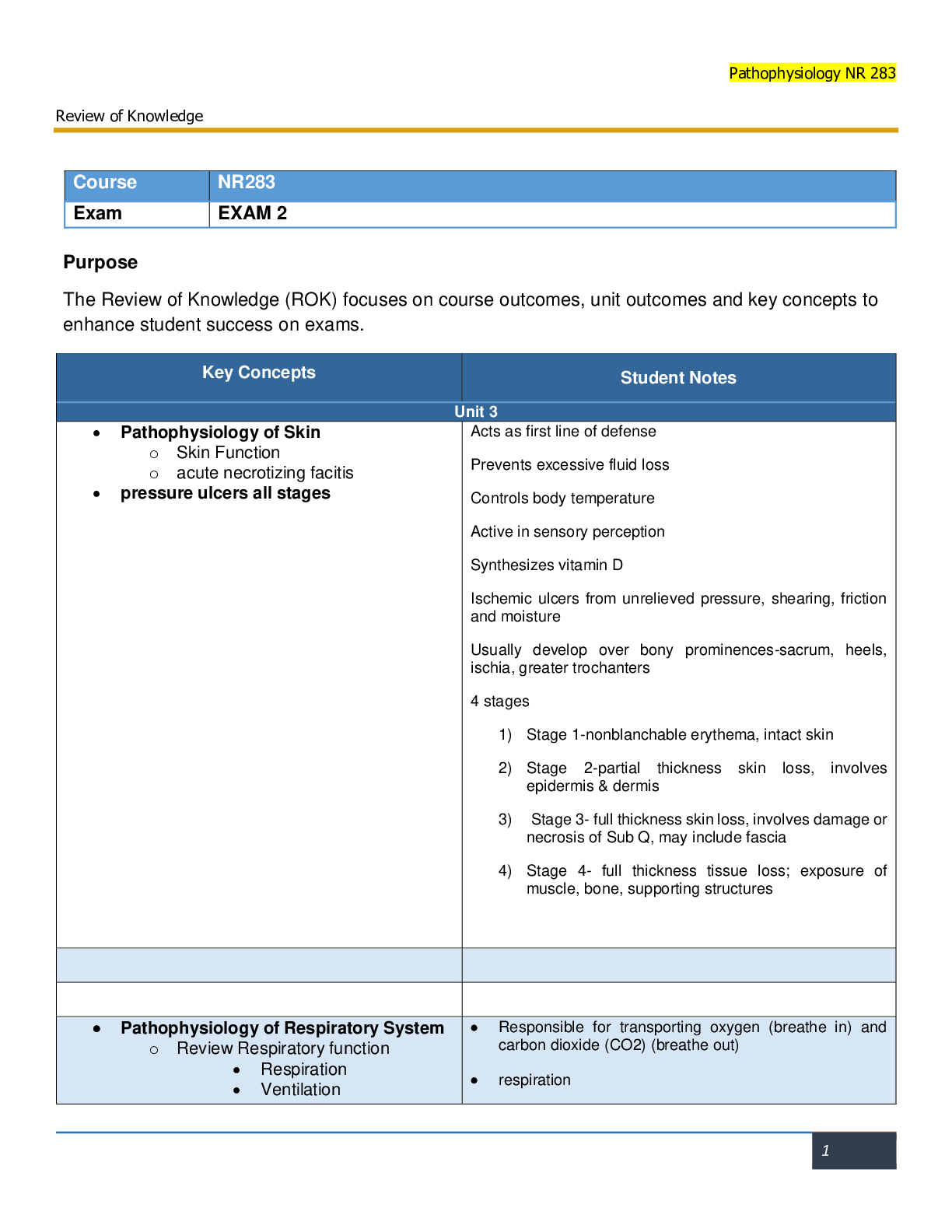
NR 283 Pathophysiology Final Exam Concept Review ***For all previous content covered on previous exams, please consult your previous concept review sheets. This is not an all-inclusive list for topi... cs to be covered. Please be sure to consult your syllabus and learning plan. This is a comprehensive final. ***Be sure to cover pathophysiology, etiology, clinical manifestations, nursing considerations, diagnostic tests for the following topics: Endocrine SIADH- Syndrome of Inappropriate Diuretic Hormone Too much ADH (antidiuretic hormone ) secretion leads to water intoxication and hyponatremia Causes include trauma, stroke, malignancies (often in the lungs or pancreas), medications, and stress S/S include signs of fluid volume overload, changes in level of consciousness and mental status changes, weight gain, hypertension, tachycardia, anorexia, nausea, vomiting, hyponatremia, concentrated urine, decreased urine output, serum osmolality decreased Nursing considerations include monitoring vital signs and cardiac and neurological status, providing a safe environment, particularly for the patient with changes in level of consciousness or mental status, monitoring intake and output and weight daily; monitoring fluid and electrolyte balance, monitoring serum and urine osmolality; restriction of fluids DI (Diabetes Insipidus)- Kidney tubules fail to reabsorb water Etiology includes stroke or trauma or may be idiopathic S/S include excretion of large amounts of dilute urine, polydipsia, dehydration (decreased skin turgor and dry mucous membranes), inability to concentrate urine, increased urine output, urine very dilute, Low urinary specific gravity, fatigue, muscle pain and weakness, headache, postural hypotension that may progress to vascular collapse without rehydration, tachycardia, hypernatremia Nursing Considerations: monitor vital signs and neurological and cardiovascular status, provide a safe environment, particularly for the patient with postural hypotension; monitor electrolyte levels and for signs of dehydration; maintain patient intake of adequate fluids; monitor intake and ouput, weight, serum osmolality and specific gravity of urine; instruct the patient to avoid foods and/or liquids that produce diuresis Hyperthyroidism- Too much thyroid hormone (T3 and T4) Characterized by an increased rate of body metabolism Common cause is Graves’ disease, also known as toxic diffuse goiter S/S include: personality changes such as irritability, agitation and mood swings, nervousness and fine tremors of the hands, heat intolerance, weight loss, smooth, soft skin and hair, palpitations, cardiac dysrhythmias such as tachycardia or atrial fibrillation, diarrhea, protruding eyeballs (exophthalmos) may be present, diaphoresis (sweating), hypertension, enlarged thyroid gland (goiter) Nursing Considerations: Provide adequate rest, provide a cool and quiet environment, provide a high-calorie diet, obtain daily weight, avoid administration of stimulants, administer sedatives as prescribed, administer antithyroid medications, administer blood pressure medication for tachycardia, prepare for thyroidectomy if prescribed Hypothyroidism- Hyposecretion of thyroid hormones (T3 and T4) Characterized by a decreased rate of body metabolism Causes: autoimmune disease, treatment for hyperthyroidism, radiation therapy, thyroid surgery, certain medications S/S: lethargy, fatigue, weakness, muscle aches, paresthesias, intolerance to cold, weight gain, dry skin and hair and loss of body hair, bradycardia, constipation, generalized puffiness and edema around the eyes and face (myxedema), forgetfulness and loss of memory, menstrual disturbances, cardiac enlargement, tendency to develop heart failure, goiter may or may not be present Hyperparathyroidism- Hypersecretion of parathyroid hormone (PTH) Causes: Tumor, Hyperplasia, Genetics; secondary causes-severe calcium or vitamin D deficiency, chronic kidney failure S/S: Hypercalcemia and hypophosphatemia, fatigue and muscle weakness, skeletal pain and tenderness, bone deformities that result in pathological fractures, anorexia, nausea, vomiting, epigastric pain, weight loss, constipation, hypertension, cardiac dysrhythmias, renal stones Nursing Considerations: Monitor vital signs, particularly blood pressure; monitor for cardiac dysrhythmias, monitor for intake and output and for signs of renal stones, monitor skeletal pain, move the patient slowly and carefully; encourage fluid intake, administer furosemide (Lasix) as prescribed to lower calcium levels, administer phosphates, which interfere with calcium reabsorption as prescribed, administer calcitonin as prescribed to decrease the skeletal calcium release and increase renal excretion of calcium, monitor calcium and phosphorus levels, prepare the patient for parathyroidectomy as prescribed Hypoparathyroidism-Hyposecretion of parathyroid hormone (PTH) Can occur following a thyroidectomy because of removal of parathyroid tissue S/S: Hypocalcemia and hyperphosphatemia, numbness and tingling in the face, muscle cramps and cramps in the abdomen or extremities, positive Trousseau’s and Chvostek’s sign, signs of overt tetany such as bronchospasm, laryngospasm, carpopedal spasm, dysphagia, photophobia, cardiac dysrhythmias, seizures; hypotension, anxiety, irritability, depression Nursing Considerations: Monitor vital signs, monitor for signs of hypocalcemia and tetany, initiate seizure precautions, place a tracheostomy set, oxygen and suctioning equipment at bedside, prepare to administer calcium gluconate intravenously for hypocalcemia, provide a high-calcium, low-phosphorus diet, instruct the patient on administration of calcium supplements as prescribed, instruct the patient on administration of vitamin D supplements as prescribed, vitamin D enhances the absorption of calcium from the GI tract, instruct the patient on administration of phosphate binders as prescribed to promote the excretion of phosphate through the gastrointestinal tract, instruct to wear a Medic-Alert bracelet Cushing’s Syndrome/Disease- is a metabolic disorder characterized by abnormally increased secretion (endogenous) of cortisol, caused by increased amounts of ACTH secreted by the pituitary gland Cushing’s syndrome is a metabolic disorder resulting from the chronic and excessive production of cortisol by the adrenal cortex or by the administration of glucocoritcoids in large doses for several weeks or longer (exogenous or iatrogenic) S/S: generalized muscle wasting and weakness, moon face, buffalo hump, truncal obesity with thin extremities, supraclavicular fat pads, weight gain, hirsutism (masculine characteristics in females), hyperglycemia, hypernatremia, hypokalemia, hypocalcemia, hypertension, fragile skin that bruises easily, reddish-purple striae on the abdomen and upper thighs Nursing Considerations: Monitor vital signs, particularly blood pressure, monitor intake and output and weight, monitor laboratory values, particularly the white blood cell count, and serum glucose, sodium, potassium, and calcium levels; provide meticulous skin care, allow the patient to discuss feelings related to body appearance, administer chemotherapeutic agents as prescribed for inoperable adrenal tumors; prepare the patient for radiation as prescribed if the condition results from a pituitary adenoma; prepare the patient for removal of pituitary tumor (hypophysectomy, transsphenoidal adrenectomy) if the condition results from increased pituitary secretion of ACTH, prepare the patient for adrenalectomy if the condition results from an adrenal adenoma; glucocorticoid replacement may be required following adrenalectomy Addison’s Disease-Hyposecretion of adrenal cortex hormones (glucocorticoids and mineralcorticoids) • Can be primary or secondary • The condition is fatal if left untreated S/S: Lethargy, fatigue and muscle weakness, gastrointestinal disturbances, weight loss, menstrual changes in women, impotence in men, hypoglycemia, hyponatremia, hyperkalemia, hypercalcemia, hypotension, hyperpigmentation of the skin (bronzed) with primary disease Nursing Considerations: Monitor vital signs, particularly blood pressure, weight, and intake and output, monitor white blood cell (WBC) count; blood glucose, potassium, sodium, and calcium levels; administer glucocorticoid or mineralcorticoid medications as prescribed; observe for addisonian crisis caused by stress, infection, trauma, or surgery Patient education: avoid individuals with an infection, diet: high protein and high carbohydrate, normal sodium intake, avoid strenuous exercise and stressful situations; need for lifelong glucocorticoid therapy; avoid over-the-counter medications, wear a medic-alert bracelet, signs and symptoms of complications such as underreplacement and overreplacement of hormones Hyperaldosteronism- Hypersecretion of mineralcorticoids (aldosterone) from the adrenal cortex of the adrenal gland Most commonly caused by an adenoma S/S: Symptoms related to hypokalemia, hypernatremia, and hypertension; headache, fatigue, muscle weakness, nocturia, polydipsia, polyuria, paresthesias; visual changes; low urine specific gravity and increased urinary aldosterone level; elevated serum aldosterone levels Nursing Considerations: Monitor vital signs, particulary blood pressure; monitor for signs of hypokalemia and hypernatremia; monitor intake and output and urine for specific gravity; Spironolactone (Aldactone) may be prescribed to promote fluid balance and control hypertension; this is a potassium-sparing diuretic and aldosterone antagonist, and patients need to be monitored for hyperkalemia, particularly those with impaired renal function or excessive potassium intake; administer potassium supplements as prescribed; prepare the patient for adrenalectomy; maintain sodium restriction, as prescribed, preoperatively; administer glucocorticoids preoperatively, as prescribed, to prevent adrenal hypofunction; monitor the patient for adrenal insufficiency postoperatively; instruct the patient regarding the need for glucocorticoid therapy following adrenalectomy; instruct the patient about the need to wear a Medic-Alert bracelet Pheochromocytoma-Catecholamine-producing tumor usually found in the adrenal medulla, but extra adrenal locations include the chest, bladder, abdomen, and brain; typically is benign tumor but can be malignant Excessive epinephrine and norepinephrine secreted S/S: paroxysmal or sustained hypertension, severe headaches, palpitations, flushing and profuse diaphoresis, pain in the chest or abdomen with nausea and vomiting, heat intolerance, weight loss, tremors Complications: hypertensive crisis, hypertensive retinopathy and nephropathy, cardiac enlargement, dysrhythmias, heart failure, myocardial infarction, increased platelet aggregation, and stroke; death can occur from shock, stroke, renal failure, dysrhythmias, or dissecting aortic aneurysm Nursing Considerations: Monitor vital signs particularly blood pressure and heart rate; monitor for hypertensive crisis; monitor for complications that can occur with hypertensive crisis, such as stroke, cardiac dysrhythmias, myocardial infarction; prepare to administer antihypertensive agents to control hypertension; monitor serum glucose level; promote rest and a nonstressful environment; provide diet high in calories, vitamins, and minerals; prepare for an adrenalectomy It is important to avoid stimuli that can precipitate a hypertensive crisis, such as increased abdominal pressure and vigorous abdominal palpation Diabetes Mellitus (DM)- A group of diseases characterized by hyperglycemia due to defects in insulin secretion, insulin action, or both Normally, a certain amount of glucose circulates in the blood. Major sources of glucose are absorption of ingested food in the GI tract and formation of glucose by the liver from food substances Diabetes is especially prevalent in the elderly; as many as 50% of people older than 65 years of age has some degree of glucose intolerance. People 65 years and older account for almost 40% of people with diabetes. Risk Factors Family history Obesity Race/ethnicity- (African Americans, Hispanics, Native Americans, Asian Americans, Pacific Islanders) Age- greater than or equal to 45 years old Impaired fasting glucose or impaired glucose tolerance Hypertension HDL/Triglyceride level- HDL less than or equal to 35 and or triglyceride level greater than or equal to 250 History of gestational diabetes or delivery of babies over 9 lbs 7 tips for managing diabetes Healthy eating Being active Monitoring Taking medication Problem solving Healthy coping Reducing risks Patho- Insulin secreted by beta cells Insulin levels increase after meals consumed During fasting periods insulin and glucagon is released Liver produces glucose through Glycogenolysis – the breakdown of glycogen to glucose which occurs in the liver & muscles Glyconeogenesis- the making of glucagon Prediabetes is classified as impaired glucose tolerance (IGT) or impaired fasting glucose (IFG) and refers to a condition in which blood glucose concentrations fall between normal levels and those considered diagnostic for diabetes Type I Diabetes-(genetics/juvenile) Insulin producing beta cells in the pancreas are destroyed by an autoimmune process, genetic susceptibility, or environmental factors Requires insulin, as little or no insulin is produced Onset is acute and usually before 30 years of age 5–10% of persons with diabetes People do not inherit type 1 diabetes itself but rather a genetic predisposition toward development of type 1 diabetes. Autoimmune: abnormal response in which antibodies are directed against normal tissues of the body, responding to these tissues as if they are foreign Environmental factors such as viruses and toxins that may initiate destruction of beta cells are being investigated Destruction of beta cells results in decreased insulin production, unchecked glucose production by the liver, and fasting hyperglycemia. In addition, glucose derived from food cannot be stored in the liver but instead remains in the bloodstream and contributes to postprandial (after meals) hyperglycemia. Because insulin normally inhibits glycogenolysis and gluconeogenesis, these processes occur in an unrestrained fashion in people with insulin deficiency and contribute further to hyperglycemia. In addition, fat breakdown occurs, resulting in an increased production of ketone bodies, which are the by products of fat breakdown. S/S- polyuria(increased urine), Polydipsia(increased thirst), polyphagia(increased hunger), weigth loss, fatigue, frequency of infections, rapid onset, insulin dependent, familial tendency, peak incidence from 10 to 15 yrs, glycosuria. Type II diabetes- Insulin resistance refers to decreased tissue sensitivity to insulin. Normally insulin binds to special receptors on cell surfaces and initiates a series of reactions involved in glucose metabolism. In type 2 diabetes, these intracellular reactions are diminished making insulin less effective at stimulating glucose uptake by the tissues and at regulating glucose release by the liver Despite the impaired insulin secretion, there is enough insulin present to prevent the breakdown of fat and the accompanying production of ketone bodies, therefore DKA(diabetic ketoacidosis) does not typically occur in type 2 diabetes, however uncontrolled type 2 diabetes may lead to hyperglycemic hyperosmolar nonketotic syndrome Because type 2 diabetes is associated with a slow, progressive glucose intolerance, its onset may go undetected for many years. More common in persons over age 30 and in the obese Slow, progressive glucose intolerance Treated initially with diet and exercise Oral hypoglycemic agents and insulin may be used CM- Polyuria, polydipsia, recurrent infections, visual changes, fatigue, low energy, skin infections, vaginal discomfort, HbAc high 6.5%, FBS high 126mg/dL, prediabetes FBS 100-125mg/dL, Metabolic Syndrome. Endocrine Control for DM- • Insulin: lowers blood glucose by facilitating glucose transport across cell membranes of muscle, liver, and adipose tissue • Glucagon: Increases blood glucose concentration by stimulation of glycogenolysis and glyconeogenesis • Somatostatin: delays intestinal absorption of glucose • Glucocorticoids (mainly cortisol): affects metabolism of all nutrients, regulates blood glucose levels, affects growth, has anti-inflammatory action, and decreases effects of stress Musculoskeletal Sprains/Strains- A sprain is a tear or injury to a ligament • A strain is a tear or injury to a tendon • An avulsion is a complete separation of a tendon or ligament from its bony attachment site • Dislocation is the complete loss of contact between 2 bones in a joint • Subluxation is a partial loss of contact • They are typically associated with bone fractures, most commonly the fingers and shoulder • However they can also be associated with other musculoskeletal disorders that affect the stability of muscles, bones, or joints. Fractures-A fracture is a break in the continuity of bone • Fractures are classified in many ways: • A complete fracture is when the bone is broken entirely: o Open – when the skin is broken and the bone pushes through o Closed – when the bone does not push through the skin, the skin remains intact o Comminuted – the bone has been broken into 2 or more fragments o Linear – the bone has been broken at a parallel to the long axis of the bone o Oblique – the bone has been fractured at an angle to the long axis o Spiral – the bone has a fracture that encircles the entire bone o Transverse – the bone has a fracture perpendicular to the long axis o Impacted – the bone has been fractured and the 2 parts get pushed together o Pathologic – the bone has fractured at a part of bone that has been weakened by an illness, such as osteoporosis • An incomplete fracture is when the bone is damaged but remains in 1 piece: o Greenstick – part of the outer portion of the bone splinters, but the inner part of the bone remains intact o Torus – the bone buckles out but does not break o Bowing – typically occurs in bones that are paired (radius and ulna) where one of the bones break, but the other bone bows out instead of breaking o Stress – small fractures that occur in bone that is subjected to repeated strain, like athletes. The key with stress fractures is that over time, repeated stress fractures to the same area will eventually cause a complete fracture of that bone A fatigue fracture is a type of stress fracture that occurs when someone undertakes a very strenuous, new activity. Muscle mass grows stronger than bone mass, so the increase in muscle puts a strain on the bone causing it to fracture Manifestations of bone fractures include: Pain and tenderness Unnatural alignment Swelling Muscle spasm Decreased mobility Healing of bone fractures-Occurs in 2 ways: • Direct: no callous formation • Indirect: callous formation and bone remodeling Multi-step Process: • Broken bone causes tissue damage and bleeding • Hematoma formation • Bone tissue death • Stimulation of inflammatory response • Osteoclasts and Callus formation • Osteoblasts function Healed bone Explanation of stages of bone healing o Direct – most often occurs with surgical fixation (pins, screws, bolts). There is no callus formation o Indirect – involves the formation of a callus and bone remodeling. This occurs most often with non-surgical treatment of a fracture such as the placement of a cast A. When a bone is broken, the periosteum, blood vessels, and surrounding tissues are disrupted, and bleeding occurs from the broken ends of the bone o B. The next step is the formation of a blood clot between the 2 fractured ends o B. At this time, the bone tissue in the fractured ends dies o C. The dead tissue stimulates the inflammatory response and an influx of the inflammatory and immune cells and osteoclasts to the site to decalcify and destroy the old bone o D. Within 48 hours a callus is formed on the broken ends of the bone o D. At this time, osteoblasts in the callus will form new bone tissue to heal the fracture o E. The new healed bone emerges Rhabdomyolysis is the rapid breakdown of muscle • It causes the release of proteins and other material from muscle cells to be released into the bloodstream • It has many causes and can result in serious complications including renal failure and metabolic acidosis • CM: Classic triad of: o Muscle pain o Weakness o Dark urine (coca cola color urine) Compartment syndrome is the result of increased pressure in the muscle compartment • Skeletal muscles are surrounded by fascia that does not expand • Increased pressure causes decreased blood flow, tissue hypoxia, and necrosis to the muscle • CM are the 5 P’s: pain, pallor, paresthesia, paresis, and pulselessness Arthritis- Gout- is a syndrome caused by incomplete metabolism of purine which results in excess levels of uric acid in the blood • There are 2 main causes for gout: o Either you are producing excessive amounts of uric acid o You are not excreting enough uric acid through the kidneys This is the most common cause • The problem with elevated levels of uric acid, is that at a certain concentration it will crystallize and deposit in either tissue or joints • When the uric acid deposits in the tissue, it leads to the development of something called tophi (singular=tophus), which is a white uric acid crystal visible through the skin • Sometimes the uric acid crystals can deposit in the joints, causing inflammation o This is known as gouty arthritis • The peak age of gout is male between the age of 40-50 • Risk factors for gout include o Male gender o Increasing age o Increased alcohol intake, red meat, and fructose RA -Rheumatoid arthritis is a chronic, inflammatory, autoimmune joint disease characterized by joint swelling, tenderness, and destruction of synovial joints • The joints that are most commonly affected are hands, feet, wrist, knees • The exact cause remains unknown • However some research believes that there is a strong genetic component that interacts with your inflammatory cells • The key genetic element found is a Human Leukocyte antigen (HLA) • Long term exposure to this antigen causes normal antibodies to become autoantibodies (antibodies that attach your own tissue) • Because these autoantibodies are found in people with RA, they are called Rheumatoid factors • The onset of RA is insidious, which means that it seems to progress slowly with few symptoms, but there is actually a lot of damage going on. The symptoms don’t appear for weeks to months • RA begins with general systemic manifestations of inflammation such as fever, fatigue, weakness, anorexia, aching and stiffness • Local manifestations appear gradually over weeks to months. • The joints become painful, tender, and stiff • That stiffness and pain typically is relieved with movement, which is the opposite of OA • An inflamed joint can lose some of its ROM which can eventually cause permanent deformities (swan neck) which lead to physical limitations Osteoarthritis- is a common age related disorder of synovial joints • It is characterized by damage and loss of articular cartilage, changes in the bone underneath the cartilage, and the formation of osteophytes or bone spurs • It is commonly found in the hands, hips, and spine • Risk factors for OA are many and include: o Increased age o Gender – females o Joint trauma o Obesity o Certain medications The exact cause of osteoarthritis is unknown, but we do know what happens The primary defect of OA is the damage and loss of articular cartilage • The surface of the cartilage becomes rough and worn, which interferes with easy joint movement • Eventually enough of the cartilage is destroyed, causing exposure of the bone • Next, osteophytes or bone spurs start to grow off of the exposed bone • Pieces of these osteophytes break off into the joint cavity, causing further irritation and joint movement interference • Eventually the joint space becomes narrow • Secondary inflammation of the surrounding tissue can also occur in response to the changes in joint movement • Manifestations: • The first and most prominent symptom is pain in one or more joints when the joint is being used • Joint stiffness, swelling, and limited ROM • Heberden and Bouchard nodules may be present as well o They are actually signs of the osteophytes in the joint cavities o Heberden are the ones in the joint closest to the fingernail Bouchard are the ones in the middle joint of the finger Ankylosing Spondylitis-(hunch back) is a chronic inflammatory joint disease characterized by stiffening and fusion of the spine and sacroiliac joints • It is similar to RA in the sense that AS is an autoimmune disease • The cause for AS is also not known, however there are strong thoughts that it may be related to the same Human Leukocyte Antigen (HLA) that is seen in patients with RA • AS is mainly found in men between the age of 15-40, but it can affect any age men and women as well • Pathophysiology: • AS begins with inflammation of the fibrocartilage in cartilaginous joints • Typically the sacroiliac joint is affected first • Inflammatory cells infiltrate the area and breakdown the cartilage • Repair of the damaged cartilage begins, but instead of normal cartilage, there is fibrous scar tissue that gets replaced • This scar tissue becomes calcified or hardened eventually causing the joint to fuse or lose flexibility • The most common CM are low back pain and stiffness, that is often worse after rest and is alleviated with physical activity • Because of the loss of cartilage and the fusing of bone, it may progress to lordosis (inward curvature of spine) and kyphosis (concavity of spine) Osteomyelitis- is a bone infection that is most often caused by S. Aureus • There are 2 types: o Exogenous – the infection came from the outside of the body Most commonly related to animal or human bites o Endogenous – the infection came from somewhere else in the body • The characteristics of osteomyelitis are similar to infections that occur anywhere in the body. In this instance, the pathogen causing the disease will cause the inflammatory response of the cells, but it will also affect the homeostasis between the osteoblasts and osteoclasts. When looking at the progression of osteomyelitis, there are big differences between it occurring in a child versus an adult • In children: • The infection enters the bone causing an accumulation of pus in one specific area of the bone • The pocket of pus causes the periosteum of the bone to pull away, causing death of blood vessels that normally feed the bone • Because that part of the bone is no longer receiving an blood, it dies • That dead pocket of bone is called a Sequestrum • Osteoblasts will sense the dead bone, so they will collect and build an area of new bone around the dead bone • That area of new bone growth is called an involucrum • This causes the malformation of bone • In adults: • Their periosteum has been fused to the bone, so it cannot get pulled away • Instead, the pus from the infection will be throughout the bone • This can cause the bone to weaken, predisposing the individual to fractures • CM vary with the area affected, the age of the person, the initiating effect, and the type of infection • Common CM are inflammation, fever, pain, malaise, and presence of abscesses Osteomalcia/Rickets-(bowed leg, no calcium and vit D deficiency, demineralization, calcification, hypocalcimia) Osteomalacia( happens mostly in men 46yrs & older Europeans decants) is a metabolic disease characterized by inadequate and delayed mineralization of osteoid (young, soft bone that has not been calcified yet) in mature bone • The remodeling cycle of bone occurs normally, but the deposit and calcification of minerals does not occur • This leads to normal looking bone that is soft and non-rigid • The biggest cause is Vitamin D deficiency • Risk factors associated with Osteomalacia include dietary deficit, malabsorption, and lack of sun exposure • This is also known as Rickets’s in children, and is commonly seen in 3rd world countries because of their dietary deficits • Manifestations include tenderness and pain of varying degrees, muscle weakness, and sometimes bowed legs Osteoporosis- is also known as porous bone • It is a complex, chronic disease that is associated with many things • This is the most common disease that affects bone and it often progresses silently for years • It is important to remember that It is not necessarily a consequence of aging • Remember that in our life bone is constantly being remodeled or remade; the old cells get destroyed and new bone cells are made • In osteoporosis, old bone is being reabsorbed faster than new bone is created • This causes the bone to have less mass or density, causing it to be thinner and more porous (think a sponge) • The loss of bone continues until the body can no longer support itself, and the individual will start to have spontaneous fractures • It is most severely seen in the femoral neck, spine, and wrists • There are many risk factors for osteoporosis including genes, hormones, bad diet, lifestyle factors, illness, and drugs and steroids. There are different types of osteoporosis, and they are typically categorized by their cause: • Regional • Confined to a segment of the skeleton • Usually associated with disuse or immobilization of a limb • Secondary • Causes by other conditions such as hormonal imbalance, medications, and certain cancers • Often times if you treat the cause, the osteoporosis is resolved • Postmenopausal • Bone loss in middle age and older woman due to estrogen deficiency • Post-menopausal changes cause an imbalance between the activity of osteoclasts and osteoblasts • Glucocorticoid induced • Caused by chronic steroid hormone use • Steroids inhibit the function and creation of osteoblasts • Age related • Exact cause is unknown but due to changes in osteoblast and osteoclast function • The key to osteoporosis is to remember that no matter what is causing it, it leads to a disruption of the homeostasis between bone reabsorption and bone formation • Most common clinical manifestations are pain and bone deformity, however it will depend on which bone is affected • Fractures are highly likely, as well as collapse of weak bones • Vertical collapse causes kyphosis (hunchback) and a decrease in height • Prevention of osteoporosis if possible is key‼ Neurological Levels of consciousness/arousal- Full consciousness is a state of awareness of yourself and the environment and the responses to that environment • Consciousness involves both arousal and awareness: o Arousal is a state of being awake and being able to respond to stimuli o Awareness is a state of being aware • Both are mediated by the reticular activating system in the brain stem, where all our vital reflexes are controlled. • When brain function from the cerebrum is lost, your RAS can maintain someone in a vegetative state • Alterations in arousal can be classified in 3 ways: • Structural causes: o Include infection, brain tumors, trauma • Metabolic causes: o Hypoxia, electrolyte changes, hypoglycemia, medications or toxins • Psychogenic causes: o Uncommon o Sign of a psychiatric disorder Locked-In syndrome- Cerebral death occurs when permanent brain damage has occurred and the person is unable to respond to any stimuli, BUT the brain stem is still functioning and is able to maintain internal homeostasis and keep the body functioning • People who have experienced cerebral death can have 1 of 4 final outcomes: o They may remain in the coma for life o They may emerge into a persistent vegetative state: The person is unaware, does not speak or follow commands But they may open their eyes and have a normal sleep cycle If the person remains in a vegetative state for greater than 12 months, it is considered a permanent state o They may progress into a minimal conscious state They may be able to follow simple commands Have simple speech, such as speaking yes and no Have some spontaneous movements such as smiling o Or they may be in what’s called a locked-in syndrome The body is fully conscious, but there is complete paralysis of the muscles The only thing they are capable of is eye movement Cerebral vascular accident-They are the leading cause of disability and the third leading cause of death in the US • Three commonly seen risk factors for a CVA include: HTN, DM, and smoking • CVA’s can be classified in different ways: • Hemorrhagic Stroke: • It is typically caused by chronic HTN problems, but can also be caused by ruptured aneurysms, bleeding tumors, or bleeding from a head trauma • The hemorrhage can be tiny or massive • As bleeding continues into the brain tissue, a mass of blood is formed • Brain tissue that is next to the bleeding area can become deformed, compressed, or displaced • This will produce edema, increased ICP, and ischemia of brain tissue • The hemorrhage will eventually resolve through reabsorption, and will leave a cavity in the brain where it was • CM: vary depending on what part of the brain is affected • A thrombotic CVA: • An occlusion of one of the arteries of the brain • Most commonly this is caused by atherosclerosis • The CVA occurs when parts of the clot detach, travel upstream, and obstruct blood flow causing ischemia • An embolic CVA: • This is caused by a piece of plaque that has broken off of a clot that is elsewhere in the body, beyond the brain • People who have embolic CVA’s are at a higher risk for developing more because the cause is still there (such as atherosclerosis of the heart) • A TIA • Is a brief episode of neurologic dysfunction due to platelet clumps or a partial obstruction • People who have TIA’s are at a higher risk for developing a CVA Traumatic Brain Injury-A major head trauma is a traumatic insult to the brain that is capable of producing many changes to an individual, including physical, emotional, social, and intellectual • Major head traumas can be caused by car accidents, falls, sports injuries, or violence • There are 2 main types of head trauma’s: o Closed When the head strikes a hard surface (wall) or an object hits the head (baseball), and the skull and meninges remain intact These are the most common o Open Commonly caused by severe accidents or bullets The skull is broken and the brain gets exposed to the environment • Brain damage from trauma can be categorized as: o Focal: affecting one area of the brain o Diffuse: affecting more than one area of the brain Hematomas-epidural/subdural/subarachnoid Extradural/Epidural hematoma/hemorrhage o Usually the result of a skull fracture that has tore an artery!!!! (Arterial bleed!, not venous) o Because arteries have a higher amount of pressure than veins do, the blood will accumulate quickly in the epidural space o The individual will lose consciousness at the time of the injury, but regain it for a period of time o This type of hematoma can be very deceiving, because the patient will feel okay, but the bleed is still occurring and is starting to accumulate o Eventually when the bleed grows big enough, the person will develop a worsening headache, vomiting, confusion, and seizures o Soon the brain will herniate, LOC will be lost, and the person may die o Epidural hematomas are considered medical emergencies Intracerebral hematomas Bleeding in the brain o Common in the frontal and temporal lobe o It can cause increased ICP, Decreased LOC, pupil and respiratory changes The third type of bleed caused by a focal injury is a subdural hematoma • It is bleeding between the dura and the brain, usually associated with a torn vein • Subdural’s can be classified as: • Acute: o Develop rapidly with CM of headache, and some type of neurological change o CM will worsen over time and quickly progress to loss of consciousness, respiratory changes, and pupillary changes and brain herniation • Chronic o Develop over weeks to months o Typically seen in the elderly because their brain has atrophied, leaving more room for the bleeding o Can be caused by what is considered a very insignificant head trauma o CM: Chronic headaches Vague symptoms – patient can have symptoms that are sometimes mistaken for dementia Based on the degree of the bleed, increased ICP and eventual herniation can occur Subarachnoid hemorrhage (SAH) is the escape of blood from a defective or injured vessel into the subarachnoid space. Individuals at risk for a subarachnoid hemorrhage are those with intracranial aneurysm, intracranial arteriovenous malformation, hypertension, or a family history of SAH, and those who have sustained head injuries. Subarachnoid hemorrhages often recur, especially from a ruptured intracranial aneurysm. CM- Early manifestations associated with leaking vessels are episodic and include headache, changes in mental status or level of consciousness, nausea or vomiting, and focal neurologic defects. A ruptured vessel causes a sudden, throbbing, “explosive” headache, accompanied by nausea and vomiting, visual disturbances, motor deficits, and loss of consciousness related to a dramatic rise in intracranial pressure. Concussion Skull Fractures Increased Intracranial Pressure- The cranium is a closed box, whatever is in there (brain, blood, CSF), is all that it can hold. If you increase the amount of something in the skull, the volume of the other content has to decrease • There are 4 stages of increased ICP that are seen: • Stage 1 o The blood vessels will compensate by constricting to decrease the volume in the brain o The ICP does not increase at this point and there are no manifestations • Stage 2 o Expansion of the contents in the cranium start to occur o This will cause the ICP to start to rise because there is no more extra room left in the brain o CM will be subtle and transient: changes in LOC: confusion or drowsiness • Stage 3 o The ICP is continuing to increase o Brain tissue will start to become hypoxic and the patient will start to deteriorate o CM: Decreased levels of arousal, bradycardia, pupil changes, hyperventilation • Stage 4 o Because of the high ICP, the brain tissue will start to shift or herniate to a compartment that has lower pressure o The part of the brain that herniates impairs blood supply to that area and can cause areas of hemorrhage o There are different types of herniation depending on where the brain tissue goes o Some places of herniation are not reversible Parkinson’s Disease- is a common movement disorder • It typically begins after the age of 40 • The cause is unknown, but it may be caused by a combination of genetics and environmental factors • The basal ganglia deteriorates and there is a loss of the neurons that produce dopamine • This results in a loss of dopamine which causes excessive excitatory activity which gets manifested as the CM of parkinsonism • Secondary parkinsonism o Presence of parkinsonism manifestations, but with a different cause o Typically caused by head injuries, infection, toxins, and medications (most common) • Classic CM of Parkinson: o Resting tremors Pill rolling tremor o Rigidity or muscle stiffness o Bradykinesia/Akinesia’s Leading to gait and movement problems o Dysarthria Impaired speech o Dysphagia Impaired swallowing o Drooling Depression is also a major CM MS-( Multiple Sclerosis) 3 specific Neurological disorders • The first is Multiple Sclerosis, which is a common acquired autoimmune inflammatory disorder involving the destruction of axonal myelin in the brain and spinal cord • It is more common in females and the average age of onset is 20-40 • MS involves an autoimmune process that develops after an individual has had a viral infection that affected the nervous system • Because of the inflammatory process, the axons of the neurons get inflamed and demyelination of the axons occurs • Scar tissue is also formed over the damaged axon which will interfere with the conduction of nerve impulses • Neurons will eventual die and brain atrophy will occur • As more and more neurons are affected, the person will have to deal with a progressive interference with functions that are controlled by the nervous system, which can be just about anything depending on which part of the brain is affected • CM: May be unpredictable, transient, or last for hours to weeks o 2 initial symptoms: Vision problems Sensory impairment (paresthesia’s) o Other CM include: Dysphagia, muscle weakness, communication difficulty, fatigue, paralysis, incontinence, tremors, slurred speech. ALS- Amyotrophic lateral sclerosis • Also known as Lou Gehrig’s Disease • It diffusely affects the upper and lower motor neurons of the brain and spinal cord • The exact cause is unknown • Thoughts are that it may be due to: o Genetics o A virus o Deficiency of nutrients supplied to the neurons o An autoimmune disorder • The pathological feature of ALS is that the motor neurons in the body get destroyed, and the other non-motor neurons start to degenerate as well • As the neurons die, the muscle fibers that they supply become atrophied • This will lead to progressive muscle weakness, eventually causing respiratory failure because of the weakness of respiratory muscle, and eventual death • The initial symptoms are related to which motor neurons are affected • CM: o The hallmark sign of ALS is muscle weakness o The muscles may develop twitching or spasms as well and eventual atrophy o Difficulty speaking and swallowing. Depression is huge MG- (Myasthenia Gravis) The last disease is Myasthenia Gravis which is an acquired autoimmune disease that affects the transmission of nerve impulses between the motor neuron and its innervated muscle cells • The cause is unknown • But it is commonly associated with other autoimmune disorders or problems with the thymus gland ( a gland located near the sternum that plays a big role in the immune response) • What happens is that acetylcholine receptors are no longer recognized as “self” (hence the autoimmune problem) and autoantibodies are produced against these receptors • The autoantibodies bind to the receptor and block the binding of acetylcholine • Eventually the receptor sites are destroyed • This will lead to a decrease in the transmission of nerve impulses and a lack of muscle depolarization • CM: muscle fatigue and weakness that is progressive o Difficulty speaking, chewing, and swallowing • In myasthenia gravis, there are 2 crisis states that can occur. We are only going to focus on one: • Myasthenic crisis: o Occurs when severe muscle weakness causes extreme paresis/paralysis, respiratory insufficiency and difficulty swallowing o This patient is in danger of going into respiratory distress Huntington’s Disease- is a rare hereditary autosomal disorder of the basal ganglia (which controls our movement along with the frontal lobe) • It is also known as chorea which is the main CM seen with Huntington • There is severe degeneration of the basal ganglia and development of protein tangles that contribute to the loss of neurons • This leads to alterations of motor and mental functioning • CM: o Progress slowly o Chorea Irregular, uncontrolled excessive movements Begins in arms and face o Athetosis Writhing movements o Ballism Flinging movements Review A&P of the brain and cranial nerves Spinal Cord Injury- Spinal cord trauma most commonly occurs due to injuries that result from: o Acceleration, deceleration, or deformation forces that are applied to the spinal cord • These forces compress the tissues, pull on the tissues, or shear tissues so that they slide against one another • Damage may be to not only the spinal cord, but to the bones, the ligaments, and the joints as well • The leading cause of spinal cord trauma is due to motor vehicle accidents, but they can also be caused by falls, sports, and violence • The most common place on the cord for injuries to occur is: o C 1, 2, 4-7 o T1-L2 o Because these are the most movable parts of the vertebra • When the injury occurs, hemorrhage and swelling will occur at the site • Cord swelling increases the degree of dysfunction o This is especially life-threatening in the cervical region because it may affect the working of the diaphragm and the brain stem • Within 24-48 hours after the injury, the hemorrhage will start to become reabsorbed • Any axons that were damaged in the area will be removed • Within 3-4 weeks of the injury, the part of the cord that was injured will be replaced with fibrous tissue and the meninges may thicken as well Spinal shock is the loss of normal body activity below the level of the spinal cord injury • This involves a complete loss of all normal reflex functions, such as: o Skeletal muscle function o Bladder and bowel function o Sexual function o Temperature control • Spinal shock typically lasts 7-20 days, but can last up to 3 months • It stops when reflex activity has reappeared • The specific CM of spinal cord injury really depend on what level the injury is at One complication of spinal cord injury is something called autonomic dysreflexia • This is a massive cardiovascular response to stimulation of the SNS • It involves sensory stimulation of the body, below the level of spinal cord injury o Most commonly seen with bladder or bowel distension • Some type of sensory stimulation occurs below the level of injury • The stimulus travels up the cord until it reaches the area of injury and it gets blocked • Because the impulse cannot reach the brain, the SNS is activated causing constriction of the blood vessels and HTN • The brain will sense the HTN, and will attempt to send messages via motor neurons to the heart and the vessels to dilate to decrease the BP • Unfortunately because the motor neuron impulse cannot pass the area of injury of the spinal cord, HTN remains and leads to problems with regulation of BP • CM: o Extremely high BP o Pounding headache o Blurred vision o Sweating o Skin flushing Seizures- A seizure is a syndrome that results from a sudden, explosive discharge of neurons • And is characterized by a sudden, transient alteration in brain function • A convulsion is a term that is sometimes applied to seizures o it usually refers to the tonic-clonic movements that are seen with some seizures • Seizures can be caused by a number of things: o Brain lesions or trauma o Biochemical disorders o Sometimes seizures can be caused by stress, fatigue, stress, drugs, loud noises, or flashing lights • Epilepsy is a condition in which there is continuous seizure activity without a known cause (everything has been ruled out but they are still having seizures) • Research has indicated that epilepsy may be due to an interaction between genetics and environmental factors • A specific group of neurons called epileptic neurons are responsible for the epileptic activity o They are hypersensitive and fire more frequently than normal Seizures are classified in many ways: partial, complex, generalized, etc Different phases of seizures- specifically those seen in the grand mal or tonic clonic seizures • Starting with the tonic phase of a seizure which is a state of continued muscle contraction due to the excessive excitation of the brain muscle • The clonic phase of a seizure is characterized by alternating muscle contraction and relaxation o This occurs when the neurons that inhibit activity start to work to stop the excess excitability to stop the seizure o This will produce intermittent contractions or seizures that gradually decrease until it stops o The post-ictal phase is the period of time immediately following the seizure • The specific CM of seizures will depend on the specific type • 2 common manifestations associated with the tonic clonic seizures: o An Aura: a partial seizure that people will describe as a peculiar sensation or dizziness or numbness that occurs right before the seizure occurs o A Prodroma: an early CM, such as malaise or a H/A, that occurs hours to days before the seizure • The most important manifestation associated with seizures is the increase in oxygen consumption o During a seizure your body is constantly contracting, so oxygen is quickly used and depleted o Once oxygen has been depleted, your body will use glucose for energy which will cause your lactic acid levels to build-up • Finally, a continued seizure activity can cause brain injury and eventual irreversible damage if it does not stop Meningitis- which is infection and inflammation of the meninges, CSF, and ventricles • There are 3 types: • Bacterial o It is primarily caused by meningococcus or pneumococcus o The infection enters the CNS causing the inflammatory response o In some cases the CSF will thicken leading to hydrocephalus and cerebral edema can occur leading to an increase in ICP o CM: S/S of systemic infection: fever, tachycardia, and chills S/S of Meningeal irritation: throbbing headache, photophobia, positive Kernig sign (when knee is bent, pain in head when leg is straightened), positive Brudzinski sign (involuntary lifting of legs when lifting up head) Neurological signs: decreased LOC, hemiparesis, seizures • Aseptic meningitis or viral meningitis o Primarily caused by a virus o CM are similar to bacterial but they are less severe o This form is self-limiting • Fungal meningitis o Chronic and less common form of meningitis o It will progress slowly o CM will mimic other neurological syndromes such as hydrocephalus or dementia Encephalitis- Encephalitis is an acute, febrile illness that affects the nervous system leading to brain inflammation • It is most commonly caused by an arthropod or mosquito borne virus (like West Nile) and HSV • But it can also result from a complication of other viral disease • It involves all of the meninges of the brain and spinal cord • It can result in neuron damage, edema, hemorrhage, increased ICP, or necrosis • CM: o Range from mild to life threatening o The most dramatic ones are: Fever Delirium Confusion progressing to unconsciousness Seizures Paralysis [Show More]
Last updated: 2 years ago
Preview 1 out of 25 pages
Buy this document to get the full access instantly
Instant Download Access after purchase
Buy NowInstant download
We Accept:

Reviews( 0 )
$20.00
Can't find what you want? Try our AI powered Search
Document information
Connected school, study & course
About the document
Uploaded On
Aug 07, 2020
Number of pages
25
Written in

Additional information
This document has been written for:
Uploaded
Aug 07, 2020
Downloads
0
Views
147