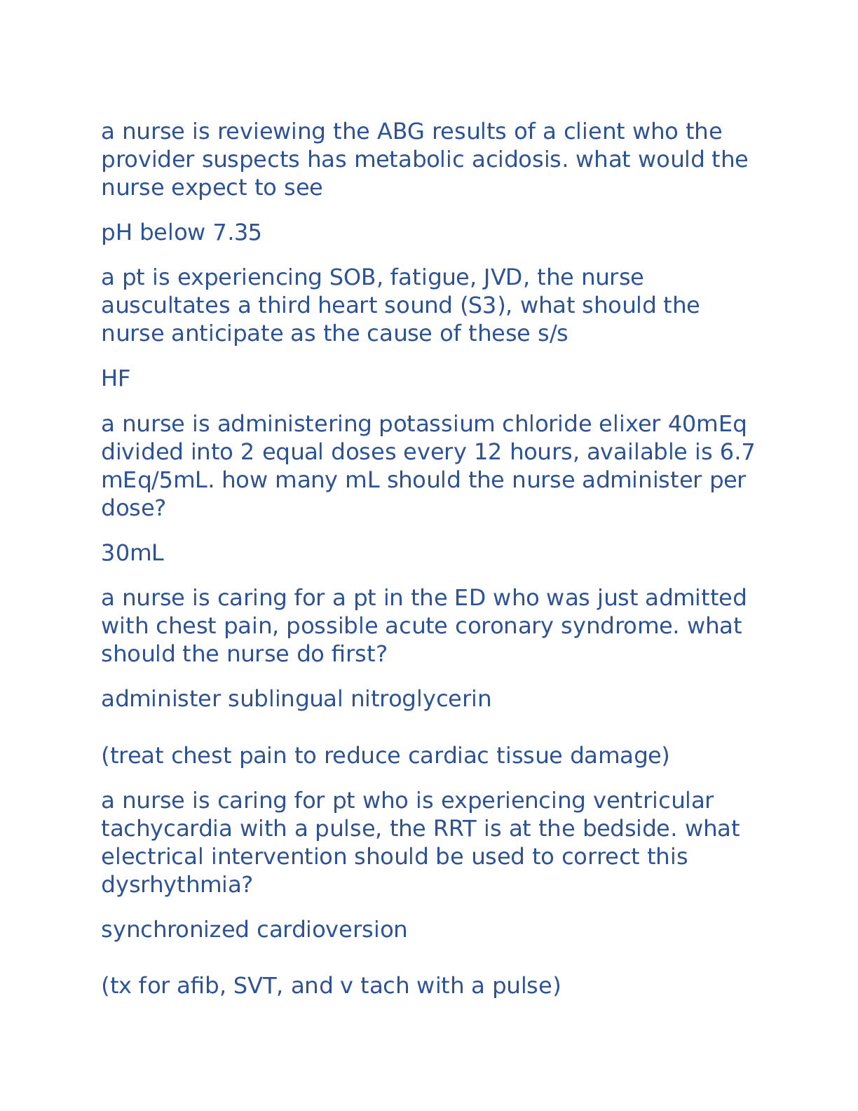
CARDIAC
Not sure where these came from…
1.The patient has chronic atrial fibrillation (AF). What action do you anticipate?
A. Monitoring the PR interval
B. Defibrillation with 360 joule
C. Teaching the patient to m
...
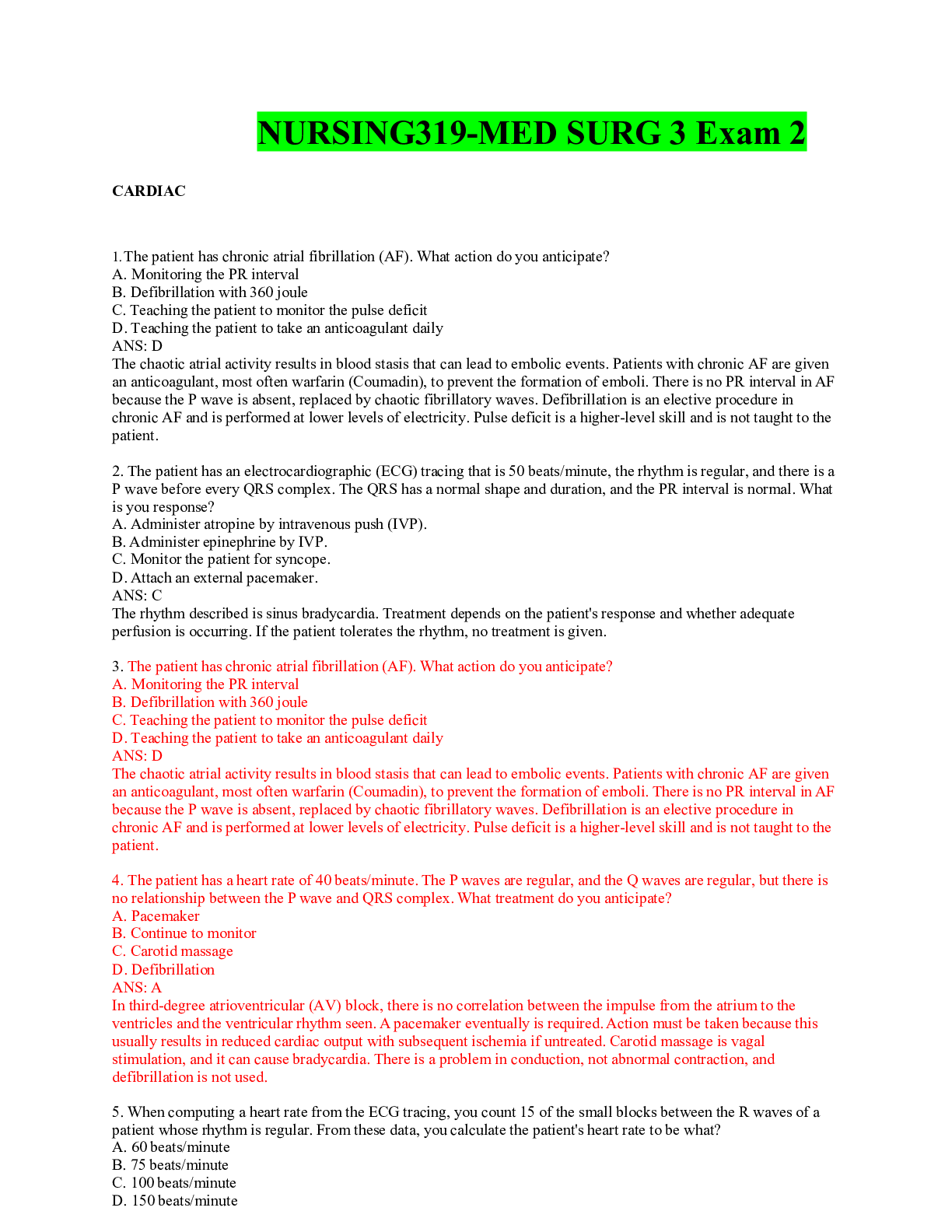
CARDIAC
Not sure where these came from…
1.The patient has chronic atrial fibrillation (AF). What action do you anticipate?
A. Monitoring the PR interval
B. Defibrillation with 360 joule
C. Teaching the patient to monitor the pulse deficit
D. Teaching the patient to take an anticoagulant daily
ANS: D
The chaotic atrial activity results in blood stasis that can lead to embolic events. Patients with chronic AF are given
an anticoagulant, most often warfarin (Coumadin), to prevent the formation of emboli. There is no PR interval in AF
because the P wave is absent, replaced by chaotic fibrillatory waves. Defibrillation is an elective procedure in
chronic AF and is performed at lower levels of electricity. Pulse deficit is a higher-level skill and is not taught to the
patient.
2. The patient has an electrocardiographic (ECG) tracing that is 50 beats/minute, the rhythm is regular, and there is a
P wave before every QRS complex. The QRS has a normal shape and duration, and the PR interval is normal. What
is you response?
A. Administer atropine by intravenous push (IVP).
B. Administer epinephrine by IVP.
C. Monitor the patient for syncope.
D. Attach an external pacemaker.
ANS: C
The rhythm described is sinus bradycardia. Treatment depends on the patient's response and whether adequate
perfusion is occurring. If the patient tolerates the rhythm, no treatment is given.
3. The patient has chronic atrial fibrillation (AF). What action do you anticipate?
A. Monitoring the PR interval
B. Defibrillation with 360 joule
C. Teaching the patient to monitor the pulse deficit
D. Teaching the patient to take an anticoagulant daily
ANS: D
The chaotic atrial activity results in blood stasis that can lead to embolic events. Patients with chronic AF are given
an anticoagulant, most often warfarin (Coumadin), to prevent the formation of emboli. There is no PR interval in AF
because the P wave is absent, replaced by chaotic fibrillatory waves. Defibrillation is an elective procedure in
chronic AF and is performed at lower levels of electricity. Pulse deficit is a higher-level skill and is not taught to the
patient.
4. The patient has a heart rate of 40 beats/minute. The P waves are regular, and the Q waves are regular, but there is
no relationship between the P wave and QRS complex. What treatment do you anticipate?
A. Pacemaker
B. Continue to monitor
C. Carotid massage
D. Defibrillation
ANS: A
In third-degree atrioventricular (AV) block, there is no correlation between the impulse from the atrium to the
ventricles and the ventricular rhythm seen. A pacemaker eventually is required. Action must be taken because this
usually results in reduced cardiac output with subsequent ischemia if untreated. Carotid massage is vagal
stimulation, and it can cause bradycardia. There is a problem in conduction, not abnormal contraction, and
defibrillation is not used.
5. When computing a heart rate from the ECG tracing, you count 15 of the small blocks between the R waves of a
patient whose rhythm is regular. From these data, you calculate the patient's heart rate to be what?
A. 60 beats/minute
B. 75 beats/minute
C. 100 beats/minute
D. 150 beats/minute
ANS: C
Because each small block on the ECG paper represents 0.04 second, 1500 of them represent 1 minute. By dividing
the number of small blocks (15 in this case) into 1500, you can calculate the heart rate in a patient whose rhythm is
regular (in this case, 100).
6. Which statement best describes the electrical activity of the heart represented by measuring the PR interval on the
ECG?
A. The length of time it takes to depolarize the atrium
B. The length of time it takes for the atria to depolarize and repolarize
C. The length of time for the electrical impulse to travel from the sinoatrial (SA) node to the Purkinje fibers
D. The length of time it takes for the electrical impulse to travel from the SA node to the AV node
ANS: C
The electrical impulse in the heart must travel from the SA node through the AV node and into the Purkinje fibers for
synchronous atrial and ventricular contraction to occur. The P wave represents atrial contraction, and the R wave is
part of the QRS complex that represents ventricular contraction. When measuring the time from the beginning of the
P wave to the beginning of the QRS (PR interval), you are identifying the length of time it takes for the electrical
impulse to travel from the SA node to the Purkinje fibers.
7. You obtain a 6-second rhythm strip, and document the following analysis: atrial rate of 70 beats/minute, regular;
ventricular rate of 40 beats/minute, regular; QRS of 0.04 second; no relationship between P waves and QRS
complexes; and atria and ventricles beating independently of each other. What is the correct interpretation of this
rhythm strip?
A. Sinus dysrhythmias
B. Third-degree heart block
C. Wenckebach phenomenon
D. Premature ventricular contractions
ANS: B
Third-degree heart block represents a loss of communication between the atrium and ventricles. This is depicted on
the rhythm strip because there is no relationship between the P waves, representing atrial contraction, and QRS
complexes, representing ventricular contraction. The atria are beating totally on their own at 70 beats/minute,
whereas the ventricle
8. You are watching the cardiac monitor, and a patient's rhythm suddenly changes. There are no P waves. Instead,
there are fine, wavy lines between the QRS complexes. The QRS complexes each measure 0.08 second (narrow),
but they occur irregularly with a rate of 120 beats/minute. You correctly interpret that this rhythm is
A. sinus tachycardia.
B. atrial fibrillation.
C. ventricular fibrillation.
D. ventricular tachycardia.
ANS: B
Atrial fibrillation is represented on the cardiac monitor by irregular R-R intervals and small fibrillatory (F) waves.
There are no normal P waves because the atria are not contracting, just fibrill
[Show More]








.png)



 Chamberlain College Of Nursing (100 Correct Q & A.png)