*NURSING > TEST BANKS > Summative Exam Questions and answers, Test bank with 100% coverage, Graded A+ (All)
Summative Exam Questions and answers, Test bank with 100% coverage, Graded A+
Document Content and Description Below
Summative Exam Questions and answers, Test bank with 100% coverage, Graded A+ An 80-year-old man was treated for ventricular arrhythmias. He presents 1 month later with joint pain. He also has ... an unusual mask-like rash over his face and body. Discontinuation of drug therapy causes the symptoms to abate. What drug was most likely administered to this patient? 1 Tocainide 2 Quinidine 3 Procainamide 4 Phenytoin 5 Propranolol - ✔✔Procainamide - often prescribed for long-term control of arrhythmias. May cause lupus like SE. A 62-year-old woman with a long-standing history of hypertension presents with severe headache; it started this morning and is rapidly worsening. During the interview, she suddenly collapses. Your brief examination shows that she responds with extensor posturing on external stimuli. Her deep tendon reflexes are 3, and you elicit Babinski bilaterally. You also notice that her breathing has a peculiar pattern: deep inspiration with a pause at full inspiration, followed by a brief insufficient release and the end-inspiration pause. How do you best describe her respiratory pattern? Answer Choices 1 Cheyne-Stokes 2 Apneusis 3 Ataxic 4 Cluster 5 Central neurogenic hyperventilation - ✔✔Her breathing pattern is apneustic. Apneustic breathing pattern characterizes *deep, gasping inspiration with a pause at full inspiration followed by a brief, insufficient release and the end-inspiration pause before expiration.* What is the mechanism of LMWH? - ✔✔Both unfractionated heparin and low molecular weight Heparin act by *forming a complex with antithrombin III.* A 5-day-old male infant has subtle, unusual facial features (i.e., a triangular face, hypertelorism, and down-slanting eyes). He also has a webbed neck and low-set ears. Suspecting a congenital disorder, you order a complete work-up, including a CBC, coagulation profile, cardiac evaluation, karyotyping, and mutation analysis. PTPN11 (protein-tyrosine phosphatase, nonreceptor-type 11) mutations are detected. Echocardiography detects a cardiac defect. What's most likely to be found on echo? - ✔✔This neonate most likely has Noonan syndrome (NS). Pulmonary stenosis is the most common cardiac defect in this condition. Noonan syndrome is a sporadic, or autosomal dominant, congenital disorder with typical phenotypical features that may not be visible to the casual onlooker. The most common facial features include hypertelorism and low-set, backward-rotated ears with a thick helix. The philtrum is deeply grooved in more than 90% of cases. Congenital cardiac defects, bleeding disorders, mental retardation, webbed neck, and a short stature are other features. A 35-year-old woman presents for follow-up. She has a 6-month past medical history of hypertension; it has responded poorly to lifestyle approaches. She denies using any medications; she does not smoke or use illicit drugs. Her review of systems is notable for muscular weakness, paresthesias, headaches, polyuria, and polydipsia. On physical exam, her blood pressure is 155/95 mm Hg. She has generalized muscular weakness that is measured in all 4 extremities. The remainder of her exam is unremarkable. Laboratory analysis reveals hypokalemia and a hemoglobin A1c level of 5.5. What dx test result is most likely? 1 Metabolic alkalosis 2 Low serum aldosterone to plasma renin activity ratio 3 Hyponatremia 4 Increased erythrocyte sedimentation rate 5 Hypoglycemia - ✔✔The correct response is metabolic alkalosis. This patient is demonstrating signs and symptoms consistent with primary hyperaldosteronism, which is most commonly caused by a unilateral adrenocortical adenoma (Conn's syndrome), but in a minority of patients, it is caused by adrenal hyperplasia. A 40-year-old man presents with atrial flutter with 2:1 atrioventricular (AV) conduction, giving him a pulse of 150 per minute, which is perfectly regular. His blood pressure is 70/40 mm Hg. He takes no medications regularly. You plan to provide him with urgent direct current cardioversion with conscious sedation. What would be an appropriate level of energy for cardioversion in order to restore sinus rhythm in this patient? 1. 10 Joules 2. 15 Joules 3. 50 Joules 4. 200 Joules 5. 300 Joules 6. 360 Joules - ✔✔Of all of the arrhythmias, both supraventricular and ventricular, atrial flutter is the easiest to cardiovert back to a regular sinus rhythm. Direct cardioversion is usually successful with low energy - 25 to 100 Joules. There is no need to apply especially high energies such as 200 Joules, 300 Joules, or 360 Joules as the initial energy for cardioversion in case of atrial flutter, as higher energies have a greater probability of causing burns or broken bones. On the other hand, 10 or 15 Joules is unlikely to result in a successful cardioversion. *50* A 48-year-old man is brought to the ER complaining of difficulty breathing, fatigue, and intermittent chest pain for the past month. On further questioning, he states that the breathing seems to worsen when lying down. On physical exam, you note elevated respiratory and heart rates and pale, sweaty skin. On auscultation, rales are noted as well as a 3rd heart sound. Which of the following is the most likely diagnosis? 1 Right Ventricular failure 2 Pulmonary Embolism 3 Mitral Valve Stenosis 4 Left Ventricular failure 5 Chronic Obstructive Pulmonary Disease (COPD) - ✔✔Left Ventricular failure The clinical picture is suggestive of left ventricular failure (LVF). Clinical presentation includes dyspnea, orthopnea, and paroxysmal nocturnal dyspnea. The patient may also have hemoptysis, chest pain, fatigue, nocturia, and confusion. On physical exam, the patient may present with cold, pale sweaty skin, tachypnea and tachycardia, rales, and 3rd and 4th heart sounds. This is diagnostically different from right ventricular failure. Right ventricular failure (RVF) has a clinical picture of shortness of breath, pedal edema, and abdominal pain. On PE, RVF will present with a S3, jugular venous distention (JVD) and may have signs of LVF. A 41-year-old woman presents for follow-up regarding elevated blood pressure. This is her third visit to the office, and her blood pressure has been elevated on multiple readings at each visit. She has a history of eczema but is otherwise healthy. Labs reveal the following: WBC: 14.5 k/uL Hgb: 13.5 g/dL HCT: 41% PLT: 152 k/uL Na: 135 mmol/L K: 2.8 mmol/L Cl: 99 mmol/L CO2: 32 mmol/L BUN: 10 mmol/L Cr: 1.02 mmol/L What lab abnormality is most likely to be causing her secondary htn? - ✔✔*Hypokalemia is correct.* Patients with primary hypertension should not have abnormalities in serum electrolytes. The patient's hypokalemia suggests a secondary cause of hypertension. *The most common cause of secondary hypertension is renal artery stenosis caused by fibrosmuscular dysplasia.* If the adult is middle-aged, then the most common secondary cause of hypertension would be *aldosteronism and a highly recommended initial diagnostic test would be an aldosterone/renin ratio.* Age is incorrect. This patient is 41 years old. Secondary hypertension is more likely in patients under 30 years old or over 50 years old. Female gender is incorrect. Primary hypertension is more common in middle-aged males than middle-aged females. The gender distribution for secondary hypertension varies with the specific cause of secondary hypertension. Hypokalemia is the most suggestive risk factor of secondary hypertension in this patient. Eczema is incorrect. Eczema is not a risk factor for secondary hypertension Leukocytosis is incorrect. Leukocytosis is not associated with secondary hypertension. A 78-year-old man with known left-sided congestive heart failure presents with a complaint of cough, worsening dyspnea with exertion, and orthopnea. What is the most direct cause of his symptoms? 1. Tricuspid insufficiency 2. Left ventricular hypertrophy 3 .Decreased peripheral vascular resistance 4. Increased pulmonary venous pressure 5. Mucus plugging - ✔✔Increased pulmonary venous pressure In left-sided congestive heart failure, the predominant feature is *low cardiac output and elevated pulmonary venous pressure, resulting in dyspnea.* As dyspnea worsens, the patient will also begin to experience shortness of breath at rest, which is worsened in the supine position. Tricuspid function is not related to CHF. Peripheral vascular resistance typically increases in CHF, which is designed to help maintain perfusion to vital organs. Mucus plugging is not associated with CHF. A 58-year-old woman presents with a 3-month history of postprandial abdominal pain. This crampy pain occurs 30 minutes after eating, every time. Due to these symptoms, the patient has lost 30 pounds and is afraid to eat. Her past medical history includes hypertension treated with enalapril, coronary artery disease for which she has undergone a right coronary artery stent, and she underwent a carotid endarterectomy for symptomatic carotid stenosis. She has smoked 2 packs of cigarettes a day for 30 years. What is the best initial test for this patient? 1 Mesenteric angiogram 2 Mesenteric duplex ultrasound 3 Computerized tomography (CT) 4 Magnetic resonance angiography (MRA) 5 Computerized tomography angiography (CTA) - ✔✔The symptoms of chronic mesenteric ischemia have a typical presentation, including a cachectic, middle-aged patient with crampy abdominal pain after eating. The risk factors for chronic mesenteric ischemia are the same as those for atherosclerosis. Treatment aimed at restoration of mesenteric blood flow is required to restore blood supply, prevent bowel necrosis, and restore normal weight. *Mesenteric duplex ultrasound is an excellent screening test to detect chronic mesenteric ischemia;* however, angiography allows for confirmation of the diagnosis. Combined B-mode and color Doppler ultrasound analyze flow though the mesenteric arteries and identifies stenosis as an elevated velocity. Limiting factors include obesity and intestinal gas, which may obstruct the ability to obtain a good ultrasound image. A 73-year-old man with no significant past medical history presents with a 1-month history of light-headedness, dizziness, and near-faintness; it has been occurring in response to sitting up and standing from a supine position. He denies chest pain, palpitations, shortness of breath, cough, loss of consciousness, vision or speech changes, nausea or vomiting, numbness, tingling, paresthesias, and focal weakness. His physical exam is noteworthy for a drop of systolic blood pressure of 24 mm Hg from a supine to standing position. What test is most helpful in identifying the cause of this patient's symptoms? 1. Hemoglobin A1c 2. Tilt-table test 3. Cardiac enzymes 4. CT scan of the head 5. Urinalysis. - ✔✔The correct response is tilt-table test. This patient is presenting with signs and symptoms consistent with orthostatic hypotension. It is defined as a reduction in systolic blood pressure of at least 20 mm Hg or diastolic blood pressure of at least 10 mm Hg within 3 minutes of standing or head-up tilt on a tilt table. It is a manifestation of sympathetic vasoconstrictor (autonomic) failure. A 62-year-old woman presents with extreme fatigue and shortness of breath. The symptoms began about 24 hours ago and have progressively worsened within the last 4 hours. Vital signs on arrival are as follows: HR 90 beats per minute; BP 165/72 mm Hg; RR16/min; SpO2 98% on 4L/min supplemental oxygen by nasal cannula. 12-lead ECG demonstrates ST-segment elevation of 2 mm in leads V4-V6. In addition to an aspirin tablet, what medication would be most appropriate in the emergency management of this patient? 1. Dobutamine 2. Dopamine 3. Morphine 4. Nitroglycerin 5. Vasopressin - ✔✔Nitroglycerin The patient's presentation is consistent with acute myocardial infarction. Emergency department management of patients with acute coronary syndromes - which include acute myocardial infarction and unstable angina - should consist of supplemental oxygen to maintain SpO2 >90%, oral aspirin 160-325 mg, and sublingual nitroglycerin unless contraindicated (for example, in the context of hypotension). A 48-year-old man with hypertension and coronary artery disease presents with protracted fever, fatigue, anorexia, weight loss, night sweats, and non-specific, non-radiating joint pains. These symptoms began insidiously following a routine dental cleaning, but they have progressed over the past 4 weeks. He denies any chills, myalgias, sore throat, palpitations, shortness of breath, pleurisy, cough, wheezing, abdominal pain, nausea, vomiting, diarrhea, peripheral edema, trauma, travel, insect bites, or sexual contact within the past year. His physical exam is remarkable for a fever of 101.3°F. His fundoscopic examination is notable for cytoid bodies and hemorrhages, while his oral mucosa displays conjunctival petechiae. There is a palpable purpuric skin rash of the lower extremities, reduced bilateral radial and ulnar pulsations, linear hemorrhages under the nails not reaching the nail margin, as well as tender, erythematous nodules occurring in the of the fingers. His cardiac exam demonstrates a soft, medium-pitched holosystolic murmur located at the apex with radiation to the axilla, while his foot exam reveals the findings in the attached image. A comparison to the patient's last physical exam reveals no abnormal physical exam findings. What pharmacotherapeutic agent is most appropriate for this patient? 1 Penicillin G 2 Rifampin 3 Linezolid 4 Doxycycline 5 Ampicillin - ✔✔This patient's presentation is most consistent with native valve endocarditis caused by Viridans group streptococci ( -hemolytic streptococci). These are a frequent cause of community-acquired native valve endocarditis. Viridans streptococci are normal residents of the oropharynx and easily gain access to the circulation after dental or gingival trauma. Adult native valve endocarditis caused by penicillin-susceptible S. viridans, S. bovis, and other streptococci should be treated with one of the following regimens: penicillin G at 12 - 18 million U/d IV by continuous pump or in 6 equally divided doses for 4 weeks, ceftriaxone at 2 g/d IV for 4 weeks, or penicillin G or ceftriaxone and gentamicin at 1 mg/kg (based on ideal body weight) every 8 hours for 2 weeks. The emergence of methicillin-resistant S. aureus (MRSA) and penicillin-resistant streptococci has led to a change in empiric treatment with liberal substitution of vancomycin in lieu of a penicillin antibiotic. A 75-year-old African-American man presents with a 5-month history of gradually progressive dyspnea that is especially pronounced when climbing stairs. He also has been noticing that his ankles and lower legs have "gotten larger" over roughly the same time period, which no longer allows him to fit into his sneakers. He denies fever, chills, chest pain, palpitations, cough, pleurisy, calf pain, abdominal complaints, sick contacts, or travel. His psychosocial history is noteworthy for chronic alcohol use. His physical exam reveals bibasilar rales, JVD of 5cm, an S3 gallop, a holosystolic murmur at the apex that radiates to the left axilla, and 2+ pitting edema to the level of the mid-calves bilaterally. A bedside echocardiogram was remarkable for biventricular enlargement. What additional physical exam finding would be expected in this patient? 1 Tachycardia 2 Fever 3 Asymmetric upper extremity blood pressures 4 Warm, moist skin 5 Acanthosis nigricans - ✔✔This patient's presentation is significant for dilated cardiomyopathy. Dilated cardiomyopathy occurs more often in African Americans than Caucasians, and it occurs in men more frequently than women. Often no cause can be identified, but chronic alcohol abuse, major catecholamine discharge, myocarditis, postpartum, and doxorubicin are frequent causes. Chronic tachycardia may also precipitate a dilated cardiomyopathy that may improve over time if rate control can be achieved. Amyloidosis, sarcoidosis, hemochromatosis, and diabetes may rarely present as dilated cardiomyopathies, as well as the more classic restrictive picture. A 62-year-old man has a 15-year history of hypertension, hyperlipidemia, myocardial infarction, and congestive heart failure. He presents with a 10-day history of shortness of breath. He finds it difficult to walk short distances due to shortness of breath. He also notes cough, orthopnea, nocturnal dyspnea, and generalized abdominal discomfort. He has been taking large doses of his furosemide without relief. He denies cough, fever, chills, diaphoresis, anxiety, chest pain, pleurisy, nausea, vomiting, diarrhea, rashes, lightheadedness, and syncope. On physical examination, the patient is acutely dyspneic at rest. He is afebrile, but tachypnic and diaphoretic. There is a diminished first heart sound, S3 gallop, laterally displaced PMI; the lungs have bibasilar rales. The abdominal exam reveals distension with hepatomegaly in the right upper quadrant. There is 2+ pitting edema of the lower extremities to the level of the mid-calf. What is the most likely expected diagnostic test result in this case? 1 Hyponatremia 2 Reduced BUN levels 3 Hyperchloremia 4 Hyperalbuminemia 5 Hyperkalemia - ✔✔his patient is experiencing an acute exacerbation of congestive heart failure. In cases of severe heart failure, prolonged, rigid sodium restriction, coupled with intensive diuretic therapy and the inability to excrete water, may lead to dilutional hyponatremia. This occurs because of a substantial expansion of extracellular and intravascular fluid volume and a normal or increased level of total body sodium. A 74-year-old man with a past medical history of diabetes mellitus, hypertension, and hyperlipidemia presents with severe chest pain and dyspnea. On examination, he is confused, agitated, pale, apprehensive, and diaphoretic. His pulse is weak and tachycardic, with a systolic blood pressure of 80 mmHg. He has a narrow pulse pressure, tachypnea, a weak apical impulse, significant jugular venous distention, and pulmonary crackles. Bedside electrocardiogram reveals ST-segment elevations in the anterior and septal leads, while a portable chest X-ray notes diffuse pulmonary congestion. What is the most appropriate step in the management of this patient? 1 Crystalloid infusion 2 Initiate intravenous β-Blocker therapy 3 Begin phenylephrine 4 Nitrates and morphine 5 Emergent percutaneous coronary intervention - ✔✔This patient's exhibits signs and symptoms of cardiogenic shock due to myocardial infarction with pulmonary edema. Treatment of cardiogenic shock includes general supportive measures of oxygen, aspirin, heparin, and "gentle" fluid challenges (250 cc) if there is no overt pulmonary edema. In cardiogenic shock, early revascularization with percutaneous coronary intervention (angioplasty) or coronary artery bypass graft is the treatment of choice. Survival from cardiogenic shock is highest with emergency coronary intervention, followed by intra-aortic balloon pump combined with thrombolytic therapy, and with thrombolytic therapy alone being least effective in reducing mortality. The greatest short-term benefit is reported in patients <75 years of age, those without previous MI, and those treated within 6 hours of symptom onset. A 32-year-old man with no significant past medical history presents to his primary care provider with a 2-month history of increased dyspnea upon exertion, which becomes apparent following walking 10 city blocks. He denies any other associated symptoms such as fever, chills, changes in weight, chest pain, abdominal pain, nausea, or vomiting. He further denies any history of cigarette smoking, occupational risk factors, sick contacts, or recent travel. His physical exam revealed normal vital signs and no distension of his jugular vein. However, there was a prominent right ventricular impulse along the lower-left sternal border associated with a palpable pulmonary artery and a midsystolic ejection murmur at the upper left sternal border that does not vary in intensity with respiration. There is a fixed split second heart sound. The remainder of his examination is normal. Following diagnostic testing, this patient was referred for surgical repair. What is the major long-term complication that requires monitoring following surgical repair? 1 Hypertension 2 Myocardial infarction 3 Mitral valve prolapse 4 Supraventricular arrhythmia 5. TIA - ✔✔This patient's presentation represents an atrial septal defect. The major long-term complication following surgical transcatheter device closure of ASD is the development of supraventricular arrhythmias, although the risk is lowered when the ASD is closed in childhood. The persistence of this risk despite relief of right-sided volume overload is thought to be related to incomplete atrial remodeling or due to the presence of the atriotomy scar. Longer follow-up is required to determine whether device closure alters the risk of atrial dysrhythmias. A 12-year-old presents with an injury of his left arm and leg. He states that he felt dizziness during the 2nd mile of the long distance run organized by the school. He fell and lost the consciousness for several seconds, but after that he felt "normal". His father has been diagnosed with Emery-Dreifuss muscular dystrophy type 1. On examination, you find a few superficial excoriations; there is also symmetric humero-peroneal weakness involving the biceps, triceps, and peroneal muscles. There is also atrophy and contractures of Achilles-heel, elbows, and posterior neck. After taking care of his injuries, what test should you order? 1 CK 2 LDH 3. EKG 4. EEG 5. CT - ✔✔Both family history and clinical presentation in this patient are consistent with Emery-Dreifuss muscular dystrophy. Cardiac disease in Emery-Dreifuss muscular dystrophy is nearly universal. It usually begins after the onset of weakness, and it manifests as syncope in the 2nd or 3rd decade of life; it can also be a cause of sudden cardiac death. Cardiac disease can take form of the bradycardia, atrial arrhythmias (including atrial fibrillation/flutter), AV conduction defect, or even atrial or ventricular cardiomyopathy. Pacemakers are often needed by the age of 30. Minimal follow up requirements in patients are annual cardiac assessment (ECG, Holter, echocardiography) and the monitoring of respiratory function. While doing rounds one morning, you come upon a 42-year-old man suspected of having an infective endocarditis and currently undergoing an extensive workup. Which of the following represents the most definitive diagnosis of Infective Endocarditis based on Modified Duke Criteria? 1. 1 positive blood culture with Staphylococcus aureus with Osler's nodes and Roth spots 2. 2 positive blood cultures with Streptococcus pneumoniae with cutaneous hemorrhages and glomerulonephritis 3. Evidence of endocardial vegetation on echocardiography with Osler's nodes 4. 2 positive blood cultures with Staphylococcus aureus and development of a new regurgitant murmur 5. Fever >100.4 degrees Fahrenheit (38 degrees Celsius) with evidence of endocardial vegetation on echocardiography and glomerulonephritis - ✔✔Clinical criteria is also known as the Modified Duke criteria and is widely utilized to establish the diagnosis of endocarditis. The criteria are classified as either Major criteria (two positive blood cultures for a microorganism that is typical to cause endocarditis; evident of endocardial involvement via an echocardiogram ((vegetation, abscess)); development of a new regurgitant murmur. Minor criteria include: vascular phenomena (skin hemorrhages, emboli, aneurysms, or pulmonary infarction), fever >100.4 degrees Fahrenheit (38 degrees Celsius), immunologic phenomenon (glomerulonephritis, Osler's nodes, Roth spots, rheumatoid factor), and positive blood cultures that do not meet the specifics of the major criteria. The correct answer is 2 positive blood cultures with Staphylococcus aureus and development of a new regurgitant murmur. Since two Major criteria are identified, a definitive diagnosis of infective endocarditis can be made with 80% accuracy. The presence of one major criterion and three minor criteria or even if there are five minor criteria listed can also qualify in this 80% accuracy diagnosis range. The diagnosis is possible but not highly likely to be infective endocarditis if the patient displays the following: one major and one minor criterion or three minor criterions are met. Any less than these should lead a healthcare provider to suspect a different diagnosis. Choice A only has one minor criterion; Choice B has one major and only two minor criteria; Choice C has only one major and one minor criterion and finally Choice D only has two minor criteria. A 4-year-old boy presents with poor weight gain, small size for his age, and dyspnea upon feeding. His mother notes that the child suffers from frequent upper respiratory tract infections. On physical exam, the child is underweight for his age. You note a precordial bulge, a prominent right ventricular cardiac impulse, and palpable pulmonary artery pulsations. You also find a widely split and fixed second heart sound as well as a mid-diastolic rumble at the left sternal border. What pharmacologic agent would be most appropriate in the medical management of this patient at this time? 1 Lasix (Furosemide) 2 Coumadin (Warfarin) 3 Procardia (Nifedipine) 4 Inderal (Propranolol) 5 Indocin (Indomethacin) - ✔✔The correct response is Lasix (Furosemide). This patient's manifestations suggest a diagnosis of an atrial septal defect (ASD). ASD with moderate-to-large left-to-right shunts result in increased right ventricular stroke volume across the pulmonary outflow tract, creating a crescendo-decrescendo systolic ejection murmur. This murmur is heard in the second intercostal space at the upper left sternal border. Patients with large left-to-right shunts often have a rumbling middiastolic murmur at the lower left sternal border because of increased flow across the tricuspid valve. Definitive therapy for ASD includes closure of the defect, which is achieved surgically or through interventional catheterization. No specific or definitive medical therapy is available; however, patients with significant volume overload or atrial arrhythmias may require specific drug therapy. For patients with large shunts and heart failure, diuretics, digoxin, and ACE inhibitors should be used before surgery. Which of the following complications is commonly associated with subarterial VSD? 1 Infective endocarditis 2 Pulmonary hypertension 3 Congestive cardiac failure 4 Cor pulmonale 5 Aortic insufficiency (AI) - ✔✔In subarterial VSD, the defect occurs in the outlet septum and is also known as the supracristal, conoseptal, or outlet VSD. It is referred to as subarterial VSD, as the aortic and pulmonary valves are in fibrous continuity with the outlet septum. Subarterial VSDs are commonly associated with aortic insufficiency. The VSDs that are complicated by AI are restrictive with high velocity shunting through the VSD. This creates a low-pressure zone, which impacts the adjacent aortic valve cusp, resulting in aortic valve prolapse (AVP), and subsequent AI. A 36-year-old woman presents with chronic dyspnea that is worse while lying prone. The patient reports progressive worsening of the symptoms. On physical examination, a heart murmur is detected upon cardiac auscultation, heard best with the bell over the apex. The murmur is a non-radiating, low-pitched diastolic rumble. A loud S1 and opening snap can also be heard in addition to an apical thrill and decreased pulse pressure. An EKG is done and shows an arrhythmia. What is the patient's most likely underlying condition? 1 Aortic regurgitation 2 Pulmonic stenosis 3 Mitral stenosis 4 Hypertrophic subaortic stenosis 5 Mitral valve prolapse - ✔✔This patient has mitral stenosis. Dyspnea and orthopnea are symptoms that can be seen with mitral stenosis. The murmur of mitral stenosis is heard best over the apex area with the bell. It is a non-radiating, low-pitched diastolic rumble. A loud S1 and opening snap is consistent with mitral stenosis. Atrial fibrillation can sometimes be seen because of dilation of the left atrium. Mitral stenosis most often occurs after an episode of rheumatic fever. A 52-year-old woman presents for a routine checkup. She has 2 children, and she attained menopause 1 year prior to presentation. Pap smears, mammogram, and DEXA bone scan are normal. She is a non-smoker. Her previous biennial checkups were always normal. Her BP is 142/96 mm Hg; pulse is 72 bpm. Her lab values are below. Fasting Blood sugar: 112 Post Prandial Blood sugar: 138 Total cholesterol: 190 LDL: 102 TSH: Normal levels What is the next best step in the management of this patient? 1 Reassurance 2 Thiazide 3 Diet and exercise 4 Statin 5 Metformin - ✔✔Diet and exercise is the correct answer. The patient has an elevated blood pressure and borderline blood sugar levels. Since all previous checkups have been normal, initiation of therapy at this stage is unwarranted. A 61-year-old male presents with a recent history of increased fatigue with mildly increased exertional dyspnea. Patient denies any significant past medical history but states that he had some heart problems as a child, though he was never clear as to what was the problem. On cardiac examination, you hear an early diastolic, soft decrescendo murmur with a high pitch quality, especially when patient is sitting and leaning forward. No thrill is felt. Based on this patient's presentation, you expect the patient to have... 1. Tricuspid stenosis 2 Aortic regurgitation 3 Mitral stenosis 4 Mitral valve prolapse 5 Pulmonic stenosis - ✔✔The correct answer is aortic regurgitation, as it presents as a soft, early diastolic, high-pitched murmur heard best when sitting and leaning forward. It is often a result of rheumatic heart disease, which may be inferred by the patient's history. A 42-year-old woman was diagnosed with deep vein thrombosis of the left leg 3 weeks ago, and therapy was initiated with heparin. History includes smoking and birth control pills for endometriosis several years ago. On exam lungs show a decrease in air entry bilaterally. Which of the following is most likely?? 1. Lupus anticoagulant 2 Oral contraceptive induced thrombosis 3 Trousseau's syndrome 4 Heparin induced thrombosis 5 Paroxysmal nocturnal hemoglobinuria (PNH) - ✔✔Trousseau's syndrome, or migratory thrombophlebitis, is a malignancy associated hypercoagulable state that is characterized by a recurrent thrombosis in a migratory pattern and involvement of superficial veins in unusual sites. It is generally associated with an occult neoplasm (50%), usually an adenocarcinoma. This patient has an extensive smoking history and possibly has an adenocarcinoma of the lung. Several mechanisms for enhanced thrombosis by tumor cells have been proposed: (1) release of a thromboplastic tissue factor, which activates the extrinsic clotting pathway (2) activated protein C resistance (3) tumor cell membranes causing direct activation of the platelets (4) tumor cells releasing a procoagulant, which activates factor X directly (5) the indirect effects on tissue factor, thrombomodulin, and other factors by cytokines derived or induced by tumor cells. Treatment is with heparin, since warfarin is ineffective, and is continued till the malignancy has been adequately eradicated. In order to test for orthostatic changes, blood pressure and pulse are measured with the patient first supine then standing. What are the criteria for a positive orthostatic change (going from supine to standing)? - ✔✔When an individual experiences a loss of blood volume, for whatever reason, he or she may become orthostatic. Causes include antihypertensive and other medications, severe dehydration or anemia, prolonged bedrest, and autonomic instability. When such a person has their blood pressure and pulse measured first in the supine position, then again while standing, both the systolic and diastolic blood pressure drop, and the pulse increases. In order to be considered significant, the systolic pressure should drop by at least 20 mm Hg, the diastolic pressure should drop by at least 10 mm Hg, and the pulse should increase by at least 20 beats per minute. A 32-year-old man with no significant past medical history presented with dyspnea, palpitations, feelings of anxiety, and dizziness, all of which occurred earlier in the morning following a brisk walk. He denied any prior episodes, illicit drug use, alcohol or cigarette use, skipping meals, or caffeine intake. He further denied fever, chills, chest pain, history of murmurs, cough, edema, rashes, syncope, headache, psychiatric, or focal neurological complaints. The physical examination demonstrated a fast, regular pulse with a constant-intensity first heart sound, but was otherwise normal. An EKG was performed, which revealed a short PR interval plus a slurred upstroke at the beginning of the QRS complex. What is the most likely mechanism responsible for this patient's presentation? 1. Conduction delay in the proximal part of the right or left branches 2. Pre-excitation occurring via an atrio-His bundle 3. Spontaneous ectopy from muscular sleeves of pulmonary veins 4. Early excitation due to accessory pathways between the atria and ventricles 5. Inappropriately enhanced automaticity of sinus node pacemaker cells - ✔✔Patients with Wolff-Parkinson-White syndrome have an additional aberrant muscular or nodal tissue connection (bundle of Kent) between the atria and ventricles. This conducts more rapidly than the slowly conducting AV node, and one ventricle is excited early. The manifestations of its activation merge with the normal QRS pattern, producing a short PR interval and a prolonged QRS deflection slurred on the upstroke, with a normal interval between the start of the P wave and the end of the QRS complex ("PJ interval"). The QRS complexes show an abnormal morphology with a width greater than the baseline QRS complex (often >0.11 second), with the characteristic initial slurring referred to as a delta wave. The paroxysmal atrial tachycardias seen in this syndrome often follow an atrial premature beat. This beat conducts normally down the AV node but spreads to the ventricular end of the aberrant bundle, and the impulse is transmitted retrograde to the atrium. A circus movement is thus established. Less commonly, an atrial premature beat finds the AV node refractory but reaches the ventricles via the bundle of Kent, setting up a circus movement in which the impulse passes from the ventricles to the atria via the AV node. What is not a common cause of aortic stenosis? 1 Rheumatic heart disease 2 Chronic intravenous drug abuse 3 Congenital bicuspid aortic valve 4 Monckeberg senile calcific changes 5 Age greater than 60 years - ✔✔Correct answer: Chronic intravenous drug abuse Explanation Intravenous drug abuse has not been described as a common cause of aortic stenosis. Common causes of aortic stenosis are: (1) Infants, children and adolescents (a) congenital aortic stenosis (b) congenital subvalvular aortic stenosis (c) congenital supraclavicular aortic stenosis (2) Young adults to middle age: (a) calcification and fibrosis of congenitally bicuspid aortic valve (b) rheumatic aortic stenosis (3) Middle aged to elderly (a) calcification of bicuspid valve (b) senile degenerative aortic stenosis (c) rheumatic aortic stenosis A 25-year-old woman with a history of a childhood murmur has chronic exertional dyspnea associated with intermittent chest pain, hemoptysis, and lightheadedness. She denies smoking, a history of travel, medication usage, fever, chills, cough, palpitations, abdominal pain, and peripheral edema. Her physical exam reveals central cyanosis, a right ventricular parasternal heave, JVD of 4 cm, and an accentuated P2 heart sound. A loud, harsh holosystolic murmur in the left third and fourth interspaces was present along the sternum, with a systolic thrill. An electrocardiogram noted right atrial enlargement, RVH, and right axis deviation. A bedside echocardiogram confirmed RVH, right atrial enlargement, and tricuspid regurgitation with a bidirectional ventricular shunt. What health maintenance recommendations should be made to this patient? 1. The patient should avoid using oral contraceptives. 2. This patient should avoid foods containing iron. 3. Daily use of aspirin is recommended. 4. Regular, strenuous aerobic exercise should be strongly encouraged. 5. Routine CBC, EKG, and echocardiograms should not be made available. - ✔✔The patient should avoid using oral contraceptives. This patient's presentation and diagnostic testing most likely reveals Eisenmenger's syndrome with a ventricular septal defect. Eisenmenger syndrome is a general term applied to pulmonary hypertension and shunt reversal in the presence of a congenital defect, including VSD, ostium primum ASD, AV canal defect, aortopulmonary window, or PDA. In the Eisenmenger VSD patient, chronic hypoxemia in cyanotic CHD results in secondary erythrocytosis due to increased erythropoietin production. Normal white cell counts with normal to decreased platelet counts are typical. Patients with decompensated erythrocytosis fail to establish equilibrium with unstable, rising hematocrits, and recurrent hyperviscosity symptoms. Iron-deficiency occurs from erythrocytosis and may be exacerbated by phlebotomy. Progressive symptoms after recurrent phlebotomy in decompensated patients with Eisenmenger's syndrome are usually due to iron depletion with hypochromic microcytosis. Iron depletion results in a larger number of smaller (microcytic) hypochromic red cells that are less capable of carrying oxygen and less deformable in the microcirculation; as such, iron-depleted erythrocytosis results in increasing symptoms due to decreased oxygen delivery to the tissues. Thus, this patient will benefit from increased iron supplementation. Higher RBCs counts relative to plasma volume will increase viscosity versus an equivalent hematocrit with fewer, larger, iron-replete, deformable cells. Owing to the risk of clot formation and vascular thrombosis due to hyperviscosity, oral contraceptives are often contraindicated for cyanotic women. A 52-year-old patient, with a known case of renovascular hypertension, presents in the with poorly-controlled hypertension. He has been treated with both enalapril and nifedipine. He had been diagnosed with unilateral left renal artery stenosis, but recent tests have demonstrated mild changes in the right renal artery also. What should be the next step in management? 1 Diuretics 2 Reduction of NaCl consumption 3 Percutaneous transluminal angioplasty 4 Add atorvastatin and observe 5 Left nephrectomy - ✔✔The correct choice would be percutaneous transluminal angioplasty because the patient is already on 2 antihypertensives, yet his hypertension is poorly controlled. Also, he is gradually developing bilateral renal artery stenosis. This will probably worsen his hypertension. If there are no contraindications for a surgical repair of the arteriosclerotic artery, it is the preferred course of action. It cures the hypertension without the need for medication. Percutaneous transluminal angioplasty is the procedure of choice in symptomatic stenosis. Additional stenting can also be done. This procedure has shown 90% success rates. A patient is presenting with substernal crushing chest pain and shortness of breath for just 15 minutes prior to arriving to the emergency department. An EKG is obtained and appears suspicious for an acute myocardial infarction. What would most likely be seen in this patient's EKG? 1 S-T segment depression 2 T-wave changes 3 Ventricular bigeminy 4 Q-wave elongation 5 Hyperacute T-wave - ✔✔The earliest presentation of an acute myocardial infarction is the hyperacute T wave. If found, this is treated the same as an ST segment elevation type of infarction (STEMI). The hyperacute T wave is considered very rare in clinical practice, as it only exists for 2-30 minutes after the onset of an infarction. Hyperacute T waves must also be distinguished from the characteristic peaked T waves, which commonly are seen with hyperkalemia. A patient is presenting with substernal crushing chest pain and shortness of breath for just 15 minutes prior to arriving to the emergency department. An EKG is obtained and appears suspicious for an acute myocardial infarction. What would most likely be seen in this patient's EKG? 1. S-T segment depression 2 T-wave changes 3 Ventricular bigeminy 4 Q-wave elongation 5 Hyperacute T-wave - ✔✔The earliest presentation of an acute myocardial infarction is the hyperacute T wave. If found, this is treated the same as an ST segment elevation type of infarction (STEMI). The hyperacute T wave is considered very rare in clinical practice, as it only exists for 2-30 minutes after the onset of an infarction. Hyperacute T waves must also be distinguished from the characteristic peaked T waves, which commonly are seen with hyperkalemia. A 68-year-old man with a past medical history of diabetes mellitus type II, hyperlipidemia, myocardial infarction 1 year ago, and congestive heart failure with left ventricular ejection fraction of 35% is rushed to his local emergency room by his wife after he collapsed and became unresponsive at their residence. He admitted to her that he had been experiencing severe chest pain and pressure, fatigue, palpitations, diaphoresis, and lightheadedness for several minutes prior to his collapse. His present medications include aspirin, atorvastatin, lisinopril, glipizide, and carvedilol. Upon physical exam, he is found to have a blood pressure of 60/palpable, is pulseless, and has gasping respirations. His troponin T level was found to be elevated at 0.2 ng/ml, and troponin I level elevated and measured to be 0.25 ng/ml. The admission ECG revealed bizarre, irregular, random waveform, no clearly identifiable QRS complexes or P waves, and a wandering baseline. Following appropriate stabilization, what is best next step for this patient? 1 Prophylactic lidocaine 2 Implanatable cardioverter-defibrillator (ICD) implantation 3 Long-term metoprolol use 4 Percutaneous coronary intervention (PCI) 5 Discontinue aspirin, atorvastatin, and lisinopril - ✔✔This patient has most likely experienced ventricular fibrillation (VF) due to a myocardial infarction. External electrical defibrillation remains the most successful treatment of VF. Patients with depressed left ventricular function at least 40 days post-STEMI are referred for insertion of an implantable cardioverter/defibrillator (ICD) if the LVEF is <30-40% and they are in NYHA class II-III or if the LVEF is <30-35% and they are in NYHA class I functional status. Patients with preserved left ventricular function (LVEF >40%) do not receive an ICD regardless of NYHA functional class. Cardiac electrophysiologists should be involved in the care of these patients. ICDs, which effectively provide early defibrillation, are used for patients at high risk for recurrent VF. Survivors of VF have a recurrence rate on the order of 20-25% per year, making ICD placement important in most patients. The annual VF rate in patients with these devices has been reduced from 25% to 1-2%. Studies indicate that patients with VF arrest who receive ICDs have improved long-term survival rates compared with those receiving only medications. However, patients with ICDs may also require oral antidysrhythmic therapy to minimize recurrent device activation. A 60-year-old man, following up after a recent myocardial infarction, presents with sharp inspiratory chest pain. Other than his recent myocardial infarction, his past medical history is significant for peptic ulcer and renal insufficiency. You suspect Dressler's syndrome after seeing diffuse ST elevations on an electrocardiogram. What is the most appropriate treatment for this patient? 1. Indomethacin 2 Ibuprofen 3 A corticosteroid taper 4 Hydrocodone 5 Diclofenac sodium - ✔✔Administering a corticosteroid taper in this patient is the safest option given his peptic ulcer history and renal insufficiency. A 60-year-old man presents to your outpatient clinic for work up after having a syncopal episode during a walk 2 days prior to presentation. He mentions generalized fatigue for several months as well as "just not feeling well". He describes a non-specific mild angina that has been coming and going for the last several months; however, he has not sought medical care. Exam reveals a harsh systolic ejection murmur that radiates to the neck and is louder when the patient leans forward. Lung auscultation is clear throughout. He has no peripheral edema or calf pain. You obtain an EKG, which reveals left ventricular hypertrophy; a chest X-ray shows cardiac enlargement with small bilateral pleural effusions. What is the most appropriate treatment plan for this patient? 1. Admit to the hospital in order to undergo further testing 2 Order an outpatient echocardiogram 3 Start the patient on anticoagulation therapy 4 Order a troponin, BNP, TSH, and chest CT; based on those result, you should decide if a referral to cardiology is necessary 5 Arrange for the patient to undergo a cardiac stress test - ✔✔The patient should be admitted to the hospital so that he might undergo further testing. The patients' history, exam, and diagnostics are consistent with a diagnosis of aortic stenosis. Due to the fact that the patient is symptomatic (syncope and angina) and there is diagnostic evidence of heart failure (pleural effusions, cardiomegaly, left ventricular hypertrophy), there is significant concern that this patient may deteriorate quickly. The best course of action at this time is hospital admission for continued diagnostic work up and treatment. A 55-year-old man presents with severe central chest pain. He is a farmer and describes the pain as tearing, saying it started suddenly and is radiating. He is only able to lie comfortably on his side. He denies any previous symptoms. He feels nauseous but has not vomited. He has no major illnesses and knows of none that run in his family. He does not use alcohol, tobacco, or illicit substances. He is allergic to sulfa drugs. On physical exam he appears to be in extreme pain, despite lying on his side. His temperature is 37.0°C, heart rate is 110 BPM, blood pressure is 180/105 mm Hg, and his respiratory rate is 20/min. Cardiac exam reveals normal S1 and S2 without rubs or gallop. The top of his internal jugular venous column is present at 2 to 3 cm above the sternal notch. Chest auscultation reveals no abnormalities. He has normal active bowel sounds tympanic to percussion. Extremity exam was normal and the lower motor and sensory function is intact. ECG shows left ventricular hypertrophy and chest X-ray shows widened mediastinum. Labs show: Hemoglobin 13.5 g/dL Leukocyte count 5,000 cells/dL Platelet count 190.000 cells/mL Urea 70 mg/dL Creatinine 2.5 mg/dL Sodium 139 mmol/dL Potassium 4.8 mmol/dL Calcium 8.9 mg/dL Bicarbonate 30 mEq/L Troponin T <0.03 Creatinine Kinase 150 IU What is the best test for diagnosing this patient's condition? 1 CT angiography 2 Magnetic resonance angiography (MRA) 3 Transesophageal echocardiography 4 Troponin 5 Chest X-ray - ✔✔This patient has an aortic dissection. Aortic dissections are more common in patients with hypertension, connective tissue disorders, congenital aortic stenosis or bicuspid aortic valve, and in those with first-degree relatives with history of thoracic dissections. Chest pain is the most common presenting symptom. The pain is usually sudden and severe and described as ripping or tearing. Magnetic resonance angiography (MRA) is currently the best test for the detection and assessment of an aortic dissection. It will produce a 3-D reconstruction of the aorta, allowing the physician to determine the location of the intimal tear, the involvement of branch vessels, and locate any secondary tears. It is a non-invasive test, does not require the use of iodinated contrast material, and can detect and quantify the degree of aortic insufficiency. A 72-year-old man with a past medical history of hyperlipidemia, inferior wall myocardial infarction 6 months ago, and congestive heart failure has presented to his local medical office with complaints of increased tiredness and fatigue upon ambulation over the past several weeks. He denies edema, syncope, lightheadedness, dizziness, chest pain, palpitations, cough, shortness of breath, abdominal pain, fever, or chills. An EKG was performed, which revealed a first degree AV block. What is the most likely physical exam finding expected in this patient? 1 Increased intensity of S1 heart sound 2 An irregular cardiac rhythm 3 Systolic murmur decreased with squatting 4 Delayed carotid upstrokes 5 Blowing apical diastolic murmur - ✔✔Blowing apical diastolic murmur This patient's EKG finding suggests a primary atrioventrucular heart block. First-degree AV block is the most common conduction disturbance and is characterized by a PR interval that is prolonged for greater than 0.2 seconds. In general, the PR interval is constant, and each atrial impulse is conducted to the ventricles; a regular cardiac rhythm is expected. First-degree AV block can be a normal variant in young or athletic individuals due to excessive vagal tone. It also occurs in elderly patients without underlying heart disease. It is also associated with myocarditis, mild digoxin toxicity, and inferior wall myocardial infarction secondary to AV nodal ischemia. The intensity of the first heart sound (S1) is decreased in patients with first-degree AV block. Patients with first-degree AV block may have a short, soft, blowing, diastolic murmur heard at the cardiac apex. This diastolic murmur is not caused by diastolic mitral regurgitation, because it reaches its peak before the onset of regurgitation. The diastolic murmur is thought to be related to antegrade flow through closing mitral valve leaflets that are stiffer than normal. A harsh, medium-pitched systolic murmur at the 3rd and 4th interspaces that is reduced with squatting and increased with strain from Valsalva or standing is consistent with a diagnosis of hypertrophic cardiomyopathy. Delayed carotid upstrokes characterized by a small and slow rise of amplitude suggests underlying aortic stenosis. A 20-year-old woman presents for counseling after being diagnosed as a carrier of Emery-Dreifuss muscular dystrophy. She manifests a mild form of the disease, with only contractures of the Achilles' heels and elbows. Both her brother and her father have been diagnosed with the disease. What test will help to change the course of the disease in this young woman? 1 Electrocardiography 2 Creatine kinase 3 Electromyography 4 Muscle biopsy 5 Antibodies to emerin - ✔✔An electrocardiogram (ECG) should be obtained in all Emery-Dreifuss muscular dystrophy patients, including female carriers. Conduction defects may occur even with minimal musculoskeletal and joint involvement. It is estimated that 10 - 20% of female carriers have atrial arrhythmias or conduction defects and need to be monitored with yearly ECG to try to prevent sudden cardiac death. Early ECG changes include low amplitude P waves and a prolonged PR interval that progress to bradycardia, absent P waves, irregular atrial rhythm, atrial fibrillation/flutter, AV-conduction defects, and a late cardiomyopathy. There is no electrical and mechanical activity of the atria; the myocardium, not the conduction system, is affected. No specific treatment for Emery-Dreifuss muscular dystrophy exists. Supportive care to preserve muscle activity and functional ability, as well as the treatment of cardiac and/or pulmonary complications, are currently the only options. Early pacemaker implantation can change the course of the disease. A 35-year-old woman presents with fatigue and yellowish coloration of her eyes and skin that started several weeks after noneventful implantation of the prosthetic mechanical heart valve. Physical examination reveals the presence of regurgitant murmur and subicterus. Laboratory results are: hemoglobin 7.0 g/dL; reticulocytes 21%; white blood cells 11,500/µL; platelets 80,000/µL; and undetected levels of haptoglobin. In lactate dehydrogenase, the direct and indirect bilirubin levels are all elevated (3,100 U/L, 2.1 and 1.2 mg/dL, respectively). Peripheral blood smear shows burr and helmet cells (schistocytes) and polychromasia. Both direct and indirect Coombs' tests are negative. You suspect microangiopathic hemolytic anemia. What is the next step in management?? 1. Hemoglobin electrophoresis 2 Glucose-6-phosphate dehydrogenase deficiency 3 Echocardiography 4 Hepatitis B panel 5 Direct agglutination test - ✔✔Anemia, reticulocytosis, and jaundice are the characteristics of hemolytic anemias. Red blood cells (RBC) survival is shortened; bone marrow increases erythroid production that results in the increased number of reticulocytes; and RBC breakdown manifests as increased unconjugated bilirubin and lactate dehydrogenase and decreased (undetectable) haptoglobin. Microangiopathic hemolytic anemia occurs when the red cell membrane is damaged in circulation, leading to intravascular hemolysis and the appearance of schistocytes (fragmented erythrocytes). Hemolytic anemia due to mechanical damage is seen with prosthetic mechanical heart valves. Your first and most important next step in the management of this patient will be to find out if there is a dysfunction of the prosthetic valve. Echocardiography will reveal the presence of significant leaking or valve dysfunction. Hemolytic anemias are generally caused by intrinsic (defects in erythrocytes membranes, enzyme systems, and hemoglobin; mostly hereditary) and extrinsic factors (immune and microangiopathic hemolytic anemias). Hemoglobin electrophoresis will be useful in the diagnosis of thalassemia or sickle cell anemia that is caused by intrinsic hemoglobin defect. Anemias in those disorders are usually hypochromic and microcytic. A 22-year-old woman presents with an 8-month history of amenorrhea. Further questioning elicits additional pertinent positives of backaches, headaches, hirsutism, and acne. Physical examination reveals a female patient with a moon-shaped facies, multiple purple striae, and significant central obesity (body mass index of 36). Considering the likely diagnosis, what other abnormality would be expected? 1 Hyperkalemia 2 Hypotension 3 Hypertension 4 Exophthalmos 5 Carotid bruit - ✔✔This patient likely is suffering from Cushing syndrome, which is also known as hypercortisolism. Consequences of excessive levels of circulating cortisol, no matter the etiology, will lead to signs and symptoms such as central obesity but thin extremities, a moon face, a buffalo hump, supraclavicular fat pads, protuberant abdomen, hirsutism; there may also be oligomenorrhea, amenorrhea, or in men, possible erectile dysfunction. Backaches, headaches, acne, purple striae, and impaired wound healing may also be found in these patients. The correct response is (secondary) hypertension; although secondary hypertension only accounts for 5 - 10% of all hypertension cases, Cushing's syndrome has been found to be a potential cause for these cases. Its main cause is via the mineralocorticoid effects of excess glucocorticoids. A 66-year-old man with a past medical history of myocardial infarction 2 years ago, angina pectoris, aortic regurgitation, congestive heart failure, atrial fibrillation, and chronic obstructive pulmonary disease is presently being monitored in the hospital status-post admission for chest pain 1 day ago. Myocardial infarction has been ruled out. An EKG performed upon admission revealed a prolonged Qt interval and significant Q waves in the anterior leads. A diagnostic echocardiogram confirms moderate aortic and mitral valve regurgitation and a left-ventricular ejection fraction of 30%. He denies any complaints upon bedside evaluation. His physical exam reveals a blood pressure of 105/70 mm Hg and tachycardia. Continuous bedside ECG monitoring notes wide, monomorphic QRS complexes with a heart rate of 160 beats per minute that spontaneously resolve within 20 seconds, reverting to the pattern identified upon admission. What antiarrhythmic agent is considered to be the pharmacologic treatment of choice in the management of this patient? 1 Lidocaine 2 Procainamide 3 Verapamil 4 Sotalol 5 Amiodarone - ✔✔The bedside electrocardiogram was remarkable for nonsustained, monomorphic ventricular tachycardia. The patient with hemodynamically stable VT in the setting of significant left ventricular dysfunction should be treated with intravenous amiodarone. When antiarrhythmic drug therapy is chosen to prevent recurrence or when VT is accompanied by hemodynamic instability, amiodarone is the treatment of choice. Patients in VT with hemodynamic compromise, congestive heart failure, chest pain, or ischemia should be treated promptly with DC cardioversion. A 27-year-old woman presents with a 3-day history of "sharp", diffuse chest pain. She states the pain is worse with movement and deep breathing. On examination, it is noted that the patient prefers to sit upright and lean forward; she states, "I feel better in this position". Vital signs include a BP of 126/72 mm Hg; HR is 82, RR is 18, O2 sat is 96% RA, and temp is 101.3? F. On exam, you appreciate a friction rub. What set of diagnostics should you order? CXR, EKG, ECHO, DDimer, and BHCG 2 CBC, BMP, BHCG, and EKG 3 CBC, BHCG, EKG, ECHO, and CXR 4 CXR, EKG, DDimer, and BHCG 5 CXR, CBC, BMP, BHCG, EKG, and ECHO - ✔✔The correct response is CXR, CBC, BMP, BHCG, EKG, and ECHO. This patient presents with acute pericarditis. Inflammation of the pericardium can occur as the result of an infectious source (including viruses, bacteria, TB, fungal, and parasitic sources), medications, systemic diseases (such as SLE), post MI (also known as Dressler's syndrome), uremia, or malignancy; there may also be no known cause.1 Pericarditis typically presents with substernal cheat pain that is aggravated by deep breathing and alleviated by leaning forward.1 The hallmark of pericarditis the finding of pericardial friction rub on exam.1 In addition to exam findings, patients with suspected pericarditis should undergo a CXR (may reveal cardiomegaly if a pericardial effusion is present), a CBC (often demonstrates leukocytosis), a BMP (to evaluate for uremia), a BHCG (necessary prior to imaging in a woman of childbearing age), EKG (may demonstrate diffuse ST segment elevation), and an ECHO (needed to evaluate for pericardial effusion and/or tamponade).1 A DDimer is not necessary in this patient; the possibility of a PE being the cause of her symptoms is low. Review of the patient's vital signs reveal a fever with a normal HR and O2 sat, making PE an unlikely cause. A 56-year-old man presents with a 1-week history of palpitations and shortness of breath. He has a long-standing history of poorly controlled hypertension. Physical examination reveals an elevated blood pressure of 190/98 mm Hg, elevated jugular venous pressure (JVP), mild hepatomegaly, bilateral pedal edema, and rales at the lung bases. Diagnostic studies reveal concentric left ventricular hypertrophy without significant valvular abnormalities on echocardiogram. What drug is beneficial in the treatment of the patient's condition by virtue of both afterload and preload reduction? 1. Loop diuretics (e.g., furosemide) 2. Angiotensin-converting enzyme inhibitor (e.g., enalapril) 3 Positive inotropic agents (e.g., digoxin) 4 Sodium channel blocker (e.g., procainamide) 5 Arterial vasodilators (e.g., hydralazine) - ✔✔Angiotensin-converting enzyme (ACE) inhibitors reduce both preload and afterload. The above signs and symptoms suggest a diagnosis of congestive heart failure (CHF), wherein the heart fails to adequately maintain the circulation of blood. The manifestations of CHF are cardiomegaly, elevated JVP, hepatomegaly, pedal edema, and pulmonary edema. The increased JVP, pedal edema, and hepatomegaly are due to the backflow pressure. The pulmonary edema is due to an imbalance between the mechanisms that keep the interstitium and alveoli dry and the opposing forces that are responsible for fluid transfer into the interstitium. Since the patient is having poorly controlled hypertension, 1 of the modalities of treatment in this patient is the administration of ACE inhibitors (e.g., enalapril). ACE inhibitors inhibit the conversion of angiotensin I to angiotensin II (AII) through ACE; this results in the favorable modification of the neurohormonal activation in heart failure. They cause favorable hemodynamic effects by causing peripheral vasodilatation, afterload, and blood pressure reduction. They also bring about reduction in the preload through the reduction of aldosterone, which in turn decreases sodium and fluid retention. A 52-year-old man is hospitalized due to an acute myocardial infarction. Cardiac enzymes are as follows: Myoglobin: Normal CK-MB: Elevated Troponin T: Elevated Troponin I: Elevated Given the above information, when did the patient's myocardial infarction most likely occur? 1. Within 2 hours of when the cardiac enzymes were drawn 2. Within 12 hours of when the cardiac enzymes were drawn 3. Within 2 days of when the cardiac enzymes were drawn 4. Within 5 days of when the cardiac enzymes were drawn 5. Within 10 days of when the cardiac enzymes were drawn - ✔✔Within 2 days of when the cardiac enzymes were drawn is the correct answer. The above patient has normal myoglobin with elevated CK-MB and troponins T and I. While myoglobin is a nonspecific marker for myocardial infarction, it typically appears within 1 - 4 hours of ischemia and normalizes by 1 day. CK-MB and troponins T and I all rise about 4 - 9 hours following myocardial infarction. CK-MB will normalize by 2 - 3 days, whereas the troponins take 1 - 2 weeks to normalize. Since the patient's myoglobin is negative, but his CK-MB is elevated, it suggests that the patient has had an acute myocardial infarction within the last 1 - 3 days. 'Within 2 days of when the cardiac enzymes were drawn' is the only answer that fits this window. A 70-year-old male presents with acute onset back pain for one hour. His past medical history includes coronary artery disease, for which he has undergone a coronary artery bypass grafting , 3 times, two years ago. He also has hypertension, and has smoked a pack of cigarettes per day for fifty years. His physical exam includes a BP of 80/76 mm Hg, pulse of 116/min, and has a palpable infraumbilical pulsatile abdominal mass with left lower quadrant fullness. What is the best diagnostic test to use in the diagnosis of this patient's condition? 1. Computed Tomography(CT) 2. Aortogram 3. Magnetic Resonance Imaging(MRI) 4. Venogram 5. Abdominal x-ray - ✔✔Computed Tomography(CT) Explanation This patient has a ruptured abdominal aortic aneurysm, which is defined as a localized dilation of the abdominal aorta greater than or equal to twice the normal diameter, with a disruption which allows blood outside the aortic wall. Abdominal aortic aneurysm is seen in 5-7% of people above age 60 years in the US. The incidence rises sharply after 55 years of age in men and 70 years of age in women. Men outnumber women by a ratio of approximately 4 to 1. Most abdominal aortic aneurysms encountered by primary care physicians are intact, asymptomatic, and found incidentally on routine physical examination, or in radiographic studies performed for other indications. Cigarette smoking, family history and hypertension are all risk factors for abdominal aortic aneurysms. Due to the high death rate from rupture (35-80%), elective surgical repair or implantation of an endovascular stent graft is advisable in appropriate patients. In general, patients with aneurysms 5cm or larger in diameter, symptomatic aneurysms, or rapidly enlarging aneurysms should be considered for repair. You are called to the emergency department at 2 P.M. to see a 44-year-old male patient. He is a 3-pack-a-day, unfiltered cigarette smoker with crushing chest pains. He has a wide-complex, rapid, regular tachyarrhythmia at 160 beats per minute. When you reach his examination room, you note his monitor also reveals evidence of "P" waves at 75 beats per minute. What type of rhythm do his symptoms show? Paroxysmal supraventricular tachycardia 2 Sinus tachycardia 3 Ventricular tachycardia 4. Ventricular fibrillation 5 Asystole - ✔✔Correct answer: Ventricular tachycardia Explanation The observation of a wide-complex tachyarrhythmia at a rate of 160 beats per minute associated with P waves at a rate of 75 beats per minute represents a total dissociation (AV Dissociation) between the atria (upper portions of the heart) and the ventricles (lower portions of the heart). A prudent provider will recognize that, in the presence of a rapid, wide complex tachyarrhythmia, AV Dissociation is the classic criterion of ventricular tachycardia (V.T.). Having diagnosed ventricular tachycardia, you will endeavor to obtain a more complete history and reasonably complete medical physical examination. It is essential that you provide timely placement of this patient in a medical intensive care unit or a coronary care unit with placement on the critically ill list and advise out-of-town family to travel to visit him. Should the patient survive, then his healthcare providers will need to document his verbalized understanding of their medical professional advice in his chronological record of medical care, which includes the recommenadation that he stop smoking cigarettes immediately and abstains from smoking cigars and pipes as well as from chewing tobacco and dipping snuff. Paroxysmal supraventricular tachycardia (PSVT) is a narrow-complex tachyarrhythmia. Sinus tachycardia is also a narrow-complex tachyarrhythmia. Ventricular fibrillation (V.F.) is a very chaotic tachyarrhythmia with no readily discernible P waves and no readily discernible QRS complexes. Asystole represents a total absence of cardiac contractions and is seen as a flat line on the monitor. Atrial fibrillation is both irregular and narrow-complex. These answers, therefore, are incorrect. A 45-year-old man is admitted to the hospital with fever, weakness, weight loss, muscle and testicular pain, and a rash on his legs. He states that his symptoms began about 1 week ago. The testicular pain began about 1 day ago and has increased significantly over the last 24 hours. He denies recent illness or injury and states that has been in good health for as long as he can remember. On physical exam, the patient was well-developed, well-nourished, and in mild physical distress. His blood pressure was elevated at 152/94 mm Hg, and a chest radiograph was negative. Laboratory analysis revealed an elevated sedimentation rate and C-reactive protein, elevated BUN, and creatinine. His red blood cell count was decreased, and his ANCA was negative. An arteriogram showed diffuse arterial saccular aneurysms and narrowing of the arteries. What is the most likely cause of this patient's symptoms? Systemic lupus erythematosus 2 Polyarteritis nodosa 3 Rheumatoid arthritis 4 Sjögren syndrome 5 Kawasaki disease - ✔✔Polyarteritis nodosa is a rare autoimmune disease that can affect medium-sized arteries in any organ of the body. The cause is unknown. It commonly affects nerves, skin, intestines, muscles, and joints. It is seen more in middle-aged people and results in decreased blood supply to affected body parts. Men and women are affected approximately equally. There has been some association made with hepatitis B. 3 criteria need to be met out a list of possible symptoms before the diagnosis of polyarteritis nodosa can be made. These include: muscle pain, a positive hepatitis B surface antigen or antibody test, elevated BUN or creatinine, livedo reticularis (a mottled purplish skin discoloration over the extremities or torso), an elevated diastolic blood pressure (greater than 90 mm Hg), weight loss of 4kg or more, a biopsy positive for vasculitis, dilated arteries or constricted blood vessels (seen by arteriogram), testicular pain or tenderness, or nerve disease. It is diagnosed by examining the tissues. This is usually achieved through a biopsy. Histological analysis usually shows focal necrotizing arteritis and a mixed cellular infiltrate within the walls of the affected blood vessels. Testing for an elevated sedimentation rate and C-reactive protein will also help with the diagnosis. Patients often have an elevated white blood cell count and a decreased red blood cell count. Urinalysis may show proteinuria and hematuria. Polyarteritis nodosa is usually treated with high doses of cortisone and immunosuppressive drugs such as cyclophosphamide or azathioprine. If the patient also has hepatitis B, antiviral medications such as interferon-alpha may be included in treatment. A 49-year-old man presents with chest pain. He has had this type of pain in the past, but it typically occurred with significant exertion, such as shoveling snow. Over the past 2 weeks, however, the pain has come on with progressively less activity, and today the pain persists despite rest. Although he has a history of hypertension, he admits to rarely taking his antihypertensive medication. On examination, his HR is 95 beats per minute, and his blood pressure is 212/100 mm Hg. An electrocardiogram is performed and reveals ST elevation in the anterolateral leads. What medication would be the most appropriate treatment? 1 Adenosine 2 Furosemide 3 Methyldopa 4 Nifedipine 5 Nitroprusside - ✔✔This patient is presenting with a hypertensive emergency, marked by severely elevated blood pressure (>180/120 mm Hg), accompanied by signs of progressive organ dysfunction; in this case, he presents with unstable angina. The patient should receive immediate antihypertensive treatment with an intravenous agent. Several drugs are available for use in this context. Sodium nitroprusside is commonly used because of its immediate onset and short duration of action. Careful medication titration is indicated, as gradual blood pressure reduction is desired to avoid compromising end-organ perfusion. A 65-year-old male presents with complaints of palpitations for 2 days, and a 3 hour history of severe, unrelenting, sharp mid-line abdominal pain. His past medical history includes hypertension, diabetes, and peripheral vascular disease. He underwent a femoral artery stent placement 3 years ago. He has also smoked 2 packs of cigarettes a day for 30 years. His physical exam consists of a blood pressure of 80/60 mmHg, pulse rate of 120 beats/ minute, and respiratory rate of 24 breaths/minute. He has minimal tenderness in the midline of the abdomen and hypoactive bowel sounds. His WBC count is 23/uL and serum lactate of 25/uL. His plain films of the abdomen are non-specific, and a CT demonstrates small bowel thickening. An aortogram and mesenteric angiogram demonstrate an abrupt cut-off of the superior mesenteric artery, just beyond its first arterial branch, with absent flow beyond this occlusion. The next treatment of this patient should include: Operative exploration 2 Thrombolysis 3 Antibiotics 4 Bowel transplantation 5 Appendectomy - ✔✔Acute mesenteric ischemia due to superior mesenteric artery thromboemboli presents as sudden onset of severe periumbilical pain, disproportionate to findings on abdominal examination. Superior mesenteric artery thrombosis usually occurs at anatomic points of narrowing. Thrombi are typically observed within 2cm of the take-off of the superior mesenteric artery and may be associated with atherosclerotic disease. Superior mesenteric artery embolism usually has a cardiac source. Typically, there is also clinical evidence of other embolic events, such as to the kidneys, and the extremities. Non-cardiac sources include arterio-arterial emboli from atherosclerosis, and aortic aneurysms and iatrogenic from intra-arterial procedures. CT scan is non-specific for acute mesenteric ischemia, however, it may demonstrate intestinal wall thickening, intestinal distension, intraabdominal fluid, pneumatosis, portal venous air, or intraperitoneal free air. If intravenous contrast is utilized, arterial occlusion may be identified. Arteriography is the gold standard to diagnose acute mesenteric ischemia due to a superior mesenteric artery embolism. Also, if surgical treatment is required, knowledge of the extent and nature of the arterial occlusion may aid in performing a mesenteric bypass. Patients with a superior mesenteric artery embolism have successfully been treated with thrombolytics; however, this is usually combined with stenting, when bowel infarction is not suspected. This method has also been combined with laparoscopy to assess bowel viability. In this particular patient, bowel infarction is probable, and requires emergent surgical exploration. An embolectomy is performed via an arteriotomy in the superior mesenteric artery, allowing thrombectomy of the proximal and distal superior mesenteric artery. The thrombosis should also be milked out by sequentially compressing the artery up to the arteriotomy. If revascularization is necessary because of poor inflow from the proximal superior mesenteric artery, then prosthetic graft or saphenous vein, in the case of infarcted bowel, should be used for bypass. A 25-year-old woman presents with intermittent palpitations that are associated with lightheadedness; she admits to a past medical history of having a self-described "hole in her heart". These seem to occur upon significant exertion and when she is "stressed out." She denies chest pain, shortness of breath, wheezing, hemoptysis, cough, syncope, abdominal pain, rashes, peripheral edema, diaphoresis, and vomiting. Her physical exam is remarkable for a mid-to-late systolic click; it is followed by a high-pitched, 'whooping' late systolic crescendo-decrescendo murmur, and it is heard best at the apex. The click occurs earlier with standing and upon Valsalva strain, and it also occurs later in the cardiac cycle with squatting and sustained handgrip. What is the most likely diagnosis? Aortic stenosis 2. Mitral valve prolapse 3 Ventricular septal defect 4 Aortic regurgitation 5 5 Mitral stenosis - ✔✔This patient's diagnosis is mitral valve prolapse (MVP). It most commonly occurs in young women and in patients with heritable disorders of connective tissue, including Marfan's syndrome, osteogenesis imperfecta, and Ehlers-Danlos syndrome. Rarely, it occurs as a sequel to acute rheumatic fever, in ischemic heart disease, in various cardiomyopathies, as well as in 20% of patients with ostium secundum atrial septal defect. MVP varies in its clinical expression, ranging from only a systolic click and murmur with mild prolapse of the posterior leaflet to severe mitral regurgitation due to chordal rupture and leaflet flail; in North America, MVP is now the most common cause of isolated severe MR requiring surgical treatment. The most important finding is the mid- or late- (non-ejection) systolic click following S1, and it is thought to be generated by the sudden tensing of slack, elongated chordae tendineae or by the prolapsing mitral leaflet when it reaches its maximum excursion. Systolic clicks may be multiple, and representing mitral valve regurgitation, they may be followed by a high-pitched, late systolic crescendo-decrescendo murmur, occasionally 'whooping' or 'honking' and heard best at the apex. The click and murmur occur earlier with standing, during the strain phase of the Valsalva maneuver, and with any intervention that decreases LV volume, exaggerating the propensity of mitral leaflet prolapse. Conversely, squatting and isometric exercises, which increase LV volume, diminish MVP; the click-murmur complex is delayed, moves away from S1, and may even disappear. Patients with MVP most frequently have symptoms of autonomic dysfunction, including easy fatigability, dizziness, and atypical chest pain. Further symptoms include palpitations, light-headedness, and syncope. A 35-year-old woman presents with 5-hour history of progressive shortness of breath, cough, and wheezing. This morning she felt that she was "catching a cold" because of sore throat and thin purulent rhinorrhea, for which she took aspirin. Her past medical history is significant for persistent rhinitis resistant to therapy. What should your patient do to prevent future asthma attacks? 1 Avoid aspirin 2 Take antihistamine 3 Inhale cromolyn 4 Take albuterol 5 Get influenza vaccine - ✔✔Your patient most probably has aspirin-exacerbated respiratory disease. It is chronic sinusitis characterized by nasal polyposis, nonallergic induced asthma, and aspirin sensitivity. The condition is sometimes called aspirin triad [SAMTER TRIAD]. It commonly starts in the third decade. Clinical symptoms of aspirin-sensitive patients are characterized by mucosal inflammation and rhinitis, severe asthma precipitated by aspirin ingestion, and aggressive bilateral nasal polyposis. The rhinitis is persistent and difficult to manage, and the rhinorrhea is thin and nonpurulent (nonallergic rhinitis with eosinophilia syndrome). Asthma usually appears an average of 2 years after rhinitis, followed by the intolerance to aspirin and the co-occurrence of nasal polyps. Severe acute asthma attack can occur within a few minutes and up to 3 hours after ingestion of aspirin. Aspirin challenge can be used to confirm a diagnosis of aspirin sensitivity in these patients. A 21-year-old woman presents with a severe headache; it is accompanied by sweating and palpitations. She was seen by her primary care physician earlier in the day, and he admitted her from the office when her blood pressure was recorded several times at 210/98 mm Hg. The patient has no known medical or surgical history, and she is currently on a daily multivitamin. She states that she has been having her symptoms episodically over the last 2 weeks, but this is the first time she has been seen by a physician. The patient is found to have elevated urinary and plasma metanephrines and catecholamines. What antihypertensive agent should be used for this patient? Lisinopril 2 Phenoxybenzamine 3 Metoprolol 4 Lasix 5 Hypertension should not be treated at this time - ✔✔The correct answer is phenoxybenzamine. The patient's young age suggests that she is suffering from a form of secondary hypertension. Given her paroxysmal headaches, diaphoresis, and palpitations accompanied by hypertension, she is likely suffering from a pheochromocytoma. Pheochromocytomas are benign tumors that arise from the adrenal glands. The tumors secrete norepinephrine, which leads to hypertension, tachycardia, headache, and diaphoresis. Diagnosis is made based on elevation in urinary and plasma metanephrines and catecholamines. and it is followed by tumor location on CT or MRI. The patient requires removal of the pheochromocytoma, but preoperative control of her blood pressure is necessary. *Alpha blockers, such as phenoxybenzamine or prazosin, are the preferred antihypertensive agents in pheochromocytoma.* A 25-year-old man presents with acute onset shortness of breath associated with right-sided chest pain. The pain is unaffected by position and is worse with inspiration. He was grocery shopping when it started. He denies chest trauma. He had an upper respiratory infection earlier in the month that had resolved without incident. He smokes 1 pack of cigarettes per day, and he has no significant past medical history. On examination, he is afebrile and BP is138/80 mm Hg; pulse is 124, respiratory rate is 24, and pulse oximetry is 94% on room air with mild respiratory distress. Trachea is midline. He has increased resonance to percussion with no breath sounds on the right anterior apex; the other lung fields are clear to auscultation. Heart is tachycardic with normal S1 and S2; no murmur, rubs, or gallops are present. What is the imaging of choice to make the diagnosis? Chest computed tomography (CT) 2 Chest radiograph 3. Chest ultrasound 4 Electrocardiogram (ECG) 5 Spiral chest computed tomography (CT) - ✔✔Correct answer: Chest radiograph Explanation The patient has a primary spontaneous pneumothorax (PSP). There is a higher incidence in men, and it usually occurs between 20 - 40 years of age. PSP is heavily associated with smoking. Patient presentation and physical exam findings will vary depending on the size of the pneumothorax. Patients typically present with unilateral chest pain, dyspnea, and cough, which occur with minimal activity, as well as tachycardia and tachypnea. A large PSP may become a tension pneumothorax, in which case the patient may have a deviated trachea, no breath sounds on the affected side with increased resonance to percussion, as well as respiratory distress and shock. Imaging of choice for a PSP is a chest radiograph, usually an upright inspiratory film. Supine or lateral decubitus views can also be used. A chest CT can be used to visualize a pneumothorax, but it is not indicated unless additional pathology is suspected. Ultrasound is user dependent, and it is not superior to a plain film in the diagnosis of PSP. A spiral CT is used to evaluate for pulmonary embolism, and an ECG is used to evaluate for pericarditis, both of which are differentials for this patient presentation, and they may have been indicated had the chest radiograph been normal. A 3-year-old girl presents with a rapid onset of high fever and noisy breathing that developed in the last 8 hours. The patient has missed standard immunizations. The child denies having a sore throat, but she tells you that she cannot eat or drink anything because of pain; when she tells you this, she points with her finger to a spot deep inside her mouth. On examination, the child has a muffled voice, drooling, fever, tachycardia, tachypnea, and she adopts a leaning forward position with the neck extended. Stridor and retractions of the chest wall are noted on inspirations, but the lungs are clear on auscultation. During the examination, the child becomes cyanotic and prostrated. What is the most appropriate next step in patient management?? Aerosol ribavirin 2. Lateral neck X-ray 3 Immediate intubation 4. Nebulized racemic epinephrine 5 Throat examination - ✔✔Epiglottitis, an infection most commonly caused by Haemophilus influenzae, is characterized by inflammation and rapidly progressive edema of epiglottis and contiguous tissue. Children of ages 2 to 7 years who missed some immunizations are prone to this infection during winter. The use of H. Influenzae vaccine caused a marked decrease in the incidence of this disease. Drooling, hoarseness, high fever, sore throat, the characteristic "sniffing dog" position and the rapidly progressive respiratory obstruction make the diagnosis a clinical one. Because of the rapid evolution to airway occlusion, a patient with suspicion of epiglottitis should receive immediate intubation under anesthesia in the operating room. Emergency cricothyrotomy is the alternative if there are no conditions to ensure a patent endotracheal airway. Intravenous ceftriaxone or cefuroxime provide an empiric antibiotic coverage. A 46-year-old man is a known asthmatic and has been on a carefully titrated dose of theophylline for 14 years without any complications. Recently, he sought medical attention for malaise, persistent cough, and weight loss. He was diagnosed with tuberculosis. During visits to the clinic for diagnosis and initiation of treatment for tuberculosis, he was also discovered to be hypertensive and to have acid peptic disease. He was put on medication for all 3 conditions. After 2 weeks of therapy, the patient had a seizure and was rushed to the emergency department. Reports showed a tearing of the bridging veins between the dural sinuses and an increase in the plasma theophylline concentration in the large cerebral veins. What drug is most likely to have caused the theophylline toxicity? 1. Atenolol 2. Cimetidine 3 Hydrochlorothiazide 4 Prazosin 5 Rifampin - ✔✔Correct answer: Cimetidine Explanation Theophylline is metabolized via the cytochrome P450 oxidative drug metabolizing system. Cimetidine is the most likely cause of the theophylline toxicity because it inhibits cytochrome P450. Theophyllines metabolism would be impaired, leading to toxic plasma levels. As the case clearly describes a supra therapeutic level of theophylline, cimetidine is the best answer as it relates to this complication. An 8-month-old infant presents with shortness of breath, wheezing, intercostal retractions, and respiratory rate of 50. What would further suggest a diagnosis of bronchiolitis? 1. Wheezing improved after nebulizer 2. Peak flow 300 prior to treatment 3. Liver and spleen palpable 4. Clubbing of the finger - ✔✔Correct answer: Liver and spleen palpable Explanation Enlargement of the liver and spleen occurs due to hyperinflation of the lungs with bronchiolitis. With this diagnosis, a nebulizer that improves wheezing is not a specific criterion, but it is more so with asthma. A peak flow of 300 is good; therefore, it would likely be lower with bronchiolitis. Clubbing of the fingers is seen with prolonged COPD in adults, not in the pediatric population. A man presents with cough, chest constriction, fever, chills, night sweats, muscle aches, and joint stiffness. Physical exam shows multiple red, non-raised lesions on the anterior aspect of the tibia. The lesions are tender to palpation. The Spherulin test is positive. What is the most likely diagnosis? 1. eosinophilia 2. Histoplasmosis 3. Pulmonary actinomycosis 4. Acute coccidioidomycosis 5. Pulmonary asbestosis - ✔✔Correct answer: Acute coccidioidomycosis Explanation Acute coccidioidomycosis is a disease caused by breathing in a fungus found in the soil in certain parts of the southwestern United States, Mexico, and Central and South America. Dark-skinned people and people with a weak immune system will have more serious infections. Infection is caused by breathing in spores of a fungus (Coccidioides immitis) found in desert regions. About 60% of infections cause no symptoms and are only recognized by a positive coccidioidin skin test. Symptoms include cough, chest pain (mild pain to severe constriction), fever, chills, night sweats, headache, muscle aches and stiffness, joint stiffness, and rash on the lower legs (erythema nodosum). Acute pulmonary eosinophilia is a self-limiting inflammation of the lungs associated with infiltration of eosinophils in the lungs and blood. The etiology includes exposure to various drugs, parasitic infestation (especially ascariasis in children), nickel exposure, recent blood transfusion, or lymphangiogram. Symptoms may include general malaise, loss of appetite, fever (greater than 2 to 3 days), productive cough (mucoid sputum), chest pain, shortness of breath, wheezing, rapid respiratory rate, headache, and muscle pain. Histoplasmosis is a chronic respiratory infection caused by inhaling the spores of the fungus Histoplasma capsulatum, which is found in bird and bat droppings common along river valleys. Most cases are mild or asymptomatic. Risk factors include travel or residence in central/eastern United States or South America, environmental or occupational exposure to droppings of chickens, bats, or blackbirds, and pre-existing COPD (chronic obstructive pulmonary disease). Immunocompromised people are also at risk. Symptoms include fever, chills, cough (with mucus or pus), skin lesions, and joint stiffness. The associated skin lesion usually presents as a lesion on the mouth or inner cheek as a papule; it may ulcerate. Pulmonary actinomycosis is an infection caused by Actinomyces israelii or actinomycete bacteria that causes disease of the chest, mouth, jaw, and pelvis. The bacterium is found in the normal flora of the mouth and gastrointestinal tract of humans. Symptoms include lethargy, weight loss, fever, productive cough, draining sinuses, night sweats, shortness of breath, and chest pain. In the chest, it results in cavities in the lung and pleural effusion, which may spread through the chest wall and produce sinuses. Pulmonary asbestosis is a respiratory disease caused by inhaling asbestos fibers, which results in pulmonary fibrosis. Asbestos-related disease includes pleural plaques (calcification), malignant (cancerous) tumors called mesotheliomas, and pleural effusion. Cigarette smoking increases the risk of developing the disease. Symptoms include shortness of breath on exertion, cough, tightness in the chest, chest pain, nail abnormalities, or clubbing of fingers. A 57-year-old man presents with progressive dyspnea on exertion and left lumbar colic. He has a history of hypertension as well as a 40-pack/year history of smoking. He denies cough, orthopnea, and paroxysmal nocturnal dyspnea. He has some mild ankle swelling; however, he has no history of congestive heart failure. The only medication he is on is amlodipine. His vital signs are as follows: temperature 99.8° F, pulse 92/min, respiration 22/min and BP 128/88 mm Hg. Of significance on physical examination is the absence of breath sounds in the left lower lung zone. Laboratory data reveals WBC 1000/μL with 70% segmented neutrophils, serum glucose 106 mg/dl, sodium 138mmol/L, chloride 102mmol/L, potassium 4.2mmol/L, bicarbonate 22 mmol/L, BUN 32 mmol/L, creatinine 1.2 mmol/L, protein 8.2g/dL, amylase 56U/dL, and LDH 250 U/mL. Thoracentesis is done and pleural fluid analysis shows: WBC 910/μL, RBC 14/μL, LDH 108U/mL, protein 2.6g/dL, glucose 82mg/dL, and creatinine 1.2 mmol/L. What test will you do to find the cause of the effusion? 1. Pleural fluid culture 2. Chest Computerized tomogram 3. Sputum AFB 4. Pleural biopsy 5. Abdominal sonogram - ✔✔Correct answer: Abdominal sonogram Explanation The correct response is abdominal sonogram. Cancer, tuberculosis, and uremia are causes of exudative effusions; since his is a transudate, they are unlikely causes of this patient's effusion. Cirrhosis typically causes bilateral effusions. Urinothorax, on the other hand, causes a unilateral effusion and is associated with obstructive uropathy. In addition to the transudative picture, a fluid to serum creatinine ratio of 1 also supports the diagnosis. An abdominal sonogram will help in identifying this as the cause. Pleural effusion occurs because of accumulation of excess amount of fluid in the pleural space. It could be transudative or exudative. Transudates have fluid protein less than 50% of the serum protein, and LDH less than 60% of serum LDH. Exudates, on the other hand, have protein more than 3g/dL and LDH greater than 200, or 50 and 60% of the serum respectively. Causes of transudative effusions include congestive heart failure, cirrhosis, nephritic syndrome, superior vena cava obstruction, pulmonary embolism, and urinothorax. Causes of exudative include cancer, tuberculosis, esophageal rupture, pulmonary embolism, connective tissue disorders, pancreatitis, and pneumonias. Pleural fluid culture and biopsy are unnecessary in transudative effusion. Chest CT will show the effusion and underlying causes such as cancer and pneumonia, which is not the case here. Therefore, it will not provide additional diagnostic benefit. Sputum AFB (acid-fast bacilli) will identify tuberculosis if the patient has active pulmonary TB. However, there is no reason to suspect tuberculosis based on this patient's symptoms. A 27-year-old accident victim with a head injury is admitted to the ICU and kept on mechanical ventilatory support. On the 7th day after admission, he is clinically diagnosed with pneumonia. Blood samples and lower respiratory secretions are submitted to the laboratory for culture, and empiric antimicrobial therapy is started. What is the most likely etiologic agent of pneumonia in this patient? 1. Streptococcus pneumoniae 2. Klebsiella pneumoniae 3. Mycoplasma pneumoniae 4. Moraxella catarrhalis 5. Haemophilus influenzae - ✔✔Correct answer: Klebsiella pneumoniae Explanation The correct response is Klebsiella pneumoniae. The patient has been under mechanical ventilation for >5 days when he develops pneumonia; therefore, he is having nosocomial late-onset ventilator-associated pneumonia (VAP). The main etiological agents of late-onset VAP consist of antibiotic-resistant exogenous organisms (e.g., multi-drug resistant Gram-negative bacilli and methicillin-resistant Staphylococcus aureus). Often more than 1 organism may be involved. The most common Gram-negative bacilli associated with late-onset VAP are Pseudomonas, Klebsiella, Enterobacter, Serratia, and Acinetobacter species. From the given list of bacteria, the most likely etiological agent of pneumonia in this patient is Klebsiella pneumoniae. Multi-drug resistant strains of Klebsiella pneumoniae occur in the hospital environment and are known to be important agents of nosocomial infections, including late-onset VAP. Streptococcus pneumoniae, Haemophilus influenzae, and Moraxella catarrhalis are generally associated with community-acquired pneumonia; however, colonization and infection by these endogenous bacteria can occur within the initial 4 - 5 days of ventilatory support (resulting in early-onset VAP). Mycoplasma pneumoniae is an important agent of community-acquired pneumonia. Recently, the possible involvement of this organism in early-onset VAP has been suggested. A 40-year-old man presents with a 5-day history of cough and purulent sputum. The patient has had recurrent attacks of cough with sputum production since his childhood. There are no other systemic complaints. The patient is febrile and has grade III finger clubbing. Rales are present all over the chest on auscultation. The chest X-ray shows a characteristic honeycomb appearance. What is the most likely diagnosis? 1. COPD 2. Tuberculosis 3. Bronchiectasis 4. Pneumonia 5. Cystic fibrosis - ✔✔Correct answer: Bronchiectasis Explanation The clinical picture along with clubbing and the honeycomb appearance on chest X-ray is diagnostic of bronchiectasis. Bronchiectasis is a disease caused by irreversible dilatation of the bronchial tree. Pathogenesis can be obstruction, congenital disorder, and infection. It can develop as a result of severe bacterial infections in childhood, often as a complication of whooping cough or measles. It can be cylindrical, varicose, or cystic, and it corresponds to the severity of degree of bronchiectasis. Typically, the patient has recurrent respiratory infections, usually cough with lots of sputum production. There will be clubbing of fingers and cyanosis (depending on the severity), and it presents at a later age compared to cystic fibrosis. Chest X-ray shows a typical honeycomb appearance. Diagnosis can be confirmed by CT scan or bronchography. Treatment is by antibiotics and assisting postural drainage. Cystic fibrosis presents with associated symptoms of malabsorption and infertility. COPD is associated with a history of smoking. It will usually have a normal X-ray, as in bronchitis, or be seen with associated features of emphysema (e.g., bullae and hyper-translucent lung field with loss of peripheral vascular markings). Pneumonia will have an acute history, and recurring infections are not characteristic. X-ray will be diagnostic. Tuberculosis will also be diagnosed on the X-ray. Bronchiectasis may develop secondary to a tuberculous hilar lymph node obstructing a major bronchus. A 3-month-old male infant presents with history of noisy breathing since birth; the noise is gradually increasing. There is no history of fever, cough, or running nose. Physical examination reveals a low-pitched, inspiratory wheeze; it is more prominent over the central airways and loudest over the trachea. Wheezing increases during crying, feeding, and when the infant is laid in supine position. There is no cyanosis, subcostal or intercostal retraction, or hoarseness of voice. Wheezing has not shown any response to bronchodilators. 1. Bronchiolitis 2. Congenital subglottic stenosis 3. Tracheomalacia 4. Tracheoesophageal fistula 5. Vocal cord paralysis - ✔✔Correct answer: Tracheomalacia Explanation Tracheomalacia is a common cause of persistent wheezing in early infancy, with male preponderance in the ratio of 2:1. In primary tracheomalacia, there is insufficient cartilage to maintain the patency of the airway throughout the respiratory cycle. It commonly occurs in premature infants. Secondary tracheomalacia occurs when trachea is compressed by structures like vascular rings or deficient cartilage due to tracheoesophageal fistula. The dominant finding is a monophonic, low-pitched wheeze most prominent over central airways. There is persistent wheeze even in the absence of any viral respiratory infection, and it is loudest over the trachea. Subcostal retractions are absent unless there is asthma or some other cause of small airway obstructions. Wheeze does not respond to bronchodilators. Diagnosis is confirmed by flexible or rigid bronchoscopy. Bronchiolitis is usually caused by viral infection. More than 50% of cases are caused by respiratory syncytial virus (RSV). Other viruses include parainfluenza virus, adenovirus, and mycoplasma. It commonly occurs in male infants who have not been breast-fed. The infant first develops a mild upper respiratory tract infection with sneezing and rhinorrhea, which is accompanied by fever up to 101 - 102 degrees Fahrenheit. Respiratory distress, cough, and wheezing appear gradually; symptoms may interfere with feeding. Apnea is more common in infants less than 2 months of age. The infant is tachypneic with nasal flaring and chest retractions. Auscultation may reveal fine crackles and wheeze. A trial with bronchodilators, albuterol, or epinephrine may be considered and continued only if there is a clinical improvement with epinephrine used in an inpatient setting. Congenital subglottic stenosis is the 3rd most common congenital anomaly of the larynx causing stridor, which is usually biphasic or primarily inspiratory. Recurrent or persistent croup occurs in these infants. The 1st symptom often occurs during an episode of respiratory infection, as edema and thick secretions cause narrowing of the already compromised airway. Esophageal atresia is a frequent congenital anomaly affecting 1/4000 neonates. Of these, over 90% have associated tracheoesophageal fistula. It typically manifests with frothing and bubbling at the mouth and nose and episodes of coughing, respiratory distress, choking, and cyanosis. These symptoms are exacerbated by feeding. Regurgitation of gastric contents through the distal fistula causes more damaging pneumonia than aspiration of pharyngeal secretions from the blind upper pouch of esophagus. Vocal cord paralysis is the 2nd most common cause of neonatal stridor. It is often associated with central nervous system lesions like meningomyelocele, Arnold-Chiari malformation, and hydrocephalus. Bilateral vocal cord paralysis typically produces airway obstruction manifested by high-pitched inspiratory stridor. Unilateral paralysis causes aspiration, coughing, and choking. Cry is weak. Other symptoms of airway obstruction are less common. Diagnosis of vocal cord paralysis is made by awake flexible laryngoscopy with ultrasonography, which is a useful adjunctive examination. A term infant is apneic at birth. After providing warmth and positioning and clearing the airway, the infant is still apneic and has central cyanosis; his heart rate is 80 beats per minute. What is the next appropriate step for resuscitation of this newborn? 1. Stimulate the infant by flicking the soles 2. Begin positive pressure ventilation 3. Start chest compressions 4 Administer epinephrine 5. Begin positive pressure ventilation and chest compressions - ✔✔Correct answer: Begin positive pressure ventilation Explanation The initial steps like providing warmth, positioning, clearing the airway, drying, removing wet linen, and evaluating the infant takes about 30 seconds. If the infant is still apneic and the heart rate is less than 100/minute, the infant's breathing should be assisted by providing positive pressure ventilation with a bag and mask. When a newborn or fetus is deprived of oxygen, there is an initial period of rapid breathing followed by a period of primary apnea during which stimulation like drying or flicking the sole will cause resumption of breathing. If oxygen deprivation continues, the infant will have gasping irregular breathing efforts and will then go into secondary apnea. At this point, stimulation will not restart the infant's breathing; therefore, positive pressure ventilation must be provided for the respiration to start. This infant will require positive pressure ventilation because he is in secondary apnea. He will not respond to stimulation by flicking the soles or free flow of oxygen. After providing PPV for 30 seconds, evaluate the heart rate. If it is below 60 beats per minute, you should support circulation by providing chest compressions (CC). Since the heart rate is 80/min, chest compressions are not required at present. Epinephrine is administered if the heart rate remains below 60/min after giving PPV with bag and mask/endotracheal tube and bag for 30 seconds, followed by PPV and CC for another 30 seconds. At present, it is not required in the above infant; however, effectiveness of PPV and chest compressions should be checked frequently. Ventilation of the lungs is the single most important and most effective step in the cardiopulmonary resuscitation of the asphyxiated newborn. Once this is accomplished, heart rate, blood pressure, and pulmonary blood flow improve spontaneously. However if oxygen levels have become very low, cardiac output may have to be supported by chest compressions and by administering epinephrine. A 67-year-old man presents with a 3-week history of increasing shortness of breath; it occurs even while he is at rest. The patient was diagnosed with congestive heart failure in the past year, and he has been well controlled on oral medication. He has no history of tobacco use. He has gained 10 pounds since his last exam 2 months prior to presentation. On physical exam, there are diminished breath sounds and decreased tactile fremitus bilaterally at the base of the lungs. Dullness to percussion is also noted in the same area. He has 3+ bilateral pitting lower extremity edema. Based on the patient's physical exam and history, what is the most likely diagnosis? 1. Lung malignancy 2. Tuberculosis 3. Empyema 4. Spontaneous pneumothorax 5. Pleural effusion - ✔✔Correct answer: Pleural effusion Explanation The correct answer is pleural effusion. This patient has a history of congestive heart failure, which is one of the most common causes of transudative pleural effusions. Pleural effusions in the setting of heart failure are usually right-sided or bilateral; in this patient's case, physical exam findings point to bilateral pleural effusion. Dyspnea is a common symptom seen in patients with a pleural effusion. Based on his pitting edema on exam, it is clear that this patient has uncontrolled heart failure. The physical exam findings are also common with pleural effusion. Lung malignancy is incorrect because he does not have a history of tobacco use, and because of his career as a post office worker, he has a low probability of being exposed to asbestos. Tuberculosis is incorrect because pleural effusions caused by tuberculosis are typically unilateral. Typically, a patient with TB would have a productive cough, night sweats, and weight loss as presenting symptoms. Empyema is incorrect because a patient would more than likely present with fever, productive cough, and pleuritic chest pain; however, a patient with empyema would have diminished breath sounds and decreased tactile fremitus. Empyema is more likely to cause a massive pleural effusion with mediastinal shift. Spontaneous pneumothorax is incorrect because it is unilateral; also, the patient complains of dyspnea and chest pain localized to the side of the pneumothorax. It is also acute onset. Physical examination shows hyperresonant percussion and decreased tactile fremitus that is localized to the side of the pneumothorax. A 69-year-old man presents with a 2-hour history of right upper extremity hemiparesis, which resolves upon presentation to the hospital. A cerebral MRI demonstrates an isolated intracranial lesion, which is consistent with metastatic disease. A chest X-ray demonstrates a right hilar mass with right lower lung atelectasis. A CT is shown below; it demonstrates right lower and middle lobe atelectasis secondary to an endobronchial cancer without lymphadenopathy. PET scan demonstrates uptake in the right lung and brain alone. Bronchoscopy demonstrates complete occlusion of the right lower lobe secondary to adenocarcinoma. The tumor encroaches upon the right middle lobe but does not occlude the orifice. What should the treatment consist of for this patient? 1. Chemotherapy alone 2. Lung resection followed by chemotherapy 3. Cranial resection followed by lung resection 4. Lung resection followed by cranial resection 5. Radiation therapy alone - ✔✔Correct answer: Cranial resection followed by lung resection Explanation Occasionally, patients present with resectable localized lung cancer and evidence of solitary metastasis. These patients should be considered for a combined approach in which the primary tumor and solitary metastasis are resected. Locations that are amenable to a combined approach include adrenal glands, brain, lung, and bone. Approximately 11% of asymptomatic and 33% of symptomatic patients with bronchoalveolar lung cancer have brain metastasis. The overall histologic incidence in order of decreasing frequency is adenocarcinoma, small cell carcinoma, squamous cell carcinoma, and large cell carcinoma. Cranial resection is done first and thoracotomy thereafter, unless symptoms from the primary tumor site dominate the clinical picture. The overall survival with this combined approach is 37.5 at 24 months with complete resections. PRIMARY TUMOR (T) TIS: carcinoma in situ T1: Tumor <3 cm in greatest dimensions, surrounded by visceral pleura, without bronchoscopic evidence of invasion more proximal than the lobar bronchus T2: Tumor with any of the following features of size or extent: >3 cm, involves main bronchus >2 cm distal to the carina, invades the visceral pleura, associated with atelectasis or obstructive pneumonitis that extends to the hilar region but does not involve the entire lung T3: Tumor of any size that directly invades any of the following: chest wall, tumor in the main bronchus <2 cm distal to the carina but without involvement of the carina, or associated atelectasis or obstructive pneumonitis of the entire lung T4: Tumor of any size that invades any of the following: Mediastinum, heart, great vessels, trachea, esophagus, vertebral body, carina; or tumor with a malignant pleural effusion or pericardial effusion, or tumor with satellite tumor nodules within the ipsilateral primary-tumor lobe of the lung REGIONAL LYMPH NODES N0: No regional lymph node metastasis N1: Metastasis to ipsilateral peribronchial or ipsilateral hilar lymph nodes and intrapulmonary lymph nodes involved by direct extension of the primary tumor N2: Metastasis to ipsilateral mediastinal or subcarinal lymph node (s) N3: Metastasis to contralateral mediastinal, contralateral hilar, ipsilateral or contralateral scalene or supraclavicular lymph node (s) DISTANT METASTASIS M0: No distant metastasis detected M!: Distant metastasis present including a different lobe from primary-tumor lobe STAGES Stage IA T1N0MO IB T2NOMO Stage IIA T1N1MO IIB T2N1MO T3N0M0 Stage IIIA T3N1MO T3N2MO T2N2MO T1N2M0 Stage IIIB T4NOMO T4N3MO Stage IV T1-4N1-3M1 A 60-year-old man with history of heavy smoking and moderately severe chronic obstructive pulmonary disease (COPD) has been feeling weak recently. He notes a 3- to 4-day history of cough, chills, pleuritic chest pain, and low-grade fever. Chest X-ray shows a small dense infiltrate in the right lower lobe. Gram stain of the patient's sputum reveals numerous Gram-negative cocci, many of which occur in pairs. What is the most appropriate treatment in this case? 1. No antimicrobial treatment 2. Tetracycline 3. Ciprofloxacin 4. Ampicillin 5.Amoxicillin and clavulanic acid - ✔✔Correct answer: Amoxicillin and clavulanic acid Explanation The correct response is amoxicillin and clavulanic acid. The history, along with the X-ray findings, suggests that the patient is suffering from lobar pneumonia. In addition to S. pneumoniae and H. influenzae, the Gram-negative coccus Moraxella (Branhamella) catarrhalis is a common cause of exacerbation of chronic bronchitis and pneumonia in patients with moderately severe COPD. Lower respiratory tract infections due to M. catarrhalis have been shown to be associated with smoking. The symptoms are typical with chills, pain, and malaise, but sometimes low-grade fever and lack of leukocytosis can be seen. The X-ray appearance can be variable, and sometimes the clinical parameters do not determine the organism causing the illness in a heavy smoker with COPD. The Gram stain in this case shows the abundant presence of Gram-negative cocci in pairs, which is typical of M. catarrhalis. M. catarrhalis is a Gram-negative, oxidase-positive diplococci, which is also called Branhamella catarrhalis. It causes bronchitis or pneumonia in children or adults with underlying lung pathology. It also occasionally causes bacteremia and meningitis, especially in immunocompromised individuals. As most strains produce beta-lactamase, penicillin, ampicillin, or amoxicillin will only be effective against the strains not producing beta-lactamase. However, since resistance to both ciprofloxacin and tetracycline has been reported, this pneumonia will require treatment with the most effective agent. Amoxicillin-clavulanic acid, 2nd and 3rd-generation oral cephalosporins, trimethoprim, and sulfamethoxazole are all effective. Therefore, the appropriate choice is the combination of amoxicillin and clavulanic acid, which will suppress the M. catarrhalis lactamase. A 64-year-old man presents with a 3-day history of insidious chest pain. He has a past medical history of hypertension, coronary artery disease, and poorly controlled left ventricular congestive heart failure due to medication noncompliance. Pain is made worse when he takes a deep breath in and when he coughs. He denies any relation of pain to position, activity, or food intake. He denies fever, chills, palpitations, sputum production, wheezing, shortness of breath, abdominal pain, nausea, vomiting, diarrhea, and peripheral edema. His physical exam reveals no respiratory distress, cyanosis, or accessory muscle usage. There are bibasilar thoracic friction rubs upon inspiration, an absence of lung fremitus, dullness to percussion, and reduced lung sounds. A chest X-ray is performed; it reveals the following image. What health maintenance approach should be recommended at this time? 1. Aggressive restriction of dietary fat intake 2. Explain the necessity of an emergent and repeated thoracentesis 3. Counseling regarding compliance with heart failure medications 4. Prophylactic use of broad-spectrum antibiotics 5. Emergent evaluation of shortness of breath is unnecessary - ✔✔Correct answer: Counseling regarding compliance with heart failure medications Explanation The patient should be counseled regarding compliance with heart failure medications. This patient's underlying condition is a transudative pleural effusion, most likely due to poorly controlled congestive heart failure. Transudative effusions are usually managed by treating the underlying medical disorder. Observation of pleural effusion is reasonable when benign etiologies are likely, as in the setting of overt congestive heart failure, viral pleurisy, or recent thoracic or abdominal surgery. Restriction of fat intake may help in the treatment of chylous effusions, which is classified as an exudative effusion characterized by elevated triglyceride levels in pleural fluid. Thoracentesis should be performed for new and unexplained pleural effusions when sufficient fluid is present to allow a safe procedure. Patients with poor performance status (Karnofsky score < 70) and a life expectancy of less than 3 months can be treated with repeated outpatient thoracentesis as needed to palliate symptoms. Antibiotics should be administered early in patients with parapneumonic effusions, empyemas, and effusions associated with esophageal perforation and intra-abdominal abscesses. Patients should be instructed to seek early assessment and intervention for shortness of breath, especially if there is chronic underlying lung disease. A 24-year-old man presents with a 1-week history of shortness of breath and a nonproductive cough. On physical exam, he is tachycardic, tachypneic, and febrile. He has lost weight without a change in dietary habits. Auscultation of this chest reveals bibasilar crackles. A chest X-ray is ordered and demonstrates diffuse interstitial infiltrates. You collect an arterial blood gas, and the results show moderate hypoxemia. A metabolic panel is ordered, and the only abnormality is an isolated elevated lactate dehydrogenase (LDH) enzyme. What is the most likely diagnosis? 1. Bowen's disease 2. Streptococcal pneumoniae 3. Mycoplasma pneumoniae 4. Stevens-Johnson syndrome 5. Pneumocystis jiroveci - ✔✔Correct answer: Pneumocystis jiroveci Explanation The clinical picture is suggestive of Pneumocystis jiroveci pneumonia. Signs and symptoms include fever, dyspnea, and a nonproductive cough. Chest examination reveals bibasilar crackles on auscultation, and X-rays reveal diffuse interstitial infiltrates. Isolated elevated LDH is often a finding on serum chemistries. The infectious agent is Pneumocystis jiroveci. This type of pneumonia is common in AIDS patients. Bowen's disease is a form of early squamous cell carcinoma. Lesions can appear as solitary or multiple, and they are pink or red in color with a slightly scaling surface, small erosions, and possible crusting. Streptococcal pneumoniae and/or Mycoplasma pneumoniae pneumonia are community-acquired pneumonias that present similarly to Pneumocystis jiroveci pneumonia, except bibasilar crackles are not present and isolated LDH is not a finding. Steven-Johnson syndrome is a mucocutaneous drug-induced or idiopathic reaction of the skin that is characterized by skin tenderness and erythema of skin and mucosa, followed by extensive cutaneous and mucosal epidermal necrosis and sloughing. You have just taken over the management of a 55-year-old man with COPD who was admitted 3 days earlier for community-acquired pneumonia. He currently feels somewhat better, and he has been afebrile for the last 24 hours. Prior to your discharging him, you decide to review his laboratory values, which are shown in the table. Leukocytes count: 5,600/μL Segmented neutrophils: 75% Hemoglobin: 19g/dL Platelets: 245,000/μL Arterial blood gas: pH 7.25 PCO2 55 PO2 57 HCO3 29 O2 saturation 88% Others are serum glucose 106 mg/dl, sodium 138mmol/L, chloride 102mmol/L, potassium 4.2mmol/L, bicarbonate 29mmol/L, BUN 18mmol/L, and creatinine 1.0mmol/L. What has been shown to improve life expectancy in a patient like this? 1. Antibiotics 2. Bronchodilator therapy 3. Home oxygen 4. Inhaled steroids 5. Acetazolamide - ✔✔Correct answer: Home oxygen Explanation The only therapy that has been shown to decrease mortality and morbidity in COPD patients with chronic hypoxemia is oxygen therapy. It is indicated in patients with COPD with PO2 or O2 saturation of less than 55 and 88 respectively or PO2 between 56 to 59 and O2 saturation of 89 if there is evidence of cor pulmonale, signs of congestive heart failure, or hematocrit of 56 or more. The patient has COPD and a PO2 57. His polycythemia qualifies him for home O2 therapy. Antibiotics are only used in acute exacerbations to treat superimposed infections, especially when there is a change in sputum quantity or color. Its long-term use has not been shown to improve mortality or morbidity. Likewise, inhaled glucocorticoids and bronchodilators, such as β-agonist and theophylline, may provide symptomatic relief to these patients but do not improve life expectancy. Acetazolamide is a diuretic that is not indicated in the management of COPD. A 29-year old patient presents with a 9-month history of recurrent hemoptysis. The patient suffered from cavitary tuberculous infection 5 years ago and was effectively treated with antituberculous drugs. A thin-walled cavity was seen over the right upper lobe when first diagnosed. At a later date, a progressive opacification of tuberculous cavity was seen. What organism is most likely responsible for such radiologic changes of a tuberculous cavity? 1. Secondary growth of staphylococci 2. Growth of Candida albicans might have filled the cavity 3. Growth of Aspergillus fumigatus might have filled the cavity 4. The cavity filled with granulation tissue and subsequent fibrosis 5. The cavity filled with desquamated epithelial cells of alveoli - ✔✔Correct answer: Growth of Aspergillus fumigatus might have filled the cavity Explanation One of the complications of tuberculous cavity is the growth of Aspergillus fumigatus within the cavity (fungal ball). Diagnosis is made by isolation of Aspergillus hyphae in the sputum. IgG antibody against Aspergillus can be demonstrated in such patients. A 72-year-old man presents to the outpatient clinic in follow-up for his dyspnea and cough. He reports shortness of breath, especially with activity, and a cough, which is non-productive. Symptoms have been present for 1 year, and they are getting worse. He initially went to the cardiologist for heart concerns, but no cardiovascular disease was found. A chest X-ray was ordered, and the patient reports it showed no masses in his chest. The patient denies any other symptoms, including fever, chills, night sweats, chest pain, and weight loss. The patient is a retired salesman; he fishes as a hobby. He lives at home with his wife; he denies use of tobacco, alcohol, and drugs. He denies any out of the country travel. On physical exam, the patient sits comfortably with normal respiratory effort. Auscultation of his lungs reveals fine crackles in both bases. A dry cough is noted a few times. Cardiovascular exam, including heart and extremities, is normal, except for clubbing of the fingers bilaterally. What test is the next most appropriate in evaluation of this patient's condition? 1. Echocardiogram 2. High-resolution computed tomography (CT) 3. Mantoux test or purified protein derivative (PPD) 4. Ultrasound of the thorax 5. Ventilation-perfusion (VQ) scan - ✔✔Correct answer: High-resolution computed tomography (CT) Explanation This patient has a likely diagnosis of idiopathic pulmonary fibrosis (IPF). This is a relatively rare disease and can be difficult to diagnose. Patients typically have symptoms for 1 - 2 years prior to definitive diagnosis. IPF is a chronic, progressive restrictive pulmonary disease of the lung parenchyma. Unlike pneumoconiosis, there are no known occupational exposures (hence the term idiopathic). IPF presents with exertional dyspnea and a non-productive cough. A high-resolution computed tomography (CT) and/or a lung biopsy are the means for definitive diagnosis. The CT may show reticular opacities, honeycombing, and perhaps a 'ground glass' appearance in the lung tissue. There are heterogeneous areas of diseased lung interspersed with healthy tissue. An echocardiogram is not helpful in diagnosing IPF. It can be assumed that evaluation by a cardiologist would have determined if an echocardiogram was necessary. This patient was cleared by a cardiologist and has only pulmonary complaints (dyspnea and cough, with findings of lung crackles and clubbing). The CT should be done next, and if not consistent with IPF, further testing with an echocardiogram should be considered. The Mantoux test or purified protein derivative (PPD) is a skin test for tuberculosis. This patient's presentation is not consistent with tuberculosis (TB), so a PPD would not be recommended. Chronic cough, fever, night sweats, weight loss, along with TB risk factors (such as nursing home setting, foreign birth or travel, etc.) would indicate PPD. Ultrasound of the thorax would not be helpful in evaluating possible IPF. In fact, ultrasound of the thoracic structures (outside of echocardiography) is not often done. A plain chest film (X-ray) is a good starting point for pulmonary complaints, followed by a CT as the next appropriate test. A ventilation-perfusion scan is a test that is primarily used in the diagnosis of pulmonary embolism (PE). This patient is not presenting with a history consistent with a PE (which may include acute dyspnea and chest pain). Routine physical examination of a 55-year-old man demonstrates marked finger clubbing. Radiography of the hand shows new bone formation beneath the periosteum. With what disorder is this finding most strongly associated? 1. Chronic renal failure 2. Colon cancer 3. Endocrine adenomas 4. Intrathoracic cancer 5. Profound anemia - ✔✔Explanation The correct response is intrathoracic cancer. Enlargement of the distal segments of the fingers and toes due to proliferation of connective tissue is known as clubbing. Clubbing may be associated with lung cancer, mesothelioma, bronchiectasis, and hepatic cirrhosis. In these conditions, the clubbing is accompanied by formation of new subperiosteal bone (hypertrophic osteoarthropathy). Chronic renal failure causes renal osteodystrophy, which may manifest as a combination of osteitis fibrosa cystica and osteomalacia. Multiple exostoses are sometimes associated with multiple colonic polyps and colon cancer in Gardner's syndrome. A variety of endocrine adenomas can be seen in polyostotic fibrous dysplasia. Profound anemia can be seen in diseases that destroy the marrow (e.g., osteopetrosis). A 33-year-old man presents with shortness of breath, wheezing, mild fever, and fatigue. He has had several similar episodes in the past, and each previous episode began after a cold that moved into his chest. Over the past several weeks, he has had a productive cough most mornings. He smokes on a social basis. What is the most likely diagnosis? 1. Chronic emphysema 2. Chronic bronchitis 3. Cor pulmonale 4. Acute asthmatic bronchitis 5. Bronchiectasis - ✔✔Explanation The clinical picture is suggestive of acute asthmatic bronchitis. Typical symptoms include persistent cough with mucus that can become thicker and more profuse, dyspnea, mild fever, and chest pains. It is commonly caused by a virus infecting the lining of the bronchial tree. Wheezing can occur for several weeks. In this particular patient, the cold (viral infection) moved to his lungs and induced acute asthmatic bronchitis. Productive cough is not seen in chronic emphysema patients. Chronic bronchitis is defined as a clinical history of productive cough for 3 months of the year for 2 consecutive years, which is not described in this patient. Cor pulmonale has similar symptoms to bronchitis and emphysema, but it also has elevated jugular venous pressures, parasternal lift, edema, hepatomegaly, and ascites, which are not seen in this patient. Bronchiectasis presents with dyspnea, wheezing, chronic cough with copious mucous production, hemoptysis, and pleuritic chest pain. Hemoptysis and pleuritic chest pain are not present in this patient. A 30-year-old woman with no significant past medical history is evaluated for a 3-month history of exertional dyspnea and increased fatigue; it is associated with chest discomfort and lightheadedness. Her physical exam reveals a right ventricular heave, a widely split S2, an accentuated pulmonic component of S2, a pulmonary ejection click, an S3 gallop, and a tricuspid regurgitation murmur. There is jugular vein distention and liver enlargement, but no peripheral edema. Pulmonary auscultation is normal. An EKG is performed; the tracing is attached. An inhaled vasodilator challenge results in a favorable response. What is the most appropriate initial therapy in this case? 1. Digoxin 2. Epoprostenol 3. Ambrisentan 4. Metoprolol 5. Nifedipine - ✔✔Explanation This patient's most likely diagnosis is (IPAH). Calcium channel blockers, such as nifedipine, have been used to treat IPAH. Calcium channel blockers (CCBs) are believed to act on the vascular smooth muscle, dilating the pulmonary resistance vessels and lowering the pulmonary artery pressure. Several studies report clinical and hemodynamic benefits from the use of long-term calcium channel blockade. Long-term treatment improves the quality of life and survival rate in patients who have a proven response to such therapy. In general, CCBs are used at high doses in patients with IPAH. Cardiac glycosides are used for prevention and treatment of supraventricular arrhythmias associated with SPAH and for patients who have concomitant left-sided heart failure. Digoxin is not useful in the treatment of right-sided ventricular failure. Only patients with an acute vasodilator response to an intravenous or inhaled pulmonary vasodilator challenge (e.g., with inhaled nitric oxide at 10 to 20 parts per million, intravenous epoprostenol (2 to 12 ng/kg/min), intravenous adenosine (50 to 350 mg/min), or inhaled iloprost [5 mg]) derive any long-term benefit from CCBs. Parenteral vasodilators such as epoprostenol are used for patients whose IPAH fails to respond to calcium channel blockers or who cannot tolerate these agents and who have New York Heart Association (NYHA) type III or IV right-sided heart failure. Ambrisentan is an endothelin receptor antagonist indicated for pulmonary arterial hypertension in patients with WHO class II or III symptoms. It improves exercise ability and decreases progression of clinical symptoms. It inhibits vessel constriction and elevation of blood pressure by competitively binding to endothelin-1 receptors ETA and ETB in endothelium and vascular smooth muscle. Because of the risks of hepatic injury and teratogenic potential, this agent is available only through the Letairis Education and Access Program (LEAP). Beta-blockers, such as metoprolol, have no established role in the treatment of IPAH. A 72-year-old man presents with an 8-month history of progressive dyspnea, which has been accompanied by a dry and persistent hacking cough. While the dyspnea now occurs at rest, he denies fever, chills, palpitations, chest pain, or peripheral edema. He states that he has worked for many years at a local chemical plant. His physical exam is remarkable for digital cyanosis and clubbing, while his pulmonary exam reveals diffuse fine, dry inspiratory crackles. His cardiac exam was positive for a prominent pulmonary valve closure sound (P2) and an elevated jugular venous pressure of 6 cm. A chest x-ray noted small lung volumes, with increased densities in the lung periphery and a honeycombing pattern; pulmonary function testing measured reductions in TLC, FEV1, and FVC with a preserved FEV1/FVC ratio. What is the best treatment for this patient at this time? 1. Erythromycin 2. Furosemide 3. Lung transplantation 4. Prednisone 5. Cyclophosphamide - ✔✔Correct answer: Prednisone Explanation This patient most likely has hypersensitivity pneumonitis (HP), also known as extrinsic allergic alveolitis. This is an inflammatory disorder of the lung involving alveolar walls and terminal airways that is induced by repeated inhalation of a variety of organic agents. The chronic form of HP typically results from low-grade or recurrent exposure over many months to years, and the lung disease may already be partially or completely irreversible. These patients are usually advised to avoid all possible contact with the offending agent. In addition to identification of the causative agent and its avoidance, institution of glucocorticoid treatment is indicated. Prednisone at a dosage of 1 mg/kg per day or its equivalent is continued for 7 - 14 days. It is then tapered to 0.25-0.5 mg/kg and is maintained at this level for an additional 4 - 12 weeks at a rate that depends on the patient's clinical status. While patients with chronic HP may gradually recover without therapy following environmental control, a trial of prednisone may be useful to obtain maximal reversibility of the lung disease. Following initial prednisone therapy (1 mg/kg per day for 2 to 4 weeks), the drug is tapered to the lowest dosage that will maintain the functional status of the patient. Many patients will not require or benefit from long-term therapy if there is no further exposure to the antigen. Improvement of lung function may continue over a few months to years. If the patient's condition continues to decline on glucocorticoids, a second agent should be introduced while lowering or maintaining the prednisone dose at 0.25 mg/kg per day. Glucocorticoid therapy is recommended for symptomatic interstitial lung disease (ILD) patients with eosinophilic pneumonias, cryptogenic organizing pneumonia, connective tissue diseases, sarcoidosis, hypersensitivity pneumonitis, acute inorganic dust exposures, acute radiation pneumonitis, diffuse alveolar hemorrhage, and drug-induced ILD. In organic dust disease, glucocorticoids are recommended for both the acute and chronic stages. A 69-year-old man presents with a 7 - 10 day history of increasing dyspnea and inspiratory chest pain. He has a 2-pack-per-day smoking history and abuses alcohol. He has hypertension, diabetes, coronary artery disease, and chronic kidney disease. Upon further questioning, you discover that he was discharged from the hospital 2.5 weeks ago; he was diagnosed with congestive heart failure after presenting with similar symptoms. The patient's breathing appears labored, and you note diminished breath sounds on auscultation and dullness to percussion in the lower half of the lung fields bilaterally. A chest X-ray reveals bilateral moderate pleural effusions. Thoracentesis reveals pleural fluid with the following characteristics: (1) turbid in appearance; (2) 2500 white blood cells/microliter; (3) glucose equal to serum levels; (4) ratio of pleural fluid protein to serum protein of 0.75; and (5) ratio of pleural fluid LDH to serum LDH of 0.72. Based on the information above, what is the most likely etiology of the pleural effusions? 1. Congestive Heart Failure 2. Nephrotic Syndrome 3. Cirrhosis 4. Malignancy 5. Bacterial pneumonia - ✔✔Correct answer: Malignancy Explanation Malignancy is the correct response. A pleural effusion is an abnormal accumulation of fluid in the pleural space. Patients with pleural effusions most often report dyspnea, cough, and pleuritic chest pain. Large pleural effusions are more likely to be symptomatic than smaller effusions. Physical findings are usually absent in small effusions. Larger effusions may present with dullness to percussion and diminished or absent breath sounds over the affected area. A diagnostic thoracentesis should be performed whenever there is a new pleural effusion and no clinically apparent cause, when there is an atypical presentation, or when an effusion fails to resolve as expected. Sampling allows visualization of the fluid in addition to chemical and microbiologic analyses to help identify the etiology of the effusion. Pleural samples should be sent for measurement of protein, glucose, and LDH, in addition to total and differential white blood cell counts. These chemistry tests are used to classify effusions as a transudate or an exudate. This distinction is important because the differential diagnosis for each is entirely different. A pleural exudate is an effusion that has 1 or more of the following laboratory features: (1) ratio of pleural fluid protein to serum protein > 0.5; (2) ratio of pleural fluid LDH to serum LDH > 0.6; and (3) pleural fluid LDH greater than 2/3 the upper limit of normal serum LDH. Additionally, samples with low glucose levels usually indicate a bacterial or significant inflammatory etiology. In contrast, transudative effusions have none of these features. Transudates are also distinguished by fewer than 1000 white blood cells/microliter and a pleural glucose level equal to serum. These types of effusions occur in the setting of normal capillary integrity and also suggest the absence of local pleural disease. The characteristics of the pleural fluid from the patient in the clinical scenario presented above indicate an exudative pleural effusion. Malignancy is the most likely etiology in this patient, as a bacterial cause would typically present with a decreased glucose level in the pleural fluid compared with the serum; there is an equal glucose level in this case. Congestive heart failure accounts for >90% of transudative pleural effusions. Nephrotic syndrome and cirrhosis with ascites can also lead to transudative pleural effusions. Bacterial pneumonia and cancer are the most common causes of exudative effusions. A 55-year-old female presents with a several-month history of increasing cough and dyspnea. She also has increased serum urea, nitrogen, and serum creatinine. A chest X-ray shows multiple bilateral small nodules. A renal biopsy shows a focal necrotizing vasculitis; her antineutrophil cytoplasmic autoantibody (ANCA) test is positive at 1:160. What additional finding would be most likely to occur? 1. Angina 2. Hemorrhagic pericarditis 3. Endocarditis 4. Hemoptysis 5. Hemothorax - ✔✔Correct answer: Hemoptysis Explanation The history of upper airway involvement (cough with dyspnea), the nodular pattern on X-ray, and the renal biopsy showing focal necrotizing vasculitis suggests the diagnosis of Wegener's granulomatosis. Pulmonary (as well as upper respiratory tract) involvement with Wegener's granulomatosis commonly leads to hemorrhage, which manifests as hemoptysis. Although the etiology is unknown, it is believed to represent an antigen-triggered immunologic reaction. Pulmonary manifestations include cough, dyspnea, chest discomfort, and hemoptysis. In most cases, the extrarenal disease precedes the onset of renal disease. Although the heart may be involved with Wegener's granulomatosis, it does not usually lead to symptoms of ischemia (angina). The vasculitis does not typically involve the pericardium extensively; therefore, hemorrhagic pericarditis would not be a finding. The renal failure could lead to a fibrinous pericarditis. Endocarditis is not a feature of Wegener's granulomatosis. The vasculitis of Wegener's granulomatosis involves very small arteries and capillaries. Hemothorax is more typical of trauma. A 41-year-old woman presents due to worsening symptoms. She was diagnosed with idiopathic pulmonary hypertension about 2 years prior to presentation; she is on home oxygen therapy. She has longstanding fatigue and dyspnea, but she is now experiencing profound dyspnea with exertion; swelling in her ankles; some discomfort in her right, upper abdomen; and the inability to breathe well when lying down. She has always been thin, but her weight has increased by 10 pounds in the last month. She denies fever and chills. She recently had an electrocardiogram (ECG), but she has not seen a healthcare provider to discuss the results. The ECG report indicates peaked p waves, right axis deviation, and tall R wave in V1. What is the most appropriate intervention for her current condition? 1. Prescribe a calcium channel blocker, such as verapamil 2. Prescribe a diuretic, such as furosemide 3. Prescribe a fluoroquinolone, such as levofloxacin 4. Prescribe a lipase inhibitor, such as orlistat 5. Prescribe a thiazolidinedione, such as actose - ✔✔Correct answer: Prescribe a diuretic, such as furosemide This patient is presenting with a progression of a primary pulmonary disease (pulmonary hypertension) into cor pulmonale, which is also known as pulmonary heart disease. Cor pulmonale, when moderate to severe, will present with signs and symptoms of right heart failure, such as the severely fluid overloaded state. In addition to treatment of the underlying pulmonary disorder, cor pulmonale is treated much like right-sided heart failure is treat. It would be most appropriate to prescribe a diuretic, such as furosemide, at this time. If possible, this patient should also be referred to a cardiopulmonary specialist. A calcium channel blocker, such as verapamil, can actually worsen right ventricular function, which is already diminished in this patient. The ECG findings indicate right ventricular hypertrophy. A fluoroquinolone, such as levofloxacin, would be an appropriate choice if this patient's dyspnea were due to bacterial pneumonia. However, she is afebrile, and her fluid overload symptoms and ECG findings are not typical in cases of pneumonia. A lipase inhibitor, such as orlistat, might be considered for weight loss in a patient with obesity and the inability to lose weight. However, this patient reports a relatively recent weight gain, which is attributable to fluids; therefore, orlistat would be inappropriate for this patient. A thiazolidinedione, such as actose, is contraindicated in a patient with symptomatic heart failure. In essence, this patient has right-sided heart failure due to her pulmonary hypertension. A 65-year-old man presents with dry mouth and difficulties rising from a chair, climbing stairs, and walking. His symptoms started about 1 year ago, are worse in the morning, and improve with exercise. He is a heavy smoker and was recently diagnosed with lung cancer. What is the most likely underlying mechanism of his weakness? 1. Infiltration of the nerves 2. Spinal cord compression 3. Spinal cord metastasis 4. Paraneoplastic 5. Nicotine poisoning - ✔✔Correct answer: Paraneoplastic Explanation Paraneoplastic syndrome (Lambert-Eaton syndrome), resembling myasthenia gravis, occurs in some people with small cell carcinoma of the lungs. It usually manifests as progressive weakness in the large muscles. Lambert-Eaton syndrome is caused by the inhibition of voltage-gated calcium channels on the presynaptic membrane of the neuromuscular junction; this prevents the release of acetylcholine. As the muscles continue to contract, acetylcholine can build up in sufficient quantities for the strength to get better; weakness improves after repetitive muscle contraction. Although the underlying mechanism is autoimmune, Lambert-Eaton syndrome in this patient is regarded as paraneoplastic because it is a consequence of a cancer, not due to a local presence of cancer cells. Tumor infiltration is a local manifestation of a tumor associated with the production of various types of extracellular matrix-degrading enzymes. Local infiltration of the nerves excludes variegated symptoms of autonomic nervous system disturbance and fluctuating weakness in several muscle groups. Secondary spinal cord tumors usually follow hematogenous spread to the vertebral bodies, epidural expansion, and/or intramedullary metastasis. Subsequent symptoms of compression (i.e., pain, radicular, or medullar symptoms) gradually worsen, not improve with exercise. Nicotine poisoning is not likely; it is impossible to overdose on nicotine through smoking alone. Smoking causes vascular disease, cancer, lung disease, peptic ulcer, and reproductive disturbances (e.g., premature birth). Nicotine may contribute to tobacco-related disease, but direct causation has not been determined because nicotine is consumed simultaneously with a multitude of other potentially harmful substances that occur in tobacco smoke. The effects of nicotine on nerves and muscles are generally dose-dependent; they also occur in nicotine-tolerant individuals. Initially, nicotine has a short-lived stimulatory phase followed by a longer inhibitory phase which leads to a neuromuscular blockade. Neuromuscular symptoms include hypotonia, decreased deep tendon reflexes, weakness, fasciculations, and paralysis of muscles (including respiratory muscles). Cholinergic effects on the autonomic nervous system, often observed initially, include diaphoresis, salivation, lacrimation, increased bronchial secretions, miosis, and later mydriasis. Nicotine acts on the sympathetic ganglia, chemoreceptors of the aorta, and carotid bodies; it affects the adrenal medulla, releasing catecholamines. Case A 22-year-old man presents with a 1-week history of fever, chills, dry or mildly productive cough, and chest wall discomfort. He is on no medications, but he has a history of mild asthma for which he does not need chronic therapy. He states that he has been under a lot of pressure at work and has not been sleeping very well. He decided to seek medical attention today because he has not been improving and is concerned he might have pneumonia. On examination, vitals include a temperature of 102.6ºF, BP: 126/76 mm Hg, P: 82 beats/min, R: 20/min. Lung exam reveals coarse rhonchi throughout lung fields with rales in the right lower lobe with egophony demonstrated over the right lower lobe. The patient is sent for PA and lateral chest X-ray; he is found to have a right lower lobe infiltration. What would be the best empiric medication for this patient's illness? 1. Amoxicillin 875 mg twice daily for 10 days 2. Azithromycin 500 mg first dose, then 250 mg daily for 4 days 3. Ciprofloxacin 500 mg twice daily for 10 days 4. Cephalexin 500 mg 3 times daily for 10 days 5. Trimethoprim/sulfamethoxazole DS twice daily for 10 days - ✔✔Correct answer: Azithromycin 500 mg first dose, then 250 mg daily for 4 days Explanation The correct answer is azithromycin. Azithromycin is a macrolide that is effective against almost all causes of community-acquired pneumonia (CAP), especially Mycoplasma pneumoniae, which may be suspected in this young patient. For empiric treatment of CAP, doxycycline and macrolides are preferable in patients under 50 and those without comorbidities. Fluoroquinolones, such as levofloxacin, are typically indicated in patients over 50 and those with significant comorbidities. Ciprofloxacin is not indicated in the empiric treatment of CAP because it does not have broad enough coverage. Amoxicillin and cephalexin are not appropriate alternatives; they are not effective against Mycoplasma sp. Trimethoprim/sulfamethoxazole is not indicated for empiric treatment. A 68-year-old man with a history of cirrhosis presents due to weight gain, increased girth, and shortness of breath. He denies fever or chills, cough, melena, hematemesis, hemoptysis, and confusion. He takes furosemide on a daily basis and has not missed any doses. On physical exam, his blood pressure is 120/80; pulse is 78, and respiratory rate is 18. He is alert and cooperative. Heart exam: regular rate and rhythm without murmurs. Lung exam: decreased breath sounds on right side. Abdominal exam: positive fluid wave, no tenderness to palpation. There is no hepatosplenomegaly appreciated. Chest X-ray shows a right-sided pleural effusion; it is free flowing, as is evidenced on a decubitus film. What is the etiology of his pleural effusion? 1. Altered permeability of pleural membrane 2. Reduced intravascular oncotic pressure 3. Increased hydrostatic pressure 4. Decreased lymphatic drainage 5. Reduced pressure in pleural space - ✔✔Correct answer: Reduced intravascular oncotic pressure Explanation Pleural effusions, an abnormal collection of fluid in the pleural space, is related to an underlying pathology causing excess fluid production or decreased absorption. Effusions are classified as transudative or exudative depending on the characteristics of the fluid. Patients with liver disease/cirrhosis have decreased protein and albumin production, leading to decreased oncotic pressure of the intravascular fluid. This contributes to ascites and dependent edema. Peritoneal ascites may travel across the diaphragm and lead to pleural effusions (hepatic hydrothorax), most commonly on the right side. Pleural effusions caused by decreased oncotic pressure are transudative. Treatment is aimed at reducing the ascitic fluid using diuretics (loops and/or spironolactone). Severe cases may require treatment with a transjugular intrahepatic portosystemic shunt (TIPS) or liver transplant. Other causes of hypoalbuminemia, such as nephrotic syndrome, also cause pleural effusions due to reduced intravascular pressure. Altered permeability of the pleural membrane causing a pleural effusion is seen with localized inflammation, malignancy, or pulmonary embolism. Decreased lymphatic drainage is commonly seen in malignancy (lung, breast, or lymphoma). The effusion is exudative and almost always unilateral on the side with the associated pathology. Pleural effusions caused by congestive heart failure are due to increased hydrostatic pressure. They are usually bilateral and transudative. Reduced pressure in the pleural space prevents full lung expansion, allowing pleural fluid accumulation. Causes of this include atelectasis (transudative) or mesothelioma (exudative). A 52-year-old woman with a history of untreated bronchogenic carcinoma presents with a 1-week history of dyspnea, facial edema, and marked neck vein distention; the distention has progressed over the past 36 hours. A chest X-ray confirms a right hilar mass with a small pleural effusion in the right hemithorax. Which of the following steps is most appropriate? 1. Consult a cardiologist for pericardiocentesis 2. Consult an oncologist for radiation therapy 3. Consult a cardiothoracic surgeon for a mediastinoscopy 4. Consult a cardiovascular surgeon for exploration thoracotomy 5. Prepare the patient for a pleurocentesis in the office - ✔✔Correct answer: Consult an oncologist for radiation therapy Explanation The patient is presenting with signs and symptoms of Superior Vena Cava (SVC) syndrome, caused by obstruction of blood flow through the SVC due to compression, thrombosis, or infiltration from a right upper hilar malignancy. Obstruction of venous drainage at the upper thorax causes venous distention. Morbidity may result from irreversible thrombosis, central nervous system damage, or pulmonary complications. The most common cause of obstruction is malignant disease, with bronchogenic carcinoma at the top of the list. Signs and symptoms of SVC syndrome include swelling and cyanosis in the face and upper trunk, dyspnea, dysphagia, neck vein distension, and headache. Cerebral edema is a potential complication. In patients who present with serious airway or central nervous system systems, the preferred management strategies are SVC stenting and/or radiation therapy. Diuretics, steroids, and thrombolytics, are sometimes used as temporizing measures, but their effectiveness is questionable. The diagnosis of pericardial effusion is not supported by the history, physical exam, and chest film; therefore, pericardiocentesis is not indicated. Direct surgical interventions are ineffective in this case. The small pleural effusion present on chest x-ray is unlikely to account for the patient's symptoms, and an in-office pleurocentesis would not be advisable. An 18-year-old man has been diagnosed with asthma since he was 12 years old. The patient states he has been controlled using his rescue inhaler but for some reason his allergies this year are really acting up. You ask him how many times a week he has been using his inhaler. The patient responds "4 times a week." He also wakes up multiple times a month due to shortness of breath. He wheezes on a regular basis. His FEV1 is above 80% predicted. You determine you need to add additional therapy at this time. Based on the stepwise approach for asthma, what should you prescribe? 1. Mometasone furoate 2. Advair 3. Serevent Diskus 4. Omalizumab 5. Prednisone - ✔✔Correct answer: Mometasone furoate [Show More]
Last updated: 2 years ago
Preview 1 out of 261 pages

Buy this document to get the full access instantly
Instant Download Access after purchase
Buy NowInstant download
We Accept:

Reviews( 0 )
$15.00
Can't find what you want? Try our AI powered Search
Document information
Connected school, study & course
About the document
Uploaded On
Aug 20, 2022
Number of pages
261
Written in
Seller

Reviews Received
Additional information
This document has been written for:
Uploaded
Aug 20, 2022
Downloads
0
Views
101
 UPDATED ON MARCH 19, 2022, BSN, R.png)


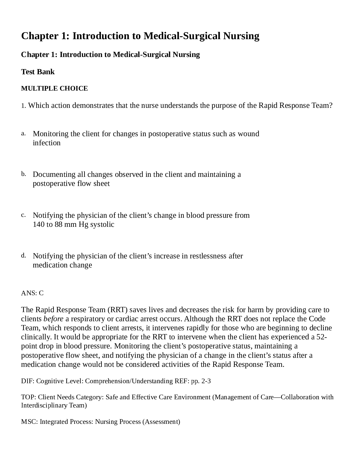


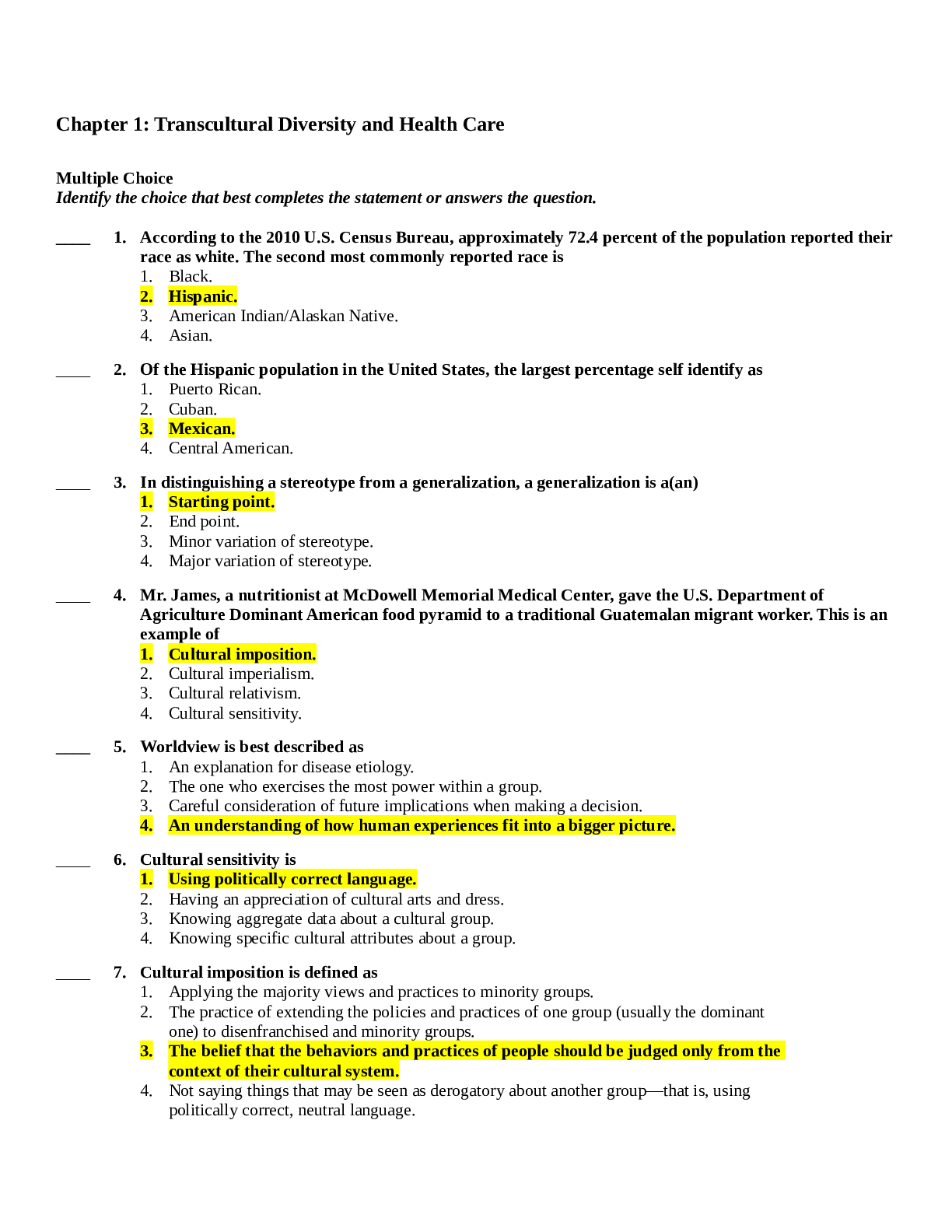


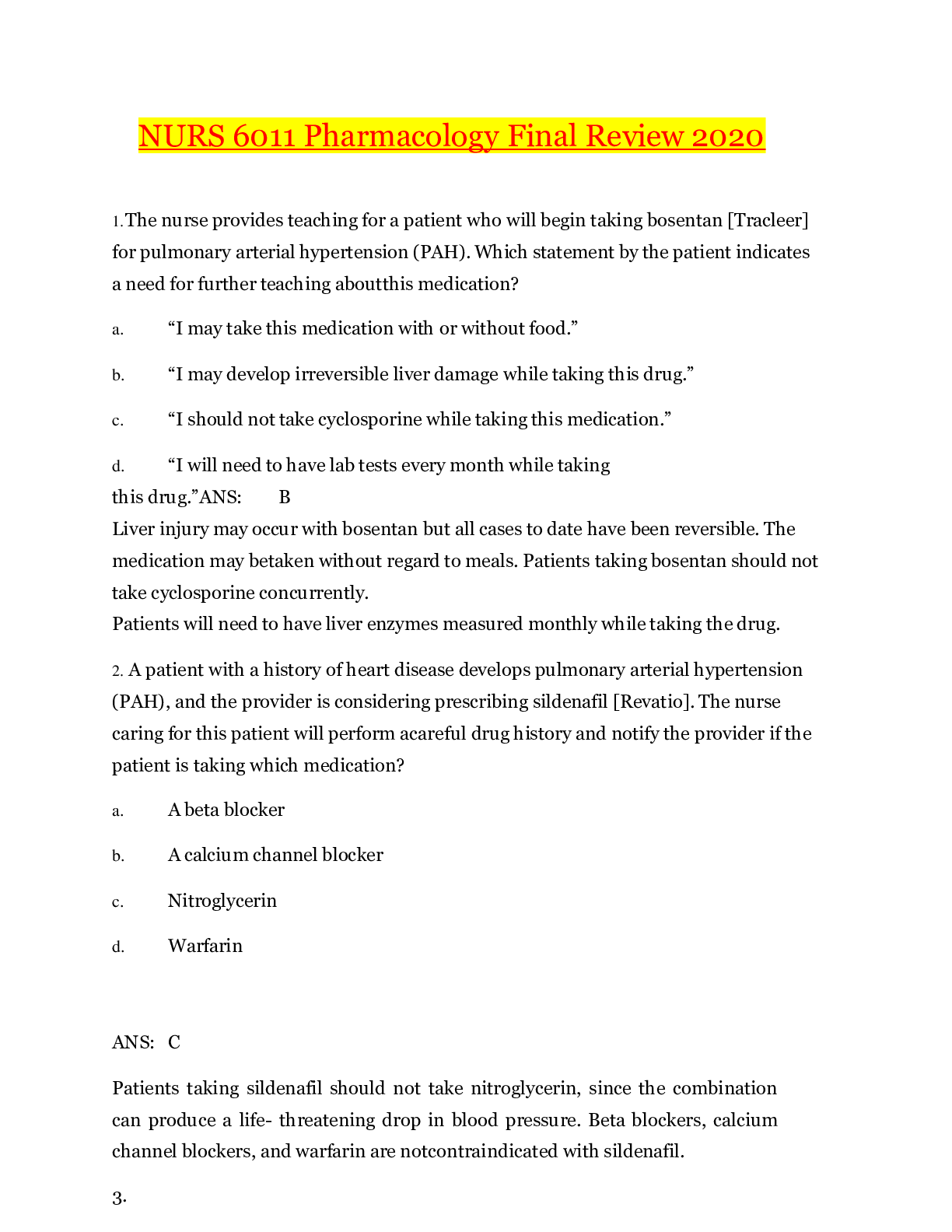



.png)

