Latest NURS 6501 Final Exam Review Guide (Weeks 7-11)
Document Content and Description Below
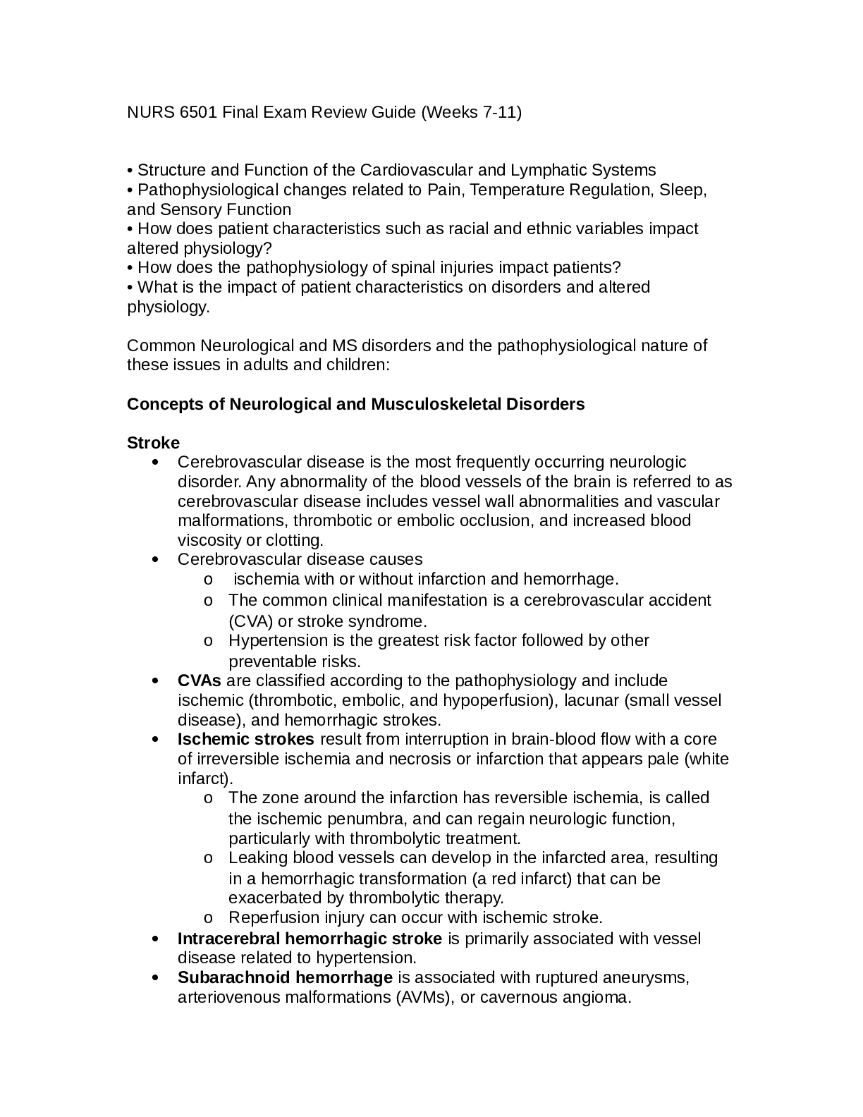
Latest NURS 6501 Final Exam Review Guide (Weeks 7-11) • Structure and Function of the Cardiovascular and Lymphatic Systems • Pathophysiological changes related to Pain, Temperature Regulation, S... leep, and Sensory Function • How does patient characteristics such as racial and ethnic variables impact altered physiology? • How does the pathophysiology of spinal injuries impact patients? • What is the impact of patient characteristics on disorders and altered physiology. Common Neurological and MS disorders and the pathophysiological nature of these issues in adults and children: Concepts of Neurological and Musculoskeletal Disorders Stroke • Cerebrovascular disease is the most frequently occurring neurologic disorder. Any abnormality of the blood vessels of the brain is referred to as cerebrovascular disease includes vessel wall abnormalities and vascular malformations, thrombotic or embolic occlusion, and increased blood viscosity or clotting. • Cerebrovascular disease causes o ischemia with or without infarction and hemorrhage. o The common clinical manifestation is a cerebrovascular accident (CVA) or stroke syndrome. o Hypertension is the greatest risk factor followed by other preventable risks. • CVAs are classified according to the pathophysiology and include ischemic (thrombotic, embolic, and hypoperfusion), lacunar (small vessel disease), and hemorrhagic strokes. • Ischemic strokes result from interruption in brain-blood flow with a core of irreversible ischemia and necrosis or infarction that appears pale (white infarct). o The zone around the infarction has reversible ischemia, is called the ischemic penumbra, and can regain neurologic function, particularly with thrombolytic treatment. o Leaking blood vessels can develop in the infarcted area, resulting in a hemorrhagic transformation (a red infarct) that can be exacerbated by thrombolytic therapy. o Reperfusion injury can occur with ischemic stroke. • Intracerebral hemorrhagic stroke is primarily associated with vessel disease related to hypertension. • Subarachnoid hemorrhage is associated with ruptured aneurysms, arteriovenous malformations (AVMs), or cavernous angioma.o Subarachnoid hemorrhage is bleeding into the subarachnoid space commonly associated with intracranial aneurysms, AVM, and hypertension. The expanding hematoma increases ICP, compresses brain tissue, reduces cerebral perfusion, disrupts the bloodbrain barrier, and causes inflammation and neuronal death. Secondary brain injury follows. Seizures and hydrocephalus can accompany neurologic deficits. Multiple sclerosis • MS is a chronic inflammatory disease involving degeneration of CNS myelin in genetically susceptible individuals. • The cause is unknown and autoreactive T and B cells recognize myelin autoantigens and produce myelin-specific antibodies triggering inflammatory demyelination with loss of oligodendrocytes and plaque formation leading to disruption of nerve conduction. • The clinical manifestations of MS involve different types: relapsingremitting, primary progressive, secondary progressive, and progressiverelapsing. Transient Ischemic Attack • A transient ischemic attack is a transient episode of neurologic dysfunction resulting from focal cerebral ischemia with risk for progressing to stroke. Myasthenia gravis • Myasthenia gravis results from a defect in nerve impulse transmission at the neuromuscular junction with generalized, ocular, or neonatal subtypes. Autoantibodies, complement deposits, and membrane attack complex destroy the acetylcholine receptor (AChR) sites, causing decreased transmission of nerve impulses, leading to muscle weakness, including ocular and systemic muscles. There can be childhood and adult onset. Headache • Migraine is an episodic disorder whose marker is headache lasting 4 to 72 hours. o Migraine is classified as a headache with and without aura and chronic migraine (migraines 15 days in a month for more than 3 months). o Migraine may be precipitated by a triggering event. o The aura is associated with cortical spreading depression, which initiates the release of neurotransmitters, particularly CGRP, that stimulate vasodilation in the trigeminal vascular system, inflammation, and sensitization of pain receptors. Glutamate is increased and serotonin is decreased. • Cluster headaches (trigeminal autonomic cephalalgia) occur in episodes several times during a day for a period of days at different times of the year, primarily in men.o The pain is unilateral, intense, tearing, and burning and associated with ptosis, lacrimation, reddening of the eye, and nausea. The cause of trigeminal activation is unknown. o There is sympathetic nervous system underactivity and parasympathetic overactivity with trigger events similar to migraine. The two forms are acute and chronic o Chronic paroxysmal hemicranias are a cluster-type headache that occurs 4 to 12 times per day for 20 to 120 minutes in both men and women. o There is sympathetic activity different from that in cluster headache, as it is relieved with indomethacin. 40. • Tension-type headache (TTH) is the most common type of headache. o Both central and peripheral pain mechanisms are associated with the etiology. o The headache is bilateral, with the sensation of a tight band around the head. The pain may last for hours or days. o There are acute and chronic forms. Seizure disorders • Seizures represent abnormal, excessive hypersynchronous discharges of cerebral neurons with transient alterations in brain function. • Seizures may be focal or generalized. • The categories of seizures include genetic, structural, metabolic, immune, infectious, and unknown. Head injury • Traumatic brain injury (TBI) is an alteration in brain function or other evidence of brain pathology caused by an external force. • Primary brain injury is caused by an impact and can be focal or diffuse with open- or closed-head injury. o Severity of TBI is graded using the Glasgow Coma Score. ▪ Focal brain injury includes coup and contrecoup, contusion (bruising of the brain), laceration (tearing of brain tissue), extradural hematoma (accumulation of blood above the dura mater), subdural hematoma (blood between the dura mater and arachnoid membrane), intracerebral hematoma (bleeding into the brain), and open-head trauma. o Open-head injury involves a skull fracture with exposure of the cranial vault to the environment. ▪ The types of skull fracture include compound fracture or perforated fracture and linear, comminuted, and basilar skull fracture. o Closed-head injuries occur in a precise location, and most are mild. More severe damage includes contusions and epidural, subdural, subarachnoid, and intracerebral hemorrhage.▪ Diffuse axonal injury (DAI) results from mechanical forces of acceleration, deceleration, and rotation that cause stretching and shearing of axons and can only be seen microscopically. The injury can be mild, moderate, or severe. ▪ Secondary neuronal injury occurs as an indirect result of primary brain injury. ▪ Systemic processes include hypotension, hypoxia, anemia, hypoglycemia, hyperglycemia, and hypercapnia or hypocapnia. ▪ Cerebral contributions include inflammation, oxidative stress, alterations in the blood-brain barrier, excitotoxicity, cerebral edema, increased intracranial pressure (IICP), decreased cerebral perfusion pressure, cerebral ischemia, and brain herniation. ▪ Complications of TBI include postconcussion syndrome, posttraumatic seizures, and chronic and traumatic encephalopathy. Spinal cord injury • Spinal cord and vertebral injuries occur most often in young men who sustain various kinds of injuries (recreational or travel-related) and older adults because of preexisting degenerative vertebral disorders. 11. • Vertebral injuries include fractures, dislocations, compressions, and penetrating bone fragments from shearing and compression force. Fractures can be simple, compressed, or comminuted. 12. o Primary spinal cord injury involves damage to vertebral or neural tissues from shearing, compression, or traction forces. 13. o Secondary spinal cord injury is related to edema, ischemia, excitotoxicity, inflammation, oxidative damage, and activation of necrotic and apoptotic cell death and begins within minutes after injury and continues for weeks. o Spinal cord injury often causes spinal shock with cessation of all motor, sensory, reflex, and autonomic functions below any transected area. Loss of motor and sensory function depends on the level of injury. o Neurogenic shock (vasogenic shock) occurs with cervical or upper thoracic cord injury above T5 and may be seen in addition to spinal shock. There is loss of sympathetic activity and unopposed vagal parasympathetic activity with symptoms of hypotension, bradycardia, and hypothermia. o Paralysis of the lower half of the body with both legs involved is called paraplegia. ▪ Paralysis involving all four extremities is called quadriplegia. ▪ Return of spinal neuron excitability occurs slowly. Reflex activity can return in 1 to 2 weeks in most people with acute spinal cord injury. A pattern of flexion reflexes emerges,involving first the toes and then the feet and legs. Eventually reflex voiding and bowel elimination appear, and mass reflex (flexor spasms accompanied by profuse sweating, piloerection, and automatic bladder emptying) may develop. o Autonomic hyperreflexia (dysreflexia) is a syndrome of sudden massive reflex sympathetic discharge associated with spinal cord injury at level T5-T6 or above and can cause life-threatening hypertension. Inflammatory diseases of the musculoskeletal system Osteoporosis • Metabolic bone diseases are characterized by abnormal bone structure. o In osteoporosis bone tissue is normally mineralized, but the density or mass of bone is reduced because the bone remodeling cycle is disrupted. o Osteoporosis is a complex, multifactorial, chronic disease that often progresses silently for decades until fractures occur. It is the most common bone disease. Multiple factors are involved including alteration in the OPG/RANKL/RANK system. • Postmenopausal osteoporosis occurs in middle-aged and older women and is caused by increased osteoclast activity (probably caused by changes in osteoprotegerin), decreased IGF levels, a combination of inadequate dietary calcium intake and lack of vitamin D, possibly decreased levels of magnesium, lack of exercise, decreased levels of estrogen, and family history. o Glucocorticoids increase RANKL expression and inhibit OPG production by osteoblasts, thus leading to lower bone density. Osteopenia • Decreased bone mass Bursitis • A trauma or overuse injury that can cause painful inflammation in the bursal sacs • The inflammation may decrease with rest, heat, and aspiration of the fluid. Tendinitis • Inflammation of tendons o Inflammatory fluid accumulates, causing swelling of the tendon and its enclosing sheath. Inflammatory changes cause thickening of the sheath, which limits movements and causes pain. o After repeated inflammations, calcium may be deposited in the tendon origin area, causing a calcific tendinitis Gout • Gout is a metabolic disorder associated with high levels of uric acid in the blood and body fluids. Uric acid crystallizes in the connective tissue of a joint, where it initiates inflammatory destruction of the joint.• Uric acid >6.8mg/dl • Three stages: o Asymptomatic hyperuricemia ▪ Serum urate level is elevated but arthritic symptomrs, tophi, and renal stones are not present; may persist throughout life o Acute Gouty arthritis: ▪ Attacks develop with increased serum urate concentrations; tends to occur with sudden or sustained increases of hyperuricemia but also can be triggered by trauma, drugs, and alcohol o Tophaceous Gout: ▪ Third and chronic stage of disease; can begin as early as 3 years or as late as 40 years after the initial attack of gouty arthritis. Progressive inability to excrete uric acid expands the urate pool until urate crystal deposits (tophi) appear in cartilage, synovial membrane, tendons, and soft tissue. Lyme Disease • Multisystem inflammatory disease caused by a spirochete borrelia burgdorferi transmitted by ixodes tick bites and is the most frequently reported vector-borne illness • Highest incidence among children • Symptoms occur in three stages however 50% are symptomless o Localized infection occurs 3-32 days after the bite with erythema migrans and a bulls eye rash (commonly appear at the tick attachment) with or without fever, malaise, myalgias, and arthralgias o Disseminated infection or secondary erythema migrans, usually with myalgias, arthralgias, and more rarely meningitis, neuritis, or carditis o Post Lyme disease syndrome or chronic lyme disease can continue for years with arthritis, encephalopathy, polyneuropathy or heart failure • Treatment: Abx doxycycline, amoxicillin, cefuroxine Spondylolysis • Spondylolysis is a structural defect in the pars interarticularis of the vertebral arch with anterior displacement (sliding) of the deficient vertebra (spondylolisthesis) and is a cause of low back pain. • Cervical spondylolysis is facet hypertrophy and disk degeneration with narrowing in the cervical spine predominantly at C5-C6 and C6-C7 and can cause radiculopathy and myelopathy with numbness and tingling in the arms, occipital headache, difficulty walking, altered sensation in the feet, and sphincter disturbances. Fractures • The most common skeletal injury is a fracture.• A bone can be completely or incompletely fractured. o A closed fracture leaves the skin intact. o An open fracture has an overlying skin wound. o The direction of the fracture line can be linear, oblique, spiral, or transverse. Greenstick, torus, and bowing fractures are examples of incomplete fractures that occur in children. o Stress fractures occur in normal or abnormal bone that is subjected to repeated stress. o Fatigue fractures occur in normal bone subjected to abnormal stress. o Normal weightbearing can cause an insufficiency fracture in abnormal bone. • Parkinson’s • PD is a common degenerative disorder of the basal ganglia (corpus striatum) involving degeneration of the dopamine-secreting nigrostriatal pathway resulting in overactivity by the subthalamic nucleus, causing tremor, rigidity, and bradykinesia. Involvement of the limbic system causes emotional lability. Progressive dementia may be associated with an advanced stage of the disease. Alzheimer’s • Dementia is an acquired impairment of intellectual function, memory, and language with alteration in behavior and can be caused by trauma, vascular disease, infection, and progressive neurodegeneration. AD is the most common chronic, irreversible dementia with accumulations of amyloid and tau protein neurofibrillary tangles in the brain. Less common forms include vascular and frontotemporal dementia. Three basic bone-formations: Osteoblasts: • Derived from fibroblasts and are responsible for construction of bone • Synthesizes collagen and proteoglycans; stimulate bone formation and are also involved in some osteoclast resorptive activity Osteocytes • Maintain bone matrix • act as mechanoreceptors • influence osteoblasts and osteoclasts Osteoclasts • Multinucleate cells of monocytic origin that remodel bone by resorption • Major role in mineral homeostasis Concepts of Psychological DisordersGeneralized anxiety disorder • GAD is characterized by excessive and persistent worries about life events. Individuals exhibit varying levels of motor disturbances, irritability, and fatigue that may be linked to fluctuations in psychosocial stress. Many GAD individuals manifest symptoms of depression. • Pathophysiologic changes in the cingulate cortex and amygdala may have prominent roles in stimulating anticipatory anxiety and attentional bias to threats in people with GAD. • Treatment of GAD usually involves a combination of behavioral therapy and drug medications, especially 5-HT/NE reuptake inhibitors. Depression • Major depression is characterized by an intense and sustained unpleasant state of sadness and hopelessness. • Environmental triggers such as psychosocial stress appear to facilitate the onset of depression in individuals with a genetic vulnerability. • A reduction in brain monoamine neurotransmission is linked to depression • Exposure to uncontrollable stress elevates secretion of the stress hormone cortisol, which increases both the secretion of proinflammatory cytokines and the risk of developing depression. Abnormalities involving thyroid hormones also are found in depression. • Stress-induced depression is accompanied by deficits in brain-derived neurotrophic factor (BDNF) and neurogenesis in the hippocampus. In animal models, stress-induced depression-like behavior and the accompanying deficits in hippocampal BDNF and neurogenesis are reversed by antidepressant treatment. • The frontal lobe and limbic system volumes are reduced in major depression and bipolar illness. • In addition, blood flow is altered in prefrontal and limbic brain regions that include the amygdala, a structure implicated in emotional behavior. • Pharmacotherapy involves the use of MAOIs, TCAs, SSRIs, and atypical antidepressants. Bipolar disorders • Psychiatric disorder characterized by alternating mania or hypomania and depression, often with periods of normal mood in between, and changes in energy and behavior according to mood • Mania characterized by: o Elevated levels of euphoria and self-esteem and feelings of grandiosity, few hours of sleep, increased energy, unorganized plans and thoughts, poor judgment, hypersexuality, excessive, rapid loud and pressured speech lasting days to months followed by depression o 50% develop psychotic symptom such as delusions, hallucinations requiring hospitalization Schizophrenia • Schizophrenia is characterized by thought disorders that reflect a break between the cognitive and the emotional sides of one’s personality.• Schizophrenic symptoms are classified into positive, negative, and cognitive categories. o Positive symptoms include hallucinations, delusions, formal thought disorder, and bizarre behavior. o Negative symptoms include flattened affect, alogia, anhedonia, attention deficits, and apathy. o Cognitive symptoms are the inability to perform daily tasks requiring attention and planning. • Schizophrenia has a strong genetic predisposition, and environmental factors (e.g., viral infection, nutritional deficiencies, prenatal birth complications, urban upbringing) may interfere with genetically programmed neural development to alter brain structure and function. • Brain imaging studies reveal structural brain abnormalities including an enlargement of the cerebral ventricles and widening of the fissures and sulci in the frontal cortex. o In addition, there is a reduction in the volumes of both the thalamus, which may disrupt communication among cortical brain regions, and the temporal lobe, which may be responsible for the manifestations of positive symptoms. o In schizophrenia, the frontal lobe shows a progressive loss in volume and a worsening of negative symptoms despite the use of antipsychotic medications. o Blood flow and metabolism are reduced in the dorsolateral prefrontal cortex, which compromises the ability to engage in goaldirected and cognitive problem-solving behavior. o Neurochemical abnormalities in dopamine and glutamate systems are found in schizophrenia. • The first generation of antipsychotic drugs blocks the dopamine D2 receptor. The second generation, called atypical antipsychotics, blocks not only D2 receptors but also dopamine, serotonin, and other neurotransmitter receptors. o Antipsychotic medications, however, are not always effective in treating schizophrenic individuals with severe negative symptoms. Talk therapies are used to increase drug compliance and to encourage coping strategies. Delirium • Acute confusional state arising from disruption of a widely distributed neural network involving the reticular activating system of the upper brainstem and is projections into the thalamus, basal ganglion, and specific association areas of the cortex and limbic areas. o Associated with autonomic nervous system hyperactivity and typically develops over 2-3 days and associated with right upper middle temporal gyrus or left temporal occipital junction disruption • Most commonly occurs in critical care units or during withdrawal from alcohol • Hospitalized older adults are most at risk• Causes: o Drug intoxication, alcohol/drug withdrawal, metabolic disorders ( hypoglycemia, thyroid storm), brain trauma/surgery, postanesthesia, febrile illnesses or heat stroke, electrolyte imbalance, dehydration, heart kidney or liver failure Dementia • Acquired deterioration and progressive failure of many cerebral functions that includes impairment of intellectual processes with a decrease in orienting, memory, language, judgment, and decision making. Declining intellectual activity may lead to alterations in behavior ie agitation, wandering, and aggression • Pathophysiology o Neuron degeneration, compression of brain tissue, atherosclerosis of cerebral vessels, and brain trauma. Genetic predisposition is associated with neurodegenerative diseases including alzheimer’s, Huntington, and parkinsons disease • Eval and treatment: o Rule out other underlying conditions that may be treatable Obsessive compulsive disease • OCD is a chronic illness characterized by irrational obsessions and ritualized acts that impair normal functioning and cause severe distress. It is a chronic disabling illness. • OCD is a time-consuming illness, which significantly impairs everyday functions, such as social relationships, job performance, and academic success. o Examples of obsessions include preoccupation with doubting, religious or sexual themes, or the belief that a negative outcome will occur if a specific act is not performed. o A pathophysiologic brain circuit consisting of the anterior thalamus, orbitofrontal cortex, dorsal anterior cingulate cortex, and especially in the basal ganglia subregions of the caudate and putamen is involved in OCD. o OCD requires long-term treatment that may include psychotherapy and pharmacotherapy. However, people with severe OCD who are resistant to these treatments may require neurosurgery to disconnect regions of pathophysiologic brain circuit to provide relief of OCD symptoms. Deep brain stimulation may be another option for uncontrollable OCD. Women’s and Men’s Health, Infections, and Hematologic Disorders Sexually transmitted diseases • Sexually transmitted diseases may be more common in certain populations related to increased physiologic risk for acquisition (such as with younger women or with men who have sex with men) or insufficientaccess to quality health care (such as with lower socioeconomic groups, racial/ethnic minorities, and marginalized groups). • Gonorrhea is a sexually transmitted communicable disease that can be local or systemic. Complications include PID, sterility, and disseminated infection. o Gonorrhea can be passed to the fetus from the mother and typically manifests as an eye infection 1 to 12 days after birth. Ophthalmic antibiotic prophylaxis alone is not sufficient to prevent vertical transmission. o Gonorrhea is rapidly becoming resistant to available antibiotics. Multidrug therapy is now recommended to decrease drug resistance. • Syphilis is an STI that becomes systemic shortly after infection. The four stages of the disease are (a) primary syphilis with a chancre at the site of infection; (b) secondary syphilis with systemic spread to all body systems; (c) latent syphilis with minimal symptoms or the development of skin lesions; and (d) tertiary syphilis, the most severe stage, with destruction of bone, skin, and soft and neurologic tissues. o Congenital syphilis contributes to prematurity of the newborn with bone marrow depression, CNS involvement, renal failure, and intrauterine growth retardation. o Syphilis is diagnosed with serologic testing and is treated with injectable penicillin. o With chancroid infection, women are generally asymptomatic and men may develop inflamed, painful genital ulcers and inguinal buboes. The incubation period is 1 to 14 days. Single-dose therapy with injectable ceftriaxone or oral azithromycin for both partners is recommended. Persons with HIV may require a longer treatment regimen. • Granuloma inguinale (donovanosis) is rare in the United States. The bacteria are gram negative and survive within macrophages. Localized nodules coalesce to form granulomas and ulcers on the penis in men and on the labia in women. Antibiotics provide effective treatment. • Bacterial vaginosis (BV) is a sexually associated condition caused by an overgrowth of anaerobic bacteria that produce aromatic amines and raise the pH of the vagina, promoting further bacterial growth (without an inflammatory response) and a fishy odor. “Clue cells” are found on the wet mount. o Metronidazole (Flagyl) provides effective treatment. BV has been associated with PID, chorioamnionitis, preterm labor, and postpartum endometritis. Treatment of male sexual partners is not recommended. • Chlamydia is the most common bacterial STI in the United States and a leading preventable cause of infertility and ectopic pregnancy. The causative organism, C. trachomatis, localizes to epithelial tissue and canspread throughout the urogenital tract or pass from the infected mother to the eyes and respiratory tract of newborn infants during birth. o C. trachomatis is susceptible to inexpensive, readily accessible antibiotics. Single-dose azithromycin is the drug of choice for infected individuals and all sexual contacts. Because of the asymptomatic nature of chlamydia and the potential sequelae of infection, widespread screening is recommended by the CDC. • Lymphogranuloma venereum is a chronic STI uncommon in the United States. The lesion begins as a skin infection and spreads to the lymph tissue, causing inflammation, necrosis, buboes, and abscesses of the inguinal lymph nodes. Primary lesions appear on the penis and scrotum in men and on the cervix, vaginal wall, and labia in women. Secondary lesions involve inflammation and swelling of the lymph nodes with formation of large buboes that rupture and drain. o A 21-day or longer course of oral doxycycline or erythromycin is needed for treatment. Treatment of sexual partners is recommended. • Genital herpes is the most common genital ulceration in the United States and is caused by either HSV-1 or HSV-2. Lesions initially appear as groups of vesicles that progress to ulceration with pain, lymphadenopathy, and fever. Herpes simplex virus can pass from mother to fetus; thus women with active lesions should give birth by cesarean section to avoid vertical transmission. o Herpes simplex virus (HSV) infection is lifelong and can result in an initial outbreak and subsequent outbreaks. Individuals are contagious during outbreaks and episodes of asymptomatic viral shedding. o Acyclovir reduces symptoms but does not cure the disease. Recurrent infections are most often attributable to HSV-2 and are generally milder and of shorter duration. • Human papillomavirus (HPV) is associated with the development of cervical dysplasia and cancer as well as condylomata acuminata. The high-risk strains of HPV (HR-HPV) that are precursors to the development of cervical cancer do not cause genital warts. Testing is available to detect HR-HPV and a vaccine is now available for the HPV types with highest risk for cervical cancer. • Condylomata acuminata (genital warts) are sexually transmitted and highly contagious. The velvety cauliflower-like lesions occur in the genital and anal areas, vagina, and cervix and are painless. They can be transmitted to the infant at birth. • Molluscum contagiosum is a benign viral infection of the skin. It is transmitted by skin-to-skin contact in children and adults. In adults, it tends to occur on the genitalia and be transmitted by sexual contact. • Trichomoniasis (T. vaginalis) causes vaginitis in women and urethritis in men. Both partners usually are infected. Women usually have a copious,malodorous, gray-green discharge with pruritus. Men usually are asymptomatic. Metronidazole is the treatment for both sexes. • Scabies is a parasitic infection that spreads by skin-to-skin and sexual contact. The scabies mite burrows through the skin, depositing eggs, causing intense pruritus, especially at night. Treatment consists of topical application of a pediculicide. • Pediculosis pubis (crabs) is commonly transmitted sexually and is caused by the crab louse, P. pubis. The lice bite into the skin for nutrition. Symptoms include mild and severe pruritus. Topical application of prescription or over-the-counter pediculicides is effective treatment. • Systemic diseases known to be sexually transmitted include AIDS (see Chapter 10), cytomegalovirus infection, and Epstein-Barr virus. • Transmission of HBV can occur through needle puncture, blood transfusion, cuts in the skin, and contact with infected body fluids. o Hepatitis B infection poses significant health risks including chronic liver disease and hepatocellular cancer. Immunization against hepatitis B is the most effective means of preventing transmission. Universal vaccination of infants and children is recommended, as well as vaccination of high-risk adults. o The risk of perinatal transmission of HBV is high for infants of HBVinfected mothers unless they receive immunoglobulin and are vaccinated. • Hepatitis C is generally transmitted percutaneously but sexual transmission appears possible. • Although normally transmitted through mosquito bites, zika virus can be transmitted through sexual contact with infected body fluids or through vertical transmission. Zika virus sequesters in fetal brain tissue, disrupting brain growth and causing persistent, lifelong microcephaly. Prostate • The prostate gland is about the size of a walnut and surrounds the urethra. Prostatic secretions are alkaline and contribute to the ejaculate. Epididymitis • inflammation of the epididymis, is usually caused by a sexually transmitted pathogen that ascends through the vasa deferentia from an already infected urethra or bladder. Factors that affect fertility • female: ovulatory disorder, abnormal semen, blockage of the fallopian tubes, endometriosis, unexplained infertility, adhesions, scarring from PID • male: hormonal disorders (thyroid or testosterone), elevations in temperature, abnormal placement of testicles, varicoceles near the testes, exposure to high temperatures in hot tubs or saunas, abnormalities in seminal tracts and sexual dysfunction that disrupts ejaculation Anemia• Anemia is defined as a reduction in the total circulating red cell mass or a decrease in the quality or quantity of hemoglobin. Polycythemias are excessive levels or volumes of RBCs. o Anemias can result from blood loss, impaired erythrocyte production, increased erythrocyte destruction, and a combination of these factors. o Total circulating red blood cell mass is reflected by changes in plasma volume caused by dehydration and fluid retention. o Anemias can be classified in several ways and a useful way is by the main underlying mechanism. o Clinical manifestations of anemia may be demonstrated in all organs and tissues (tissue hypoxia) throughout the body. Decreased oxygen delivery to tissues causes fatigue, dyspnea, syncope, angina, compensatory tachycardia, and organ dysfunction. o Posthemorrhagic anemia is a normocytic-normochromic anemia caused by acute blood loss. A major cause of acute blood loss is trauma, a rising global problem. o Anemia from chronic blood loss occurs if the loss is greater than the replacement capacity of the bone marrow. If iron stores are depleted, iron deficiency anemia can occur. o Macrocytic (megaloblastic) anemias are characterized by larger than normal erythroid precursors (megaloblasts) in the bone marrow that mature into large erythrocytes. They most commonly are caused by deficiency of vitamin B12 or folate. o PA results from inadequate vitamin B12 absorption because autoimmune gastritis impairs the production of IF, which is required for vitamin B12 uptake from the gut. o Folate deficiency anemia is caused by inadequate dietary intake of folate. Both anemias respond to replacement therapy. o Microcytic-hypochromic anemias are characterized by abnormally small erythrocytes with insufficient hemoglobin content. The anemias result from disorders of (a) iron metabolism (IDA), (b) porphyrin and heme synthesis (SAs), or (c) globin synthesis (thalassemia). o IDA is the most common type of nutritional disorder worldwide. It is usually a result of dietary deficiency. Other major causes are impaired absorption, increased requirement, and chronic blood loss. IDA usually develops slowly, with a gradual insidious onset of symptoms, which include fatigue, weakness, dyspnea, alteration of various epithelial tissues, and vague neuromuscular complaints result. ▪ Individuals at highest risk for developing IDA include older adults, women, infants, teenagers eating poor diets, and those living in poverty. Once the source of blood loss isidentified and corrected, oral iron replacement therapy can be initiated. o ACD, also called anemia of inflammation, results from decreased erythropoiesis and impaired iron utilization in people with chronic systemic disease or inflammation. ACD is common among hospitalized individuals. ▪ Examples of mechanisms associated with ACD include (1) decreased erythrocyte life span, (2) reduced production of erythropoietin, (3) ineffective bone marrow response to erythropoietin, and (4) iron sequestration in macrophages. In particular, the proinflammatory cytokine IL-6 increases hepatocyte release of hepcidin which suppresses ferroportin transport of iron out of macrophages. o AA is a critical condition characterized by a reduction or absence of all three blood cell types (pancytopenia). Unless the cause is determined, bone marrow aplasia results in death. o Hemolytic anemia is a result of excessive destruction of erythrocytes and may be acquired or hereditary. Common, acquired forms are autoimmune reaction (immunohemolytic) and druginduced hemolysis. o AIHAs include (a) warm reactive antibody type, (b) cold agglutinin type, and (c) cold hemolysin type (paroxysmal cold hemoglobinuria). ITP • Immune thrombocytopenic purpura (ITP) is a major cause of platelet destruction, often affecting females, and results in hemorrhaging that ranges from petechiae to bleeding from mucosal sites. TTP • Thrombotic thrombocytopenic purpura (TTP) causes platelet aggregation leading to microcirculatory occlusion DIC • DIC is a complex syndrome that results from a variety of clinical conditions that release tissue factor, causing an increase in fibrin and thrombin activity in the blood and producing augmented clot formation and accelerated fibrinolysis. Sepsis is often associated with DIC. • DIC is characterized by a cycle of intravascular clotting followed by active bleeding caused by the initial consumption of coagulation factors and platelets and diffuse fibrinolysis. • Diagnosis of DIC is based on dysfunctional coagulation activity. • Treatment is complex, nonstandardized, and focused on removing the primary cause, restoring hemostasis, and preventing further organ damage.Thrombocytopenia • Thrombocytopenia is characterized by a platelet count less than 150,000/mm3 of blood; a count less than 50,000/mm3 increases the potential for hemorrhage associated with minor trauma. 2. • Thrombocytopenia may be congenital or acquired and primary or secondary to other acquired or congenital conditions. Acquired thrombocytopenia is associated with autoimmune diseases, viral infections, nutritional deficiencies, chronic renal failure, bone marrow hypoplasia, radiation therapy, and bone marrow infiltration by cancer. • Most common forms of thrombocytopenia are the result of increased platelet consumption Pediatrics • Shock, Multiple Organ Dysfunction Syndrome, and Burns in Children • Shock in children is present when there are signs of poor systemic perfusion, regardless of blood pressure. o Hypovolemic shock is the most common type of shock in children and most frequently results from dehydration and trauma. Hypovolemic shock also may result from expansion of the vascular space, producing inadequate intravascular volume relative to the vascular space. o Hypotension is a sign of severe (preterminal), decompensated shock, referred to as hypotensive shock. ▪ Clinical manifestations of hypovolemic shock include inadequate systemic perfusion associated with intravascular fluid loss. Adrenergic compensatory mechanisms can produce tachycardia, redistribution of blood flow, peripheral vasoconstriction, cool extremities, delayed capillary refill, and oliguria. o Neurogenic shock is caused by a loss of vasomotor tone after severe injury to the spinal cord. ▪ Clinical manifestations of neurogenic shock include warm skin, hypotension with a low diastolic blood pressure, and poor systemic perfusion. Tachycardia is not present. o Cardiogenic shock, with decreased cardiac output, is observed most commonly after cardiovascular surgery or with inflammatory diseases of the heart, such as cardiomyopathy and myocarditis. It is also found in children with obstructive congenital heart disease and those with drug toxicity or severe electrolyte or acid-base imbalances. ▪ Clinical manifestations of cardiogenic shock include inadequate systemic perfusion despite adequate intravascular volume. Cardiac output is typically low. Adrenergic compensatory mechanisms, including peripheralvasoconstriction and decreased urine volume, are similar to those found in hypovolemic shock. o Once septic shock is present, immediate treatment is urgently needed. Therapy in the first hour includes aggressive fluid resuscitation (typically 60 to 80 mL/kg administered in the first hour of therapy, and approximately 200 to 240 mL/kg in the first 8 hours of therapy). If the child does not respond to volume administration alone, vasoactive support must be initiated within the first hour of treatment. Antibiotics also must be administered within the first hour. Goals of therapy are to rapidly normalize the heart rate and blood pressure for age and to normalize capillary refill to less than 2 seconds. The child’s shock index (heart rate/systolic blood pressure) should fall during the first hour of management if therapy is effective. Fluid and vasoactive therapy should support high cardiac output and oxygen delivery, maintaining the SvO2 at approximately 70%. o Sepsis is a systemic response to infection. It is present when manifestations of SIRS are observed. SIRS is present when the child demonstrates two or more of the following as an acute change from baseline values: altered temperature, altered heart rate, altered respiratory rate, and alteration in the WBC count. The newborn often develops hypothermia rather than fever as a sign of infection and may develop bradycardia instead of tachycardia. o Severe sepsis is present when there is evidence of SIRS and signs of organ dysfunction, hypoperfusion, or hypotension. ▪ The development of septic shock is heralded when the child with severe sepsis develops signs of cardiovascular dysfunction. The child may become hypotensive despite adequate fluid resuscitation or require vasopressors to maintain blood pressure. ▪ Reperfusion and inflammatory injury stimulate free oxygen radicals that can damage cell membranes, denature proteins, and disrupt chromosomes. This process likely affects endothelial cells and the microvasculature, causing MODS. ▪ Lactic acidosis (i.e., rise in serum lactate) may be the most sensitive indicator of inadequate systemic perfusion in children; effective shock therapy should eliminate lactic acidosis. ▪ The general goals of treatment for shock are maximization of oxygen delivery and minimization of oxygen demand. This requires support of airway, oxygenation, and ventilation. Support of cardiovascular function requires support of appropriate heart rate and rhythm, adequate intravascular volume, good myocardial function, and appropriate vascularresistance and distribution of blood flow. The child should be kept warm, but fever must be treated promptly. ▪ The signs of shock should lessen or disappear if management of shock is effective. The warmth of the child’s extremities, briskness of capillary refill, quality of peripheral pulses, level of consciousness and responsiveness, urine volume, oxygenation, ventilation, and acid-base status should improve throughout shock therapy. • Burns o Burns in children are often the result of inadequate supervision, curiosity, inability to escape the burning agent, or nonaccidental trauma. o Scald injuries are commonly seen in young children and result from exposure to hot water, grease, or other hot liquids, whereas flame burns are more prevalent among older children. o A child’s skin is thinner and thus more susceptible to injury than adult skin. The kitchen and bathroom are common sites of burn injury. o Approximately 8% to 12% of all forms of child abuse cases in the United States result from burn injury. o Flame burns involving flammable liquids, most notably gasoline, are more common in older children. Risk-taking behaviors in young males can lead to electrical burns. Children may be exposed to chemical injury by swallowing caustic agents at home. o Use of the standard Rule of Nines results in inaccurate calculation of the percentage of TBSA in children. A modified Rule of Nines deducts 1% from the head and adds 0.5% to each leg for each year of life after 2 years of age. o Major burn trauma involves all body systems, and the consequences of injury include shock, infection, hypermetabolism, organ failure, and functional limitations. These effects can be magnified in the pediatric population as a result of physiologic immaturity and age-related variation in treatment modalities. o Infection, trauma, or applying ice to the burn area may convert a partial-thickness injury to a full-thickness one, especially in young children, who have thinner, more delicate skin. o Marked reduction in cardiac output occurs immediately after injury and is accompanied by an initial increase in systemic vascular resistance. o The inefficient and labile peripheral circulation of the infant complicates management of the burn shock phase of treatment. A higher risk of chest constriction and impairment of respiratory excursion may result because of the increased pliability of the rib cage, especially in very young children. Younger children are also more susceptible to increased intraabdominal pressure.o The leading cause of death in children after burn injury, as in adults, is inhalation injury. o Children require fluid resuscitation for smaller burns than does the adult population as a result of limited physiologic reserves. Colloid replacement, although controversial, may be required in the very young child who fails to respond to fluid replacement. o Children younger than 2 years lack the ability to concentrate urine because of the immaturity of the renal system and are therefore at increased risk for dehydration. Because children have a relatively larger body surface area in relation to weight than adults, they require proportionately increased fluid during burn shock resuscitation to compensate for evaporative water losses. o Some children exhibit immunosuppression for a prolonged period after wound closure. o A biphasic pattern of physiologic responses is evident in the burninjured child. The initial ebb phase occurs during the immediate postburn period and continues for 3 to 5 days. This phase is characterized by reduced oxygen consumption, impaired circulation, and cellular shock. After this phase and the restoration of volume, the metabolic response shifts to a catabolic, or flow, phase. This phase is characterized by hypermetabolism with an increased oxygen consumption and elevation of catecholamines, glucocorticoids, and glucagon. o Glycogen stores are limited in children, making it hard for them to meet the increased energy demands of the burn. This prolonged metabolic dysfunction may lead to loss of lean body mass and increased morbidity. o Although age was not found to be a predictor of hypertrophic scarring, children have greater skin tension and an accelerated rate of collagen synthesis. o Children require specialized management to ensure optimal functional and cosmetic results. Long-term scar and contracture management is necessary because of changes in body composition as the child grows and matures. • Pediatric Disorders Cancer in Children • Cancer in children and adolescents is rare, but it is still the leading cause of death from disease in this population. • Leukemias and brain tumors account for 61% of cancer in children from birth to 14 years of age, with neuroblastoma and soft tissue or bone sarcomas less common. • The most common cancers among the adolescent and young adult populations (15 to 39 years of age) are Hodgkin lymphoma, leukemia,germ cell tumors (particularly testicular), central nervous system (CNS) tumors, non-Hodgkin lymphoma, thyroid cancer, melanoma, sarcomas, and breast, cervical, liver, and colorectal cancers. • Etiology o The interaction of many factors most likely produces cancer in children and adolescents, a concept referred to as multiple causation or multifactorial etiology. o Oncogenes and tumor-suppressor genes have been associated with childhood and adolescent malignancies. o Chromosomal aberrations or single-gene defects including aneuploidy, amplifications, deletions, translocations, and fragility are associated with the development of childhood cancer. o Wilms tumor and retinoblastoma are pediatric malignancies that are linked in a familial manner. o Childhood exposure to ionizing radiation, drugs, or viruses has been associated with the risk of developing cancer. • Prognosis o Nearly 85% of children and adolescents diagnosed with cancer are cured. o Mortality rates have declined significantly in the past 45 years largely because of advances in treatment and increased participation in clinical trials. o Young children are particularly prone to long-term sequelae of cancer therapy. The development of more effective, targeted therapies with fewer side effects is imperative. Growth and development • A child's growth and development can be divided into four periods: o Infancy o Preschool years o Middle childhood years o Adolescence • Soon after birth, an infant normally loses about 5% to 10% of their birth weight. By about age 2 weeks, an infant should start to gain weight and grow quickly. By age 4 to 6 months, an infant's weight should be double their birth weight. During the second half of the first year of life, growth is not as rapid. Between ages 1 and 2, a toddler will gain only about 5 pounds (2.2 kilograms). Weight gain will remain at about 5 pounds (2.2 kilograms) per year between ages 2 to 5. Between ages 2 to 10 years, a child will grow at a steady pace. A final growth spurt begins at the start of puberty, sometime between ages 9 to 15. • The child's nutrient needs correspond with these changes in growth rates. An infant needs more calories in relation to size than a preschooler or school-age child needs. Nutrient needs increase again as a child gets close to adolescence. A healthy child will follow an individual growth curve. However, the nutrient intake may be different for each child.• Provide a diet with a wide variety of foods that is suited to the child's age.Healthy eating habits should begin during infancy. This can help prevent diseases such as high blood pressure and obesity • INTELLECTUAL DEVELOPMENT AND DIET o Poor nutrition can cause problems with a child's intellectual development. A child with a poor diet may be tired and unable to learn at school. Also, poor nutrition can make the child more likely to get sick and miss school. o Breakfast is very important. Children may feel tired and unmotivated if they do not eat a good breakfast. The relationship between breakfast and improved learning has been clearly shown. There are government programs in place to make sure each child has at least one healthy, balanced meal a day. This meal is usually breakfast. Programs are available in poor and underserved areas of the United States. Scoliosis (ortho) • Scoliosis is a lateral curvature of the spinal column that can be caused by congenital malformations of the spine, neuromuscular disease, trauma, extraspinal contractures, bone infections, metabolic bone disorders, joint disease, and tumors. Kawasaki • Kawasaki disease is an acute systemic vasculitis that may result in the development of coronary artery aneurysms and thrombosis. Alterations in children Congenital (heart syndrome) • Most congenital cardiovascular defects have begun to develop by the fourth week of gestation, and most have many causes, both environmental and genetic. • Environmental risk factors associated with the incidence of CHD typically are maternal conditions. Among these are viral infections, diabetes, drug intake, alcohol intake, metabolic disorders, and advanced maternal age. • Genetic factors associated with CHD include, but are not limited to, trisomy 21 or Down syndrome, trisomy 13, trisomy 18, cri du chat syndrome, and Turner syndrome. It now appears, however, that most genetic mechanisms of causation are multifactorial. • Classification of CHDs is based on whether they (a) cause blood flow to the lungs to increase or decrease, (b) obstruct ventricular blood flow patterns, or (c) cause mixing of unoxygenated and oxygenated blood. Symptoms of HF are usually the result of CHDs that increase blood volume and pressure in the pulmonary circulation, or myocardial failure. Clinical manifestations are almost the same as the manifestations of HF in adults, with the addition of FTT in children.• Cyanosis, a bluish discoloration of the skin, indicates that the tissues are not receiving fully adequate oxygenated blood. Cyanosis can be caused by defects that (a) reduce pulmonary blood flow; (b) overload the pulmonary circulation, causing pulmonary hypertension, pulmonary edema, and respiratory difficulty; and (c) cause large amounts of unoxygenated blood to shunt from the pulmonary to the systemic circulation. • Congenital heart defects that maintain or create direct communication between the pulmonary and systemic circulatory systems cause blood to shunt from one system to another, mixing oxygenated and unoxygenated blood and increasing blood volume and pressure on the receiving side of the shunt. • The direction of shunting through an abnormal communication depends on differences in pressure and resistance between the two systems. Flow is always from an area of high pressure to an area of low pressure. The resistance to flow determines the volume of the shunting. • Acyanotic CHDs that increase pulmonary blood flow consist of abnormal openings (PDA, ASD, VSD, AVC defect, or truncus arteriosus) that permit blood to shunt from left (systemic circulation) to right (pulmonary circulation). Cyanosis does not occur because the left-to-right shunt does not interfere with the flow of oxygenated blood through the systemic circulation. • If the abnormal communication between the left and right circuits is large, volume and pressure overload in the pulmonary circulation leads to HF. • In truncus arteriosus the main trunk fails to divide longitudinally into the aorta and PA. All blood from both ventricles enters the truncus so that mixed blood is delivered by both circulatory systems, causing varying degrees of cyanosis and HF. • In CHDs that decrease pulmonary blood flow (TOF, tricuspid atresia), myocardial hypertrophy cannot compensate for restricted right ventricular outflow. Flow to the lungs decreases, and cyanosis is caused by mixing of systemic and pulmonary venous return. • Obstruction of ventricular outflow commonly is caused by PS, AS, COA, or interrupted aortic arch. o Despite obstruction, ventricular output remains normal for a long time because of compensatory ventricular hypertrophy stimulated by increased afterload and, in postductal COA, development of collateral circulation around the coarctation. • Complex CHDs that depend on mixing of the pulmonary and systemic circulations for survival during the postnatal period include TGA, HLHS, and TAPVC. This mixing results in desaturated systemic blood flow and cyanosis. o In TGA the circulatory systems are not connected serially or through a shunt so that oxygenated blood remains permanently in the pulmonary circulation and unoxygenated blood remains in the systemic circulation. Survival depends on patency of the ductusarteriosus; in the absence of patency, surgical intervention is mandatory. o TAPVC is caused by abnormal pulmonary vein development and the lack of direct pulmonary venous return to the LA. All blood from the pulmonary and systemic circulations enters the RA. Mixed blood enters the LA through an ASD; it then flows into the systemic circulation and causes cyanosis. o Tricuspid atresia [left] and HLHS [right] are types of single-ventricle defects that commonly require three staged palliative surgical procedures. • Treatment for all hemodynamically severe CHDs is surgical or interventional palliation of the anomaly and management of cyanosis and HF • Patent Ductus Arteriosus • Unclosed hole in the aorta that allows blood to skip the circulation to the lungs Sudden Infant Death Syndrome (SIDS) • SIDS is a diagnosis of exclusion after thorough investigation and autopsy following sudden death of an infant younger than 1 year of age. Usually the event occurs during nighttime sleep. • The cause is unknown. However, some known risk factors are avoidable, such as maternal smoking, prone sleeping, using soft bedding surfaces, and overheating of the infant. • The incidence of SIDS has decreased significantly since public health campaigns have encouraged the supine sleeping position for babies. Asthma • Asthma is a chronic inflammatory disease characterized by bronchial hyperreactivity and reversible airflow obstruction; it usually occurs in response to an allergen and has episodes of acute respiratory symptoms (cough, wheeze, dyspnea) and intermittent or chronic subacute symptoms. • It is the most common chronic condition in children and results from genetic susceptibility and environmental factors with varying phenotypes. • Environmental triggers cause inflammatory cell infiltration, mucosal edema, mucus plugging of airways, and epithelial damage with obstruction to airflow and long-term remodeling of airways. • Lead poisoning and effects on neurological functioning • Lead encephalopathy and is responsible for serious and irreversible neurologic damage • Children less than 72 months of age are at greatest risk along with pica or those living in lead-contaminated environment• Lead intoxication also may occur from long-term exposure to smelters, sniffing of lead-containing solvents, exposure to lead-based paint, and ingestion of airborne lead or contaminated food and water • Developmental delays and encephalopathy with ataxia, stupor, coma, seizures, and death Sickle cell • Sickle cell disease (SCD) is an autosomal recessive condition resulting in defects in the β-globin units of the hemoglobin molecule. Conditions resulting in decreased oxygen tension result in polymerization of the abnormal hemoglobin, causing the RBC to take on the characteristic sickled shape. SCD is most common among Africans, blacks, and those of Mediterranean descent. Hemophilia • Hemophilias A and B are characterized by hereditary deficiencies in coagulation factors resulting in a decreased ability to form blood clots in response to injury. Because transmission is X-linked recessive, hemophilia occurs almost exclusively in males. [Show More]
Last updated: 2 years ago
Preview 1 out of 24 pages
Buy this document to get the full access instantly
Instant Download Access after purchase
Buy NowInstant download
We Accept:

Reviews( 0 )
$27.00
Can't find what you want? Try our AI powered Search
Document information
Connected school, study & course
About the document
Uploaded On
Feb 18, 2021
Number of pages
24
Written in
Additional information
This document has been written for:
Uploaded
Feb 18, 2021
Downloads
0
Views
66


