*NURSING > STUDY GUIDE > NURSING 3SS3 Maternity Readings Part 2_Latest,100% CORRECT (All)
NURSING 3SS3 Maternity Readings Part 2_Latest,100% CORRECT
Document Content and Description Below
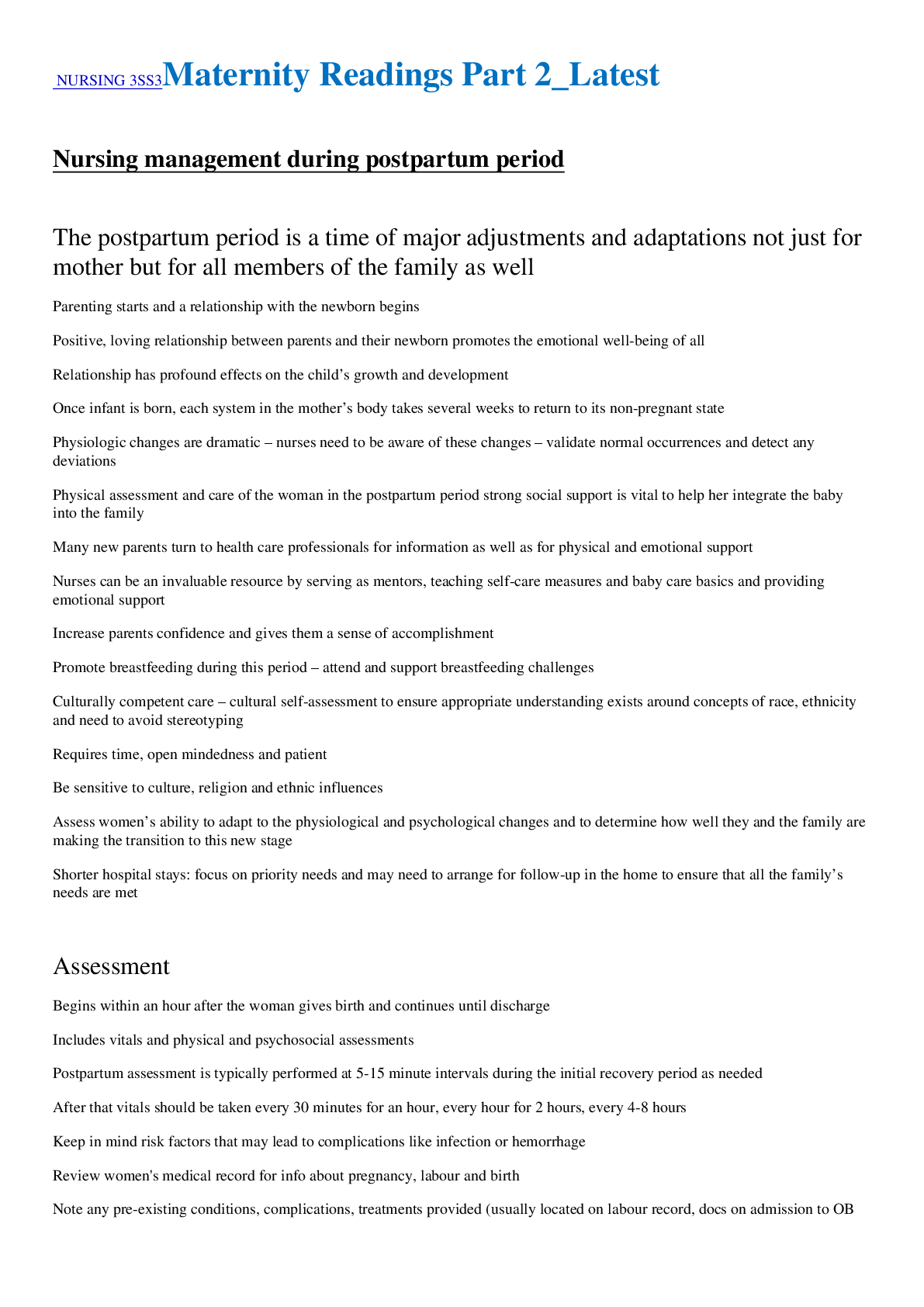
NURSING 3SS3Maternity Readings Part 2_Latest Nursing management during postpartum period The postpartum period is a time of major adjustments and adaptations not just for mother but for all mem... bers of the family as well - Parenting starts and a relationship with the newborn begins - Positive, loving relationship between parents and their newborn promotes the emotional well-being of all - Relationship has profound effects on the child’s growth and development - Once infant is born, each system in the mother’s body takes several weeks to return to its non-pregnant state - Physiologic changes are dramatic – nurses need to be aware of these changes – validate normal occurrences and detect any deviations - Physical assessment and care of the woman in the postpartum period strong social support is vital to help her integrate the baby into the family - Many new parents turn to health care professionals for information as well as for physical and emotional support - Nurses can be an invaluable resource by serving as mentors, teaching self-care measures and baby care basics and providing emotional support - Increase parents confidence and gives them a sense of accomplishment - Promote breastfeeding during this period – attend and support breastfeeding challenges - Culturally competent care – cultural self-assessment to ensure appropriate understanding exists around concepts of race, ethnicity and need to avoid stereotyping o Requires time, open mindedness and patient o Be sensitive to culture, religion and ethnic influences - Assess women’s ability to adapt to the physiological and psychological changes and to determine how well they and the family are making the transition to this new stage - Shorter hospital stays: focus on priority needs and may need to arrange for follow-up in the home to ensure that all the family’s needs are met Assessment - Begins within an hour after the woman gives birth and continues until discharge - Includes vitals and physical and psychosocial assessments - Postpartum assessment is typically performed at 5-15 minute intervals during the initial recovery period as needed - After that vitals should be taken every 30 minutes for an hour, every hour for 2 hours, every 4-8 hours - Keep in mind risk factors that may lead to complications like infection or hemorrhage - Review women's medical record for info about pregnancy, labour and birth - Note any pre-existing conditions, complications, treatments provided (usually located on labour record, docs on admission to OB unit or antenatal record) - Postpartum assessment (PPA): VS, pain, review of systems, BUBBLE-EE, parent & family assessment (attachment and bonding with newborn) - Be alert for danger signs – see pages 469 boxes Vital signs - Obtain vitals and compare to previous for deviations; can be an early indicator of complications - Temperature o Use a consistent measurement technique to get accurate readings o Typically,within normal range but some women experience fever as a result of exertion from labour and possible dehydration o Should be normal after 24h with replacement of fluids lost during labor, if not may be indicative of infection o Temp over 38 at any time may indicate infection - Pulse o Because of changes in blood volume and CO after delivery, bradycardia may be noted; may range from 50-70 bpm as a decreased CO takes hold following the expulsion of the placenta and the body adjusts to decreased circulating blood volume and increased SV o Usually stabilizes to Pre-pregnancy levels within 10d o Tachycardia may suggest anxiety, excitement, fatigue, pain, excessive blood loss, infection of underlying cardiac problems - Respirations o Should be within normal range (16-20) o Change may indicate resp infection, pulmonary edema, atelectasis or pulmonary embolism; lungs should be clear - Blood pressure o Elevations might suggest preg induced hypertension (DBP 90-95) o Dec may suggest dehydration or excessive blood loss o Assess in the same position every time, be alert for orthostatic hypotension (especially sig after childbirth d/t changes in body fluid volume and blood loss) o Mobilize women with assistance of nurse in case of fainting - Pain - 5th VS o Type, location, severity, 0-10, the goal is to have pain fall into 0-2 range at all times especially after breastfeeding o Warm showers, deep breathing, reassurance, and a calm environment can also be helpful o Pre-medicate women for afterbirth pains rather than wait for her to experience them – ask about pain freq o If there is severe pain in the perineal region despite use of physical comfort measures, check for hematoma by inspecting and palpating; if found, notify HCP Breasts - Inspect size, contour, asymmetry, engorgement, erythema - Check nipples for cracks, redness, fissures, or bleeding (indications that the baby is not latching properly – not positioned properly on breast) - Palpate to see if they are soft, engorged (hard, tender, taught), filling (firmer) - Palpate for nodules, masses, areas of warmth o May indicate plugged duct that can lead to mastitis - Discharge from the nipple (colostrum: creamy yellow; foremilk: bluish white) - Inquire about nipple discomfort - Nipples erect, flat or inverted: Flat or inverted nipples can make breastfeeding a challenge - For women who are not breastfeeding using a gentle, light touch to avoid stimulation of milk production Uterus - Assess fundal height (top of uterus) to determine the degree of uterine involution; use a two-handed approach, supine position, bed flat and palpate abdo gently, feeling for top of uterus, other hand is placed on lower segment of stomach to prevent uterine prolapse - Once fundus is located, place index finger on fundus and count the # of finger widths between the fundus and umbilicus; - Fundus is typically between the umbilicus and symphysis pubis 1-2h after birth; normally progresses downwards at a rate of 1 finger width/day after birth; on 1st day postpartum top of fundus should be located 1 finger below the umbilicus (recorded as U-1) o Second day – 2cm under belly button – U2 o 6-12 hours –usually fundus is at the level of the belly button - Fundus should be midline and firm - Boggy/relaxed uterus is a sign of uterine atony, can be the result of bladder distention which displaces the uterus up and to the right or retained placental fragments and both predispose women to hemorrhage - If not firm, gently massage using a circular motion until it becomes firm - Have woman empty bladder before fundal assessment - Get women to pre-medicate herself before palpation of fundus Bladder - Considerable diuresis (AKA “puerperal” diuresis) is as much as 3000 mL and may flow for several days after birth as mom’s body adjusts its fluid balance to pre-preg state o Begins in first 12-24h - Many postpartum women do not sense need to void even if the bladder is full - Risk for bladder distention and difficulty voiding if received regional anesthesia until sensation returns within several hours after - May have trouble voiding d/t trauma during birth (especially if the baby was large, forceps were used, or there was a prolonged second stage of labour) - Urethral edema should be considered - Be alert for signs of infection: infrequent or insufficient voiding, fever, chills, nausea, discomfort, burning, urgency, foul-smelling urine - Assess for distention after voiding and adequate emptying after attempts to void - Palpate over symphysis pubis (should not be palpable if empty – rounded mass suggests bladder distension - Full bladder dull to percussion - If bladder is full, lochia drainage will be more than normal d/t inability of uterus to contract to suppress bleeding - Review ins and outs including blood loss to evaluate for dehydration - After woman voids palpate and percuss area to determine adequate emptying o If still distended women ma be retaining urine in bladder – may need catheter - Ask women: gone to b-room yet? Difficulty urinating? Burning or discomfort? Feel bladder is empty? Signs of infection? Can you squeeze your muscle to control flow? Do you leak urine when you cough laugh or sneeze? Bowels - BMs may not occur for 2-3d after birth d/t decrease in muscle tone in the intestines or because of bowel excavation during delivery - Inspect abdomen for distention, auscultate for bowel sounds, palpate for tenderness - Abdomen should be soft, non-tender, non-distended and have bowel sounds present - Ask about flatus and BM status (common problem especially after c/s) - Women who have lacerations or an episiotomy or c/s may be reluctant to bear down to achieve a BM; may benefit from stool softeners Lochia - Amount, colour, odour, and change with activity and time, presence of clots - Ask how many pads used in past 1-2h and how much drainage was on each (saturated completely, half the pad, etc?) - Pads can be weighed, 1g = 1mL of blood - Amount decreases daily - Lochia typically has a musky scent like period blood with no large clots - Foul smelling – suggest infections and large clots suggest poor uterine involution - Flow will inc when woman stands up from sitting (blood pools in vagina and uterus while laying) and when breastfeeding (oxytocin release causes uterine contraction) - Scant: 2.5-5 cm lochia stain on pad, Light/small: approx. 10cm, Moderate: 10-15cm, Large/heavy: saturated within 1h - Total vol of lochia about 240-270ml (500-1000ml constitutes a hemorrhage) - Report heavy, bright red, foul-smelling, large tissue fragments - If heavy bleeding occurs, massage fundus until it is firm to reduce flow of blood - Women who have a c/s have less lochia than those who delivered vaginally but stages and colour changes remain the same o Higher risk of hemorrhage in c/s pts – assess for excessive bleeding o Palpate fundus and assess lochia to ensure they are in normal range - On discharge: info about lochia and expected changes o Red lochia returns after serosa – indicates sub involution or women is to active and needs to rest more - Lochia excellent medium for bacterial growth – frequent pad changes, use peri bottle to rinse peri area, handwash before and after pad changes Episiotomy and perineum - Position women on side with top leg flexed upward at the knee o May need penlight for lighting o Lift upper buttock o Inspect episiotomy for irritations, ecchymosis, tenderness or hematomas and hemorrhoids - Early on peri tissue is swollen and slightly bruised but the episiotomy site should not have redness, discharge or edema - Majority of healing takes place in first 2wks, may take 4-6mo to heal completely - Assess episiotomy and lacerations q8h - Lacerations acquired during birth are classified as: 1st degree (involves only skin and superficial structures above muscle), 2nd degree (extends through perineal muscles), 3rd degree (extends through anal sphincter), and 4th degree (continues through anterior rectal wall) - Pelvic or vulvar hematomas: bluish skin, large swollen areas, severe pain - Infection: redness, swelling, increasing discomfort or purulent drainage - White line the length of the episiotomy is a sign of infection same with swelling or discharge - Peri hematoma: ecchymosis, peri discoloration and sever intractable pain - Ice can be applied for pain and edema, sitz baths to promote comfort and peri healing Extremities - Mother is in a hypercoagulable state during pregnancy and this protects against excessive blood loss during childbirth and placental separation - Increase risk of DVT, pulmonary embolism (blood clot from extremities goes to pul artery and obstructs blood flow to lungs) - May report lower extremity tightness or aching when ambulating that is alleviated with rest and elevation of the leg, edema in the affected leg, warmth, tenderness - Diagnosis: client history, phys exam, venous ultrasound - Those at risk should weight compression socks, encourage ambulation; may use heparin for DVT - Increase risk of clots: stasis (compression of large veins d/t pregnant uterus), altered coagulation, and localized vascular damage (may occur during birth) - Risk factors associated with thromboembolic conditions: anemia, DM, smoking, obesity, preeclampsia, HTN, varicose veins, pregnancy, OCs or hormone replacement, c/s, previous thromboembolic disease, multiparty (i.e. twins), inactivity, late maternal age (35+) Emotional status - How mom interacts with family, her level of independence, energy levels, eye contact with infant, posture and comfort level while holding the newborn and sleep and rest patterns - Be alert for mood swings, irritability or crying episodes Bonding and attachment - mother from different cultures may behave differently from what is expected in own culture; don’t assume different behaviour is wrong - Bonding is the close emotional attraction to a newborn by parents that optimally develops during the first 30-60mins after birth; unidirectional from parent to infant; requires period of close contact; skin-to-skin is encouraged soon after birth Nursing management of labor and birth at risk - See pages 126-133 - Many complications occur with little or no warning ad present challenges for the perinatal health care team and family - Nurses need to identify problem quickly and immediately intervene - Several conditions occurring during labor and birth that may increase risk for an adverse outcome for the mother and fetus - Procedures during birth for woman who develops a condition that increases her risk or that may be needed to reduce the woman’s risk for developing a condition o Promote optimal maternal and fetal outcomes Dystocia - Abnormal or difficult labor is influenced by many mom and fetus factors - Progress of active labor deviates from normal – slow and abnormal progression - Occurs in about 10% of hospital deliveries - Labour: starts with regular uterine contractions that are strong enough to result in cervical effacement and dilation o Early in labor uterine contractions are irregular and cervical effacement and dilation occur gradually o Dilation reaches 4cm and contractions become more powerful active labor - Dystocia cannot be predicted or diagnosed so we use the term failure to progress - Lack of progressive cervical dilation (over 4 hours with less than .5 cm dilation) and lack of descent of fetal head (over 1 hour of pushing with no descent) - Adequate trail of labor is needed to say failure to progress - Can be prevented through prenatal education, allowing for spont onset of labor and providing continuous labor support, free movement in labor, no routine interventions and appropriate non pharm and pharm pain relief measures - Minimize risk to mom and fetus are the goals - Risk factors: epidural, excessive analgesia, multiple gestation, maternal exhaustion, ineffective pushing, occiput posterior position, unripe cervix with induction, first birth, mom short stature, big baby, obesity of mom, fetal malpresentation, mom over 35, more than 41 weeks pregnant, ineffective uterine contraction, high fetal station Problems with powers - Expulsive forces of the uterus becomes dysfunctional - Hypertonic uterine dysfunction (hypertonic contractions) o Uterus may never fully relax between contractions o Contractions are erratic and poorly coordinated because more than one uterine pacemaker is sending signals for contraction o Placental perfusion becomes compromised – reducing O2 to the fetus o Exhaust mom – frequent, intense, painful contractions with little progression o Placing the fetus in jeopardy or relax too much - Hypotonic uterine dysfunction (hypotonic contractions) o Causing ineffective contractions o During active labor when contractions becomes poor in quality and lacks sufficient intensity to dilate and efface the cervix o Risk factors: over distension, malposition of fetus, excessive analgesia o Major risk is hemorrhage after giving birth because uterus cannot contract effectively to compress blood vessels - Precipitous labor o Uterus contracts so frequently and with so much intensity that a very rapid birth will take place o Completed in less than three hours o Women typically have soft peri tissues that stretch readily permitting fetus to pass through the pelvis quickly and easily o Maternal complications are rare if maternal pelvis is adequate and the soft tissues yield to a fast-fetal descent, but peri lacerations and hemorrhage can occur o Fetal complications: head trauma like intracranial hemorrhage or nerve damage and hypoxia due to rapid progression of labor Problems with the Passenger - Any presentation other than occiput anterior or a slight variation of the fetal position or size increases the probability of dystocia o Can affect contractions or fetal descent through maternal pelvis - Problems: occiput posterior position, breech presentation, multifetal pregnancy, excessive size as it relates to cephalopelvic disproportion and structural anomalies - Persistent occiput posterior is one of the most common malposition’s o Slightly larger diameters to the maternal pelvis slowing fetal descent o Fetal head not flexed enough may be responsible o Poor uterine contractions may not push fetal down into pelvic floor to extent that the fetal occiput sinks into it rather than being pushes to rotate in an anterior direction o More painful labor, prolonged labor, and dystocia - Face and brow presentations are rare and associated with fetal malformation - Breech in 3-4% is frequently associated with high parity with uterine relaxation, previous breech delivery, uterine anomalies, multi-fetal pregnancies, placenta previa, preterm births, fetal anomalies like hydrocephaly o Perinatal mortality is increased wit breech, regardless of delivery method o Offer cephalic version – attempt to turn fetus to cephalic presentation and is performed under carefully controlled clinical conditions - Shoulder dystocia—obstruction of fetal descent and birth by the axis of the fetal shoulders after the head has been delivered o Failure of shoulders to deliver spontaneously places both the woman and the fetus at risk for injury o Postpartum hemorrhage secondary to uterine atony or vag lacerations is the major complication to the mother o Brachial plexus palsies and clavicular or humeral fractures are most common fetal injuries o Failure to deliver whole body within 6 minutes has been shown to increase the incidence of acidosis, asphyxia, perm CNS impairment, and death o Risks: history of shoulder dystocia, macrosomia, maternal diabetes, excessive weight gain, maternal obesity, post-term preg, fast 2nd stage, operative delivery with vacuum or forceps, and long second stage o McRobert’s manoeuvre or suprapubic pressure can reduce severity of injuries to the mother and newborn - Multiple gestations – twins, triplets, or more infants in a single preg (page 677 box) o Incidence is increasing due to the result of infertility treatment and an increased number of women giving birth at an older age o Most common complication is hemorrhage resulting from uterine atony - Excessive fetal size and abnormalities can also contribute to labor and birth dysfunctions o Macrosomia: newborn weights more than 4000g (over 8 pounds) o 10% of all preg higher incidence in aboriginal women o Fetal abnormalities: ascites, large mass on head or neck o Complications: dysfunctional labor, increased incidence of instrumental delivery and C-section, increased hospital stay, feto-pelvic disproportion, soft tissue laceration, fetal injuries or fractures, asphyxia, lower Apgar scores, NICU Problems with the Passageway - Pelvis and birth canal - Contraction of one or more of the three planes of the maternal pelvis: inlet, midpelvis and outlet - Midpelvis more common than inlet and typically causes an arrest of fetal descent - Obstructions in maternal birth canal such as swelling of the soft maternal tissue and cervix – termed soft tissue dystocia can hamper fetal descent and impede labour progression outside moms bony pelvis Problems with the Psyche - Array of emotions: fear anxiety, helplessness, being alone and weariness - Psychosocial stress can cause dystocia Nursing Assessment - Review client’s history to look for risk factors for dystocia - Also include the mother’s frame of mind like fear, anxiety, stress, lack of support, and pain which can interfere with uterine contractions and impede labor progress - Help woman relax – promote normal labor progress - Assess vitals – elevations in temp (infection), changes in HR or BP (hypovolemia) - Evaluate uterine contractions for freq and intensity – ask about changes in contraction pattern like an increase or decrease in freq and intensity - Assess fetal heart rate and pattern - Assess fetal position to identify deviations - Vag exam to determine cervical dilation, effacement, and engagement of the fetal presenting part - Evaluate for evidence of membrane rupture and report malodorous fluid Nursing Management - Patience, physical and emotional support - Final outcome: depends on size and shape of maternal pelvis, quality of uterine contractions, and size presentation and position of the fetus - Dystocia – diagnosed after labor has progressed for a time - Promoting the progress of labor o Nurse needs to help determine the progress of labor o Continue to assess the woman, frequently monitor cervical dilation and effacement, uterine contractions, and fetal descent and document this o Eval. progress in active labor by using a partogram o Membranes rupture – observe for color, odour and visible cord prolapse o Assess fetal well-being and document FHR o Assess woman’s fluid balance – check skin turgor and mucous membranes, monitor ins and outs o Monitor bladder for distension every 2 hours an encourage her to empty bladder often and monitor bowel status (full bladder/rectum can impede descent) o Fetus in breech or presenting part too high – look for cord prolapse and decelerations in heart rate o Be prepared to administer a labor stimulant like oxytocin if ordered to treat hypotonic labor contractions o Anticipate need to assist with manipulations if shoulder dystocia is diagnosed o Prepare women for possibility of a c-section - Providing physical and emotional comfort o Promotes relaxation and reduces stress o Warm blankets and a backrub to reduce muscle tension o Environment conductive to rest – women to conserve energy lower lights and reduce external noise o relaxation – warm shower or bath o pillows to support woman in a comfy position, change position every 30 minutes to reduce tension and enhance uterine activity and efficiency o offer fluids/food to moisten mouth and replenish energy o encourage mom to ambulate or assume diff positions to promote fetal rotation o upright positions – facilitate fetal rotations and descent o encourage woman to visualize descent and birth of the fetus o occiput post position: backrub, provide counter-pressure o assess pain and degree of distress, administer analgesics PRN o eval level of moms fatigue like verbal expression of feeling exhausted, inability to cope in early labor or inability to rest or calm down between contractions o praise women for her efforts and provide empathetic listening to increase clients coping ability - Promoting empowerment o Educate client an family about dysfunctional labor, explain interventions, encourage them to participate in decision making about interventions o Let them express their fears and anxieties, provide encouragement, support in coping efforts, inform them of progress and advocate for them Preterm labor - Occurrence of reg uterine contractions accompanied by cerv effacement and dilation before the end of the 37th week of gestation - Contributing factors: infection, prior preterm birth, periodontal disease, genetic influence, working during preg, lifestyle like smoking illicit drug us inadequate weight gain anxiety or stress, and ethnicity (aboriginal women and Africans) - Preterm birth – biggest contributors to perinatal morbidity and mortality in the world - Late pre-term deliveries (between 34-37 weeks) occurring in level 3 centre have a 99-100% survival rate with infant appearing the same as a term baby o Obs and interventions to maintain thermoreg and glycemic control - One of the most common obstetric complications - Risk for resp distress syndrome, resp failure, CNS hemorrhage, infections, thermoreg problems which can lead to acidosis and weight loss, GI complications like necrotizing enterocolitis and feeding difficulties and long-term cognitive, visual, motor, hearing, growth and behavioural problems. - Leading cause of infant death in Canada Therapeutic management - Predict risk only valuable if available intervention - Factors that influence the decision to intervene: prob of progressive labor, gestational age, and risks of treatment (accurate dating of fetus is essential) - Treatment for preterm labor: tocolysis and varying degrees of activity restriction o Antibiotics to treat presumed or confirmed infections o Steroids to enhance fetal lung maturity between 24-34 weeks gestation Nursing assessment - History to determine the estimated date of birth - Many women are unsure of the date of their LMP so the date given may be unreliable - Many dates are still misdated – accurate gestational dating is essential - Antepartum assessment for a post-term preg typically includes daily fetal movement counts done by the woman, non-stress tests twice weekly, amniotic fluid assessments and weekly cervical exams to evaluate for ripening - Assess client’s understanding of the various fetal well-being tests, clients stress and anxiety, clients coping ability and support network Nursing management - Once postdate status is confirmed, monitoring fetal well-being becomes critical - First decision is whether to deliver baby or wait - Wait – fetal surveillance is key - If decision is to have the woman deliver, labor induction is initiated Providing support - Intense surveillance is time consuming and intrusive adding to anxiety and worry - Let women discuss her feelings and provide reassurance - Validate woman’s stressful state Educating the woman and her partner - Teach them about testing required and reasons for these tests - Describe methods for cervical ripening if indicated - Explain the possibility of induction if labor isn’t spontaneous or if a dysfunctional labor pattern occurs - Prepare for c/s if fetal distress occurs Providing care during the intrapartum period - Continuously assess and monitor FHR to identify potential fetal compromise early – atypical or abnormal FHR - Monitor woman’s hydration status to ensure max placental perfusion - Membranes rupture: assess amniotic fluid characteristics – color amount and odor to identify previous hypoxia and prepare for prevention of meconium aspiration - Report meconium stained amniotic fluid immediately - Anticipate need for amnioinfusion to minimize risk for meconium aspiration by diluting meconium in the amniotic fluid expelled by hypoxic fetus - Monitor labor pattern – dysfunctional patterns are common - Answer questions, provide support, presence, information and encouragement Women requiring labor induction and augmentation - Many women need help to initiate or sustain the labor process - Labor induction involves the stimulation of uterine contractions by medical or surgical means to produce delivery before the onset of spontaneous labor - Medical induction of labor increases the risk of c/s - Labor induction isn’t an isolated event – brings about a cascade of other interventions that may or may not produce a favorable outcome o IV therapy, fetal monitoring, sig discomfort from stimulating uterine contractions, increased use of analgesia and prolonged stay on labor unit - Labor augmentation enhances ineffective contractions after labor has begun – continue fetal monitoring (FHR) - Multiple reasons for inducing labor – most common being posterm gestation o Pre-labor rupture of membranes, hypertensive disorders, renal disease, chorioamnionitis, intrauterine fetal demise, and pre-existing diabetes - Contraindications to labor induction the same as contraindications to labor and vag delivery and may include complete placental previa, transverse fetal lie, prolapsed umbilical cord, prior classic uterine incision that entered the uterine cavity, previous myomectomy, previous uterine rupture, vasa previa, invasive cervical cancer, active genital herpes infection and abnormal FHR pattern - Labor induction is indicated when the benefits of birth outweigh the risks to the mom or fetus for continuing the pregnancy Therapeutic management - Decision to induce labor based on a thorough eval of maternal and fetal status - Assessment to evaluate fetal size, position, and gestational age, non-stress test to evaluate fetal well-being, nitrazine paper and/or fern test to confirm ruptured membranes, CBC, vaginal exam to evaluate cervix for inducibility - Accurate dating very important to prevent a preterm birth - Placental perfusion decreases as placenta ages and becomes less efficient at delivering oxygen and nutrients to the fetus - Amniotic fluid volume begins to decline by 40 weeks gestations, increasing fetus risk for oligohydramnios, meconium aspiration and cord compression Cervical ripening - Cervix is unfavorable or unripe, a successful vaginal birth is less likely - Ripe cervix is shortened, centered, softened and partially dilated - Unripe cervix is long, closed, posterior, and firm - Cervical ripening usually begins prior to onset of labor contractions and is necessary for cervical dilation and the passage of the fetus - Bishop score commonly used today (page 140) and helps identify women who would be most likely to achieve a successful induction o Duration of labor inversely correlated with the bishop score o Score of 8 indicates a successful vaginal birth o Less than 6 – cervical ripening method should be used before induction - Non-pharm methods: less freq used o Herbal agents, castor oil, hot baths, enemas o Sex and breast stimulation – promote release of oxytocin which stimulates uterine contractions – not validated tho o Human semen bio source of prostaglandins - Mechanical methods o Used to open cervix and stimulate the progression of labor o Application of local pressure stimulates the release of prostaglandins to ripen the cervix o Compared to pharm methods – simplicity or preservation of the cervical tissue or structure, lower cost and fewer side effects o Risks: infection, bleeding, membrane rupture, and placental disruption o Indwelling catheter can be inserted into endocervical canal to ripen and dilate the cervix Direct pressure stimulates the release of prostaglandins o Hygroscopic dilators like luminaria (dried seaweed) absorb endocervical and local tissue fluids They enlarge and expand endocervix and provide controlled mechanic pressure Absorption of water – expansion of dilators and opening of the cervix - Surgical methods o Stripping of membranes and performing an amniotomy o Stripping of membranes: inserting a finger through the internal cervical os and moving it in a circular direction which causes membrane to detach o Manual separation of the amniotic membranes from the cervix is thought to induce cervical ripening and the onset of labor o An amniotomy involves inserting a cervical hook through the cervical os to deliberately rupture the membranes which promotes pressure of the presenting part on the cervix and stimulates an increase in the activity of prostaglandins o Risks associated with these include umbilical cord prolapse or compression, maternal or neonatal infection, FHR deceleration, bleeding and client discomfort o Both kinds: amniotic fluid characteristics – whether it is clear, bloody or meconium is present and FHR pattern monitored closely - Pharm agents o use of prostaglandin to attain cervical ripening has been found to be highly effective in producing cervical changes independent of uterine contractions o some cases women will go into labor, requiring no additional stimulants for induction o offers advantage of promoting both cervical ripening and uterine contractility o limitation: ability to induce excessive uterine contractions which can increase maternal and perinatal morbidity o prostaglandin analogues like dinoprostone and misoprostol o misoprostol a synthetic PGE1 analogue is a gastric cytoprotective agent used in the treatment and prevention of peptic ulcers o intravaginally or orally to ripen cervix or induce labor o Misoprostal not used in Canada with a live fetus o Contraindicated for women with prior uterine scars and shouldn’t be used for cervical ripening in women attempting a VBAC Oxytocin - ¬potent endogenous uterotonic agent used for both artificial induction and augmentation of labor and is most common induction agent used worldwide - produced naturally by posterior pituitary gland and stimulates contractions of the uterus - low bishop scores cervical ripening is initiated before oxytocin is administered - once cervix is ripe, oxytocin is the most popular pharm agent used for inducing or augmenting labor - unfavorable cervix may be admitted the evening before induction to ripen her cervix with one of the prostaglandin agents - induction begins with oxytocin the next morning if she hasn’t already gone into labor - response to oxytocin varies widely – some women are very sensitive to small amounts - most common adverse effect: uterine hyperstimulation, leading to fetal compromise and impaired oxygenation - the response of the uterus to the drug is closely monitored throughout labor so that the oxytocin infusion can be titrated - oxytocin also has an antidiuretic effect resulting in decreased uterine flow that may lead to water intoxication - symptoms to watch for include headache and vomiting - oxytocin is administered via IV infusion pump and piggybacked into main IV line - dose titrated – to achieve stable contractions every 2-3 minutes lasting 40-60 seconds - uterus should relax between contractions if not uteroplacental insufficiency and fetal hypoxia can result - super important to continually monitor FHR - oxytocin has many advantages – potent and easy to titrate, short half-life (3-10 min) and is generally well tolerated - side effects – water intoxication, hypotension and uterine hypertonicity - drug doesn’t cross placental barrier – no direct fetal problems Nursing assessment - thorough history and physical exam - review women’s history for relative indications for induction or augmentation like diabetes, hypertension, postern status, dysfunctional labor pattern, prolonged ruptured membranes and maternal or fetal infection - contraindications like placenta previa, over distended uterus, active genital herpes, fetopelvic disproportion, fetal malposition or severe fetal distress - assist with determining gestational age - assess fetal well-being - evaluate cervical status, including cervical dilation and effacement and station via vaginal exam and determine bishop score Nursing management - Explain induction/augmentation procedure clearly - Ensure there is informed consent Administering oxytocin - Prepare oxytocin infusion by diluting 10 units of oxytocin in 1000mL of RL - Use infusion pump on a secondary line connected to primary infusion - Start oxy infusion at initial dose is 1-2 - Anticipate increasing the rate in increments of 1-2 every 30 min to a max dose of 20 - Maintain the rate once the desired contraction freq has been reached - To ensure adequate maternal and fetal surveillance – 1:1 Nurse to client ratio - During induction or augmentation – monitoring fetal and moms status is essential - Obtain the mothers VS and the FHR every 15 minutes - Evaluate and doc contractions (freq, duration and intensity) and resting tone and adjust the oxytocin infusion rate accordingly - FHR: baseline rate, baseline variability and decelerations – to determine whether the oxytocin rate needs adjustment - Discontinue oxy if uterine hyperstimulation or an abnormal FHR pattern occurs - Vag exam to determine cervical dilation and fetal descent - Monitor FHR continuously and document it every 15 minutes during the active phase of labor and every 5 minutes during second stage and assist with pushing during 2nd stage - Ins and outs to prevent excess fluid volume and encourage to empty her bladder every 2 hours to prevent soft tissue obstruction Providing pain relief and support - Assess level of pain and ask freq to rate pain and provide pain management PRN - Offer position changes and non-pharm methods - Monitor her need for comfort as contractions inc - Freq assure women and partner about fetal status and labor progress - Freq updates, support and encouragement - Assess ability of mom to cope Intrauterine fetal demise - Unborn life suddenly ends with fetal loss - Sudden loss of expected child is tragic and the families’ grief can be very intense - Variety of mental health challenges - Fetal death can be due to numerous conditions such as infection, hypertension, advanced maternal age, maternal obesity, multiple pregnancy, diabetes, congenital anomalies, umbilical cord accident, placental abruption, hemorrhage or coagulopathies or it may go unexplained - Early pregnancy loss before 20 weeks may be through spontaneous abortion (miscarriage), an induced abortion (therapeutic abortion) or a ruptured ectopic pregnancy - Wide spectrum of feelings from relief to sadness to despair - Stillbirth can occur at any gestational age after 20 weeks and typically there is little or no warning other than reduced fetal movement - Period following a fetal death is extremely difficult for the family - Feelings of loss can be intense - Relationship can become trained and healing can become hampered unless appropriate interventions and support provided - Fetal death also affects the health care staff Nursing assessment - History and physical exam are of limited value in the diagnosis of fetal death since only history tends to be recent absence of fetal movement - Inability to obtain fetal heart sounds on exam suggests fetal demise but an ultrasound is necessary to confirm the absence of fetal cardiac activity - Once fetal demise is confirmed, induction of labor is indicated Nursing management - Give accurate understandable info to the family - Encourage discussion of the loss and venting of feelings of grief and guilt - Provide family with baby mementos and pictures to validate the reality of death - Allow unlimited time with stillborn to validate the death – see touch and hold the infant - Use appropriate touch such as holding a hand or touching a shoulder - Inform the chaplain or the religious leader of the families denomination about the death and request his or her presence - Assist parents with the funeral arrangements or disposition of the body - Provide families with brochures offering advice how to talk to other siblings about the loss - Refer the family to a local support group – lost infant through abortion, miscarriage, fetal death, stillbirth, or other tragic circumstances - Make community referrals to promote a continuum of care after discharge Women experiencing an obstetric emergency - Obstetric emergencies are challenging because of the increased risk or adverse outcomes to mom and fetus - Need quick clinical judgement and good critical decision making to increase positive outcomes for both mom and fetus Umbilical cord prolapses - Protrusion of the umbilical cord alongside or ahead of the presenting part of the fetus - Risk is increased further when the presenting part doesn’t fill lower uterine segment as is the case with incomplete breech presentations, premature infants and multiparous women - 50% perinatal mortality rate – one of the most catastrophic events - Patho: prolapse usually leads to total or partial occlusion of the cord o Fetus only lifeline so fetal perfusion decreases rapidly o Complete occlusion – fetus helpless and oxygen deprived o Fetus will die if cord compression isn’t relieved - Nursing assessment: prevention is key and we need to identify clients at risk for this condition o Cord prolapse is more common in preg involving mal-presentation, prematurity, ruptured membranes with a fetus at high station, polyhydramnios, grand multiparity, and multifetal gestation o Assess client and fetus often to detect changes and to eval effectiveness of any interventions performed - Nursing management o Prompt recognition of a prolapsed cord is essential to reduced the risk for fetal hypoxia o When membranes are artificially ruptured, assist with verifying that presenting part is well applied t the cervix and engaged into pelvis o If pressure or compression of the cord occurs assist with measure to relieve the compression o Sterile gloved hand into vagina and hold presenting part of umbilical cord until delivery o Change woman’s position to a modified Sims, Trendelenburg or knee-chest position also helps relieve cord pressure o Monitor FHR, bed rest and administer O2 as needed o If moms cervix isn’t fully dilated, prepare the woman for an EMRG c/s Placental abruption - Refers to premature separation of a normally implanted placenta from the maternal myometrium - Risk factors include hypertensive disorders, intrauterine growth restriction, prolonged rupture of membranes, chorioamnionitis, advanced maternal age, seizure activity, uterine rupture, trauma, smoking, cocaine use, coagulation defects, previous history of abruption, trauma (domestic violence) and placental pathology o These conditions may force blood into the underlayer of the placenta and cause it to detach - Management depends on the gestational age, the extent of the hemorrhage and maternal-fetal oxygenation perfusion - Treatment based on circumstances - Typically, once diagnosis is established the focus is on maintain the CV status of the mom and developing a plan to deliver the fetus quickly - A c/s if fetus is alive, vaginal if fetal demise Uterine Rupture - Catastrophic tearing of the uterine wall into the abdominal cavity - Its onset often marked by sudden fetal bradycardia - Treatment requires rapid surgery for good outcomes - From time of diagnosis to delivery, only 10-37 minutes are available before clinically sig fetal morbidity occurs - Can also result in catastrophic hemorrhage, fetal anoxia or both - Nursing assessment o Risk conditions such as previous uterine surgery (including c/s), prior rupture, trauma, prior invasive molar preg, history of placenta previa or increta, malpresentation, labour induction with excessive uterine stimulation, dystocia, over distended uterus (multiple gestation or polyhydramnios) and crack cocaine o First and most reliable symptom of uterine rupture is sudden fetal distress o Other signs: decreased baseline uterine pressure, loss of uterine contractility, ab pain with or without an epidural, hemorrhage, irregular ab wall contour, loss of station in the fetal presenting part, and hypovolemic shock in women fetus or both o Timely management of uterine rupture depends on prompt detection o Difficult to prevent uterine rupture or predict which women will experience it so we need to be constantly prepared o Screen all women with previous uterine surgical scars and continuous fetal monitoring is very important - Nursing management o Presenting signs may be nonspecific so initial management same as that for any other cause of acute fetal distress o Urgent delivery by c/s o Monitor maternal VS and observe for hypotension and tachycardia which might indicate hypovolemic shock o Insert a Foley if one isn’t in place already o Remain calm and provide reassurance o The life-threatening nature of uterine rupture is underscored by the fact that the maternal circ system deliveries about 500 mL of blood to the term uterus every minute o Maternal death a real possibility without rapid intervention o Newborn outcome after rupture depends largely on speed with which surgical rescue is carried out Amniotic fluid embolism - Bleeding that keeps going and bruising or petichieae appears – suspect DIC - AFE a rare and often fatal event characterized by the sudden onset of hypotension, hypoxia and coagulopathy - Amniotic fluid containing particles of debris like hair skin or meconium enters maternal circulation and obstructs pul vessels, causing resp distress and circ collapse - Caring for a woman receiving an amnioinfusion o Explain the need for the procedure, what it involves and how it may solve the problem o Inform mom that she’ll need to remain on bed rest during procedure o Assess moms VS and discomfort and ins and outs o Assess duration and intensity of uterine contractions freq to identify over distension or increased uterine tone o Monitor FHR pattern – improving fetal status o Prepare mom for a possible c/s if FHR doesn’t improve after procedure Forceps or vacuum assisted birth - Used to apply traction to the fetal head or to provide a method of rotating the fetal head during birth - Forceps are stainless steel instruments, similar to tongs with rounded edges that fit around fetus head - Outlet forceps are used when fetal head is crowning, and low forceps are used when fetal head is at +2 station or lower but not yet crowning - Forceps are applied to side of the fetal head - Forceps have a locking mechanism that prevents the blades from compressing the fetal skull - Vacuum extractor is a cup shaped instrument attached to a suction pump used for extraction of the fetal head - Suction cup is placed against the occiput of the fetal head. - Pump used to create negative pressure, then use traction until fetal head emerges from vagina - Indications for use of either method are similar: prolonged second stage of labor, abnormal FHR pattern, failure of the presenting part to fully rotate to the occiput anterior position, presumed fetal jeopardy or fetal compromise, maternal heart disease, maternal cerebrovasc malformations and maternal fatigue - Use of these things poses the risk for tissue trauma to mom and newborn - Maternal trauma may include lacerations of the cervix, vagina or perineum, hematoma, extension of the episiotomy incision into the anus, hemorrhage and infection - Potential newborn trauma: ecchymosis, facial and scalp lacerations, facial nerve injury, cephalhematoma, intracranial hemorrhage - Prevention key to reducing use of these techniques – freq changing clients position, encourage upright posture and ambulation, freq reminding client to empty bladder to allow max space for birth and provide adequate hydration - Assess moms VS, contraction pattern, fetal status and the maternal response to procedure(thoroughly examine procedure and rationale for use) - Reassure mom that marks or swelling on newborns head or face will disappear without treatment within 2-3 days - Observe for bleeding or infection related to genital lacerations C-section - Deliv of fetus through an incision in the abdomen and uterus - Classical vertical or low transverse incision (more common today) may be used - Number of c/s has risen steadily in Canada - Increase use: Widespread use of continuous electronic fetal monitoring which identifies fetal distress early, reduced number of forceps assisted birth, maternal obesity, older maternal age, reduced parity, more nulliparous women having infants, convenience to pt and doctor, elective repeat c/s, elective c/s for breech, attitudes of client, nurse and doctors about birth - c/s major surgical procedure with 4x increase in mortality vs vag birth - risk for complications: infection, hemorrhage, aspiration, venous thromboembolism, bowel and urinary tract trauma, feta injury, resp difficulty for baby - spinal/epidural or general anesthesia is used for c/s o epidural less risk and most women want to be awake and aware of birth Nursing assessment - history for indications associated with a c/s and complete a physical - any condition that obstructs or prevents safe passage of fetus through the birth canal or that seriously compromises maternal or fetal well-being may indicate c/s - ex: genital herpes, fetopelvic disproportion after a trial of labor, prolapsed umbilical cord, placental abnormality (placental previa), previous classic uterine incision or scar, HIV in women who haven’t had therapy or who choose an elective c/s and dystocia - fetal indications: malpresentation, congenital anomalies like neural tube defects and fetal compromise Nursing management - obtain diagnostic tests as ordered: CBC, blood type and cross-match so that blood is available for transfusion if needed and an ultrasound for fetal position and placental location - focus must remain on the women not on the equipment surrounding mom - provide education, use touch, eye contact, therapeutic communication and caring Providing pre-op care - varies depending on whether the c/s is planned or unplanned - major difference is time allotted for prep and teaching - in an unplanned c/s – quick measures to ensure best outcomes for mom and fetus - informed consent and allow for discussion of fears and expectations - provide teaching and explanations - assess understanding of surgery and reinforce reasons for surgery - explain what to expect post-op - pain management throughout procedure - ask woman about time she last had anything to eat or drink – time and what was consumed and assess mom and fetal status freq - pre-op teaching to reduce risk for post-op complications - demonstrate use of incentive spirometer and deep-breathing and leg exercises - instruct women on how to splint incision - complete pre-op procedures: prep surgical site as ordered, start IV infusion for fluid replacement therapy as ordered, insert a Foley and inform client about how long it will remain in place (24 hours usually), administer pre-op meds as ordered Providing post-op care - similar to care of those after a vag delivery - assess VS and lochia flow every 15 min for first hour 30 in the next hour and then 4 if stable - assist with perineal care and instruct client and inspect ab dressing and doc description - assess uterine tone to determine fundal firmness - check patency of IV lines ensuring its flowing at correct rate and inspect site for redness - assess LOC and ensure safety precautions until woman is fully alert and responsive - regional anesthetic was used monitor for the return of sensation to the legs - assess for evidence of ab distention and auscultate bowel sounds - assist with early ambulation to prevent resp and CV problems and promote peristalsis - monitor ins and outs at least every 4 hours at first and then every 8 hours - encourage women to cough, perform deep breathing exercises and use incentive spirometer every 2 hours - administer analgesics as ordered and provide comfort such as splinting the incision and pillow for positioning - assist client to move in bed and turn side to side to improve circ - encourage woman to ambulate to promote venous return from the extremities - encourage early touching and holding of the newborn to promote bonding - assist with breastfeeding initiation and offer support - suggest alternate positioning techniques to reduce incision discomfort while feeding - Verbalize feelings, assist with positive coping - Teach women about need for adequate rest, activity restrictions such as lifting and signs and symptoms of infection Vaginal birth after C-section - Women who have a c-section are candidates for vag birth and should be offered a trial of labor for subsequent preg - Choice of vag or repeat c/s can be offered to a woman who had a lower ab incision - Argument against this: risk for uterine rupture and hemorrhage -- rate of fetal mortality in the event of a uterine rupture is very high - Contraindications: prior classical uterine incision, prior hysterotomy or myomectomy, previous uterine rupture or another contraindication to labor - Most women do a trial of labor to see how they progress, performed in env capable of providing a continuous electronic fetal monitoring and c/s if needed - Prostaglandin E2 and misoprostol increases risk for uterine rupture and isn’t recommended for VBAC patients - Woman considering induction of labor after previous c/s needs to be informed of increased risk for uterine scar dehiscence with an induction than with spont labor - Women primary decision makers about birth method but need education of VBAC - Management similar to women experiencing labor - Consent – fully informed consent. Advise client of risks and benefits, understand ramifications of uterine rupture - Documentation - Surveillance: abnormal fetal monitor tracing in a woman undergoing a trial of labor after a c/s should alert nurse to the possibility of uterine rupture o Terminal bradycardia must be considered an EMRG and prepare for EMRG deliv - Readiness for EMRG: previous c/s the doctor, anesthesia provider and operating room team must be available – woman and fetus at risk otherwise - Be able to read fetal monitoring tracings and identify atypical or abnormal patterns [Show More]
Last updated: 2 years ago
Preview 1 out of 24 pages
Buy this document to get the full access instantly
Instant Download Access after purchase
Buy NowInstant download
We Accept:

Also available in bundle (1)

NURSING 3SS3 Maternity Readings Part 1& 2_Latest,100% CORRECT
NURSING 3SS3 Maternity Readings Part 1_Latest/NURSING 3SS3 Maternity Readings Part 2_Latest
By securegrades 4 years ago
$23.5
2
Reviews( 0 )
$16.00
Can't find what you want? Try our AI powered Search
Document information
Connected school, study & course
About the document
Uploaded On
Mar 10, 2021
Number of pages
24
Written in
Additional information
This document has been written for:
Uploaded
Mar 10, 2021
Downloads
0
Views
69

.png)


