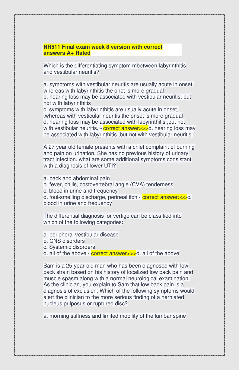
NR511-midterm-study-guide.docx
Dermatology
1. Actinic keratosis most common precancerous skin lesion in light skinned patients, more common in patients
50 years or older (most common in Celtic, Irish, and Scottish de
...
NR511-midterm-study-guide.docx
Dermatology
1. Actinic keratosis most common precancerous skin lesion in light skinned patients, more common in patients
50 years or older (most common in Celtic, Irish, and Scottish descent)
Found in sun exposed areas
Caused by skin cells that accumulate from repeated sun exposure
Pathophys: continued sun damage from UV radiation damages the DNA in epithelial cells
Primary lesions: macules or plaques, poorly circumscribed
Secondary lesion: erythematous and scaly
(May feel like sandpaper when touched)
Not an aggressive form of cancer if/when it changes to squamous cell unless on the lip
Patient complaints subjective: irritated, rough or scaly rash, pruritus, tenderness or stinging sensation
Objective findings: reddened, scaly, rough, or uneven surfaces. Hard or spiny lesion. Sandpaper like texture.
Diagnostic tests: fluorescence using photosensitizing drug (methyl ester of 5-aminolevulinic acid) over area of
concern will have a pink fluorescence with the wood’s lamp
Treatment: no evidence to support removal of lesion as most will not turn cancerous however it is standard to
REMOVE the lesion(s)
Topical Therapy:
5-fluorouracil (5-FU) cream (Efudex, Carac) applied in a thin layer over the lesion BID for 3 weeks, avoid
eyelids, lips, and folds of the nose. This treatment causes red, raw, and painful skin in the areas applied
which may lead to noncompliance. Exposure to sunlight makes this worse
Imiquimod 5% cream used for face and scalp lesions. Applied 3x weekly for 8 weeks.
Diclofenac 3% in 2.5% hyaluranon gell (Solaraza) applied BIF for 60 to 90 days
Adapalene 0.1%to 0.3% (topical retinoid)applied daily for 4 weeks and then increased to BID
Side effects of these treatments include redness, itching, rash, and dry skin
Topical chemotherapy combined with phototherapy with blue or red wavelength have better cosmetic
results than cryosurgery. 2 day course
Cryosurgery tissue is destroyed by freezing using liquid nitrogen. Hypopigmentation may occur at site of previous
lesion
Surgical curettage or shave excision are not considered first line treatments for actinic keratosis
Surgical biopsy is the only way to obtain an intact sample to be analyzed as a way to confirm diagnosis
If treatment does not work no matter the choice always refer to dermatologist
Education is centered around prevention, avoidance of excessive sun exposure, use of protective clothing, and use
of sunscreen.
Should teach patients ABCDE mnemonic
A= asymmetry
B= border irregularity
C= color change
D= Diameter larger than a pencil eraser
E= elevation from a flat lesion to a raised or evolving lesion
2. Dermatitis
DERMATITIS
ATOPIC DERMATITIS
• Atopic dermatitis (eczema) is not considered a distinct disease entity but is a descriptive term for a group of skin
disorders characterized by pruritus and inflammation, whose distinct cause is unknown. • Eczemais a more
general term that is often used collectively to describe skin of an erythematous and inflamed appearance,
reflective of a superficial pathological process. Currently, the terms eczema and dermatitisare often used
synonymously in the clinical arena in a nonspecific sense.
• The use of the term eczematous rash, although also indistinct, may be helpful both diagnostically and
therapeutically, because eczematous dermatitis may be classified into two major etiological categories—contact
dermatitis and atopic dermatitis.
• Early in its presentation, atopic dermatitis is erythematous in appearance, with papulovesicular lesions that ooze
and crust. At its later stages, the rash becomes a red-purple color, dries, and develops scaling and lichenification,
which is exacerbated by itching resulting from its highly pruritic nature.
Epidemiology and Causes
• Atopic dermatitis is a constitutional and inherited reaction, which usually begins in infancy. • Children born to
older women are more likely to develop eczema than children born to younger women.
• Prevalence of atopic disease is now estimated at 1 in 18 or 5.5%, which amounts to 15 million people in the United
States. About 10% of the U.S. population will have atopic dermatitis at some point in their lifetime.
• Atopic dermatitis presents more severely in childhood. Onset during the first year of life occurs in up to 50% of all
patients; in 85%, onset is before age 5 years. Up to 5% of all children are affected by atopic dermatitis. Most cases
(40%) resolve by adulthood, however. The remainder of patients with atopic dermatitis are affected with a chronic
course of the disease that is characterized by acute exacerbation
(often during times of stress) and intermittent remissions.
• No ethnic predisposition has been found for atopic dermatitis, and it occurs equally in both sexes. • The
cause of atopic dermatitis is unknown.
• Family history is positive for atopy in two-thirds of all cases. Genetic predisposition may be the most important
etiological factor in all-atopic conditions. A personal or family history of all or part of the
“atopic triad”—asthma, allergic rhinitis, and eczema—is often present.
• It has been proposed that individuals with any of these three conditions have preferential production of allergen-
specific immunoglobulin E (IgE) and that the presence of such antibodies should be a mandatory criterion for the
diagnosis of atopic dermatitis. Such a diagnostic test, however, only establishes the diagnosis of atopic syndrome,
not atopic dermatitis. Any patient with a history of hives
(urticaria), hay fever, or rashes should be considered to have an atopic history.
• All atopic individuals seem to have itchier skin, yet what seems to be unique about the atopic patient's skin is its
hypersensitivity.
• Many factors that do not make non-atopic individuals itch will make the atopic person feel itchy. Atopic patients are
known to itch seconds after experiencing a stressful event. This type of reaction is thought to be caused by
neuropeptide-induced vasodilation, which produces a rise in skin temperature and erythema.
• Symptoms are triggered or exacerbated through the interaction between genetic predisposition and environmental
factors. Environmental factors that trigger atopic dermatitis include dust mites, animal dander, pollen, microbes,
pollutants, climate, and emotional stress.
• Excessively hot or cold climates or excessively dry or moist environments are particularly suitable for setting the
stage for the atopic process. Anything that dries the skin can aggravate symptoms: • Common triggers include
excessive bathing, hand washing, lip licking, sweating, or swimming. • Contact with irritants such as solvents,
detergents, deodorants, tobacco, cosmetics, soap, and woolen and synthetic fabrics can precipitate an
exacerbation of atopic dermatitis. Heat and sweat may also be aggravating factors for atopic dermatitis.
• Factors that generate an increase in body temperature include hot showers or baths, overdressing, use of heating
pads, and electric blankets.
• Patients with atopy are intolerant of heat, have difficulty with thermal sweating, and are more likely to develop
heat exhaustion. It is thought that perspiration retention might be a complicating factor in atopic patients.
• Excessive humidity is, therefore, a problem, because it interferes with normal evaporation of sweat from the body.
Improperly fitting clothes can create friction and irritate the skin, and contact with certain fabrics, most notably
wool, can precipitate a flare-up. Other skin conditions or infections can also lead to an exacerbation of atopic
dermatitis (eczema).
Pathophysiology
The inflammatory process in eczema causes erythema of the skin as a result of dilated blood vessels that are
surrounded by inflammatory cells that migrate into the epidermis, resulting in edema both inside and in between
the epidermal cells (spongiosis). The epidermal cells malfunction as a consequence, resulting in thickening of the
epidermis (acanthosis), excess production of keratin, and scaling. The outer epidermal layer of the skin, the stratum
corneum, normally forms an impermeable barrier that protects the living cells beneath from environmental
irritants and toxins. In atopic dermatitis, this outer barrier is impaired. There is an increase in the water loss and a
decrease in water binding, which leads to a brittle outer barrier. This condition is made worse by environmental
factors such as physical trauma from scratching, cycles of wetting and drying, and the chemical erosion that is
caused by detergents and solvents. In addition, superinfection of eczematous skin by bacterial (e.g., Staphylococcus
aureus) or fungal (e.g., Malassezia furfur) species and irritation from dust mites and their dung is an important
factor that worsens atopic dermatitis by potentiating the immune response. Superinfection is also much more likely
in atopic dermatitis than in other forms of dermatitis such as psoriasis. Thus, infection may be thought of as both a
trigger and a complication of atopic dermatitis.
Immunological abnormalities are key to the pathophysiology of the atopic response. These abnormalities can
include elevated serum IgE levels, which are seen in 85% of affected individuals; hypereosinophilia; reduced cell-
mediated immunity and antibody-dependent cellular cytotoxicity; slowed chemotaxis of neutrophils and
monocytes; relative increase in the number of CD4-positive (CD4+) Th2 helper T cells that secrete interleukin-4 (IL-
4); and a decrease in CD4+ T helper cells that secrete interleukin-2 (IL-2). Interestingly, however, in later stages of
the immune reaction, Th1 helper T-cell activity, which enhances cell-mediated immunity, appears to play an
increasing role. Th17 cells and their associated cytokines have also been implicated in this disease process,
including in the protection against infection/colonization with superficial skin fungi and bacteria (e.g.,
Staphylococcus) containing superantigens that are thought to trigger dysregulated immune responses, resulting in
eczematous lesions. However, reports in the literature are conflicting and have implicated Th17 cells in both pro-
inflammatory and anti-inflammatory roles. Impairment of essential fatty acid metabolism has also been implicated
as a causative factor of atopy.
Clinical Presentation Subjective
• Atopic dermatitis is characterized by an extremely low threshold for pruritus and has been referred to as
“the itch that rashes.” Almost always, the itch occurs before the rash appears, and scratching the rash only
worsens it clinically.
• The cardinal sign of atopic dermatitis is severe pruritus, which is often extremely distressing in both the
acute and chronic stages. In turn, the diagnosis of atopic dermatitis cannot be made without a history of
pruritus, and if pruritus is absent, alternate diagnoses should be sought.
• The patient may report a personal or family history of other atopic conditions (asthma, allergic rhinitis).
• The patient usually reports a history of episodic exacerbation of similar symptoms or of a childhood rash
or eczema.
• The clinician should inquire about any exposure to known or unknown common antigens and irritants,
regardless of the history. Individuals with atopic dermatitis are not immune to contact dermatitis; in fact,
they are more susceptible to irritant reactions because of their impaired epidermal barrier layer. Often,
the rash is reported as better in the warmer months and worse in the fall and winter. Objective
• Atopic dermatitis usually begins as infantile eczema, with lesions affecting the cheeks, face, and upper
extremities. Erythema is often seen before pruritus.
• The acute lesions are often excoriated, maculopapular, and inflamed. In infancy and early childhood,
oozing and crusting usually characterize the erythema. As the child becomes older, the disease can go into
remission or change to a flexural distribution (antecubital fossae and neck area).
Flexural eczema usually lasts until about age 4 to 10 years but may continue into adulthood.
• In adults, eczema presents with symmetrical lesions that are crusting and excoriated. I
• n the early stages, lesions may be erythematous, papulovesicular, edematous, and weeping. Later the rash
becomes crusted, scaly, thickened, and lichenified. Intergluteal involvement is uncommon and should raise
suspicion of another diagnosis.
• The classic locations for lesions are noted to correspond to areas that are most accessible to rubbing and
scratching. In addition, the typical flexural sites are more susceptible because they are areas that are more
likely to be hot and moist.
Diagnostic Tests
• Laboratory tests are usually not useful in the diagnosis of atopic dermatitis, but they can be helpful in
ruling out other disorders or to confirm that a patient is prone to atopy (allergic reactions). • If a viral
etiology (e.g., HSV) is suspected, a viral culture should be done on the exudate and moist parts of the rash.
• If atopy (allergy) is suspected, a radioallergosorbent test (RAST) may be done on serum to quantify levels
of allergen-specific IgE. The RAST test is usually available to primary-care clinicians, whereas the scratch
(skin prick) tests are typically done only by trained allergists. However, interpretation of RAST test results
requires specialized knowledge of the specificity and sensitivity of the assay, because false-positive results
are not uncommon. Thus, RAST tests should not be ordered arbitrarily or as a general atopic screening
tool; instead, they should be directed by a detailed patient history. RAST panels often include not only
antigen-specific IgE levels but also antigen-specific IgG and IgM levels, which are not helpful in the
diagnosis of atopic disease (hypersensitivity) and are, therefore, prone to misinterpretation.
Distribution
• Infants: Trunk, face, extensor surfaces, scalp
• Children: Antecubital fossae, popliteal fossae
• Adults: Face, neck, upper chest, genital area, hands
Stages
Acute
• Erosions with serous exudate
• Intense pruritus
• Papules and vesicles on an erythematous base
• Pain, heat, tenderness
Subacute
• Scaly, excoriated
• Pruritus (may be intense)
• Papules or plaques over an erythematous base
• Secondary infection possible
Chronic
• Lichenification, pigmentary changes (increased or decreased)
• Pruritus
• Excoriated papules and nodules
• Dryness, fissuring
Other Clinical Manifestations
• Keratosis pilaris (“chicken skin”): Asymptomatic follicular papules, particularly on the posterolateral
aspects of the upper arms and lateral thighs • Lichenification of the skin: predilection for flexural
creases
• Ichthyosis vulgaris: Hyperlinear palms and soles and fishlike scales, especially on the lower legs
• Dennie's sign/Morgan line: Infraorbital fold
• Excessive fissuring under the earlobes, palms, soles, and fingers
• Pityriasis alba: Hypopigmented asymptomatic areas on the face and shoulders
• Allergic “shiners”: Facial pallor and infraorbital darkening
• Anterior capsular cataracts
• Keratoconus: A cone-shaped cornea may develop in the second or third decade of life (in severe cases)
• Facial erythema, dry skin, history of wool intolerance, nonspecific hand dermatitis, and a tendency for skin
infection (commonly impetiginization of excoriated skin
CONTACT DERMATITIS
• Contact dermatitis is a common condition categorized as either irritant dermatitis or allergic dermatitis.
Although both of these conditions can have similar presentations, the etiology of each disease is what
differentiates the two dermatitides.
• Allergic contact dermatitis is immunologically mediated, whereas irritant contact dermatitis is the result of
repeated “insults” to atopic skin from caustic, irritant, or detergent-type substances.
Epidemiology and Causes
• Almost any substance may induce a cutaneous reaction depending on its concentration, the duration of
contact, and the condition of the contacted skin.
• The etiology of allergic contact dermatitis may be from antimicrobials such as neomycin, antihistamines,
anesthetics such as benzocaine, hair dyes, preservatives, latex, or adhesive tape. • The etiology of
irritant contact dermatitis may be from soaps, detergents, or organic solvents.
Irritant contact dermatitis accounts for about 80% of all cases of contact dermatitis.
Delayed-type hypersensitivity reactions are immunological responses to contact allergens that occur in sensitized
individuals.
[Show More]


.png)


.png)
