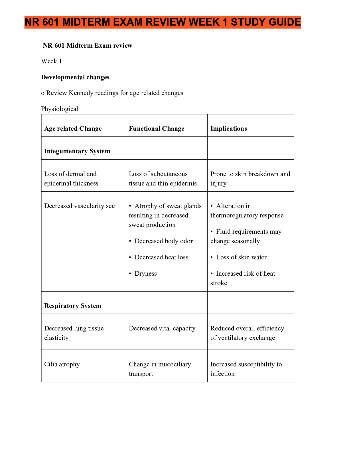
NR 601 Midterm Exam review November 2019 Week 1 Developmental changes o Review Kennedy readings for age related changes Physiological Age related Change
Functional Change Integumentary System
Loss of dermal and epiderm
...
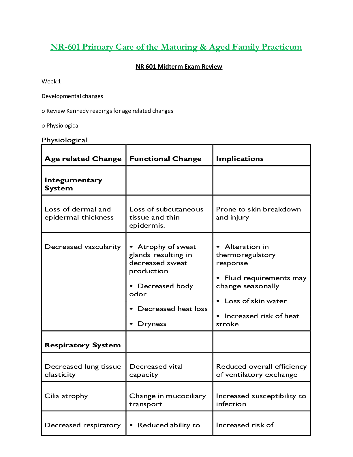
NR 601 Midterm Exam review November 2019 Week 1 Developmental changes o Review Kennedy readings for age related changes Physiological Age related Change
Functional Change Integumentary System
Loss of dermal and epidermal thickness
Decreased vascularity see
Loss of subcutaneous tissue and thin epidermis.
• Atrophy of sweat glands resulting in decreased sweat production
• Decreased body odor • Decreased heat loss • Dryness
Respiratory System
Decreased lung tissue elasticity
Cilia atrophy
Decreased respiratory muscle strength
Decreased vital capacity
Reduced overall efficiency of ventilatory exchange
Change in mucociliary transport
• Reduced ability to handle secretions and reduced effectiveness against noxious foreign particles
• Partial inflation of lungs at rest
Increased susceptibility to infection
Increased risk of atelectasis
Prone to skin breakdown and injury
• Alteration in thermoregulatory response
• Fluid requirements may change seasonally
• Loss of skin water
• Increased risk of heat stroke
ImplicationsCardiovascular System
Heart valves thicken and
become fibrotic
Fibroelastic thickening of the
sinoatrial node; decreased
number of pacemaker cells
Decreased baroreceptor
sensitivity (stretch
receptors)
GI
Liver becomes smaller
Decreased muscle tone
Reduced stroke volume,
cardiac output; may be
altered
Slower heart rate
Decreased responsiveness to
stress
Increased prevalence of
arrhythmias
Decreased sensitivity to
changes in blood pressure
Prone to loss of balance,
which increases the risk for
falls
Decreased storage capacity
Altered motility
Increases risk of constipation,
functional bowel syndrome,
esophageal spasm,
diverticular disease
Decreased basal metabolic
rate (rate at which fuel is
converted into energy)
NE CONDE)
Lab results
Lab Test
UA
Protein
May need fewer calories
Normal
Changes with age
Comments
0-5mg/100ml
Rises slightly
May be due to kidney
changes with age,
urinary tract infection,
renal pathology
Specific Gravity
1.005-1.020
Lower max in elderly
1.016-1.022
Decline in nephrons
impairs ability toconcentrate urine
Hematology
ESR
Men: 0-20
Women: 0-30
Iron Binding
50-160mcg/dl
230-410mcg/dl
Hemoglobin
Men: 13-18g/100ml
Women: 12-16g
Hematocrit
Men: 45-52%
Women 37-48%
Significant increase Neither sensitive nor
specific in aged
Slight decrease
Decrease
Men: 10-17g
Women: None noted
Slight decreased
speculated
Leukocytes
4,300–10,800/mm3 Drop to 3,100–
9,000/mm3
Anemia common in the
elderly
Decline in
hematopoiesis
Decrease may be due to
drugs or sepsis and
should not be
attributed immediately
to age
Lymphocytes
00–2,400 T
cells/mm3 50–200
B cells/mm3
T-cell and B-cell
levels fall
Platelet
Blood Chemistry
Albumin
150,000–350,000/ No change in
number
Infection risk higher;
immunization
encouraged
3.5–5.0
Decline
Related to decrease in
liver size and enzymes;
protein-energymalnutrition common
Globulin
2.3–3.5
Total serum protein 6.0–8.4 g
Slight increase
No change
Decreases may indicate
malnutrition, infection,
liver disease
Blood urea nitrogen Men: 10–25
Women: 8–20 mg
Creatinine
0.6–1.5 mg
Creatinine clearance 104–124 mL/min
Increases
significantly up to 69
mg
Increases significantly
up to 69 mg
Increases to 1.9 mg Related to lean body
mass decrease
Decreases
10%/decade after
age 40 years
Glucose tolerance
62–110 mg/dL after
fasting; >120 mg/dL
after 2 hours
postprandial
Alkaline
phosphatase
13–39 IU/L
Slight increase of 10
mg/dL/decade after
30 years of age
Increase by 8–10
IU/L
Used for prescribing
medications for drugs
excreted by kidney
Diabetes increasingly
prevalent; drugs may
cause glucose
intolerance
Elevations >20%
usually due to disease;
elevations may be
found with bone
abnormalities, drugs
(e.g., narcotics), and
eating a fatty meal
o Atypical disease presentations
1. Acute abdomenAbsence of symptoms or vague symptoms, acute confusion, mild discomfort and
constipation, some tachypnea and possibly vague respiratory symptoms, appendicitis pain
may begin in right lower quadrant and become diffuse
2. Depression
Anorexia, vague abdominal complaints, new onset of constipation, insomnia
hyperactivity, lack of sadness
3. Hyperthyroidism
Hyperthyroidism presenting as “apathetic thyrotoxicosis,” i.e., fatigue and weakness; weight
loss may result instead of weight gain; patients report palpitations, tachycardia, new onset
of atrial fibrillation, and heart failure may occur with undiagnosed hyperthyroidism
4. Hypothyroidism
Hypothyroidism often presents with confusion and agitation; new onset of anorexia, weight
loss, and arthralgias may occur
5. Malignancy
New or worsening back pain secondary to metastases from slow growing breast masses
Silent masses of the bowel
6. Myocardial
Absence of chest pain infarction (MI), vague symptoms of fatigue, nausea, and a decrease in
functional and cognitive status; classic presentations: dyspnea, epigastric discomfort,
weakness, vomiting; history of previous cardiac failure, higher prevalence in females versus
males Non-Q-wave MI
7. Overall infectious diseases process
Absence of fever or low-grade fever, malaise
8. Sepsis
Without usual leukocytosis and fever, falls, anorexia, new onset of confusion and/or
alteration in change in mental status, decrease in usual functional status
9. Peptic ulcer disease
Absence of abdominal pain, dyspepsia, early satiety, painless, bloodless, new onset of
confusion, unexplained, tachycardia, and/or hypotension
10. PneumoniaAbsence of fever; mild coughing without copious sputum, especially in dehydrated patients;
tachycardia and tachypnea; anorexia and malaise are common; alteration in cognition.
11. Pulmonary edema
Lack of paroxysmal nocturnal dyspnea or coughing; insidious onset with changes in
function, food or fluid intake, or confusion
12. Tuberculosis (TB)
Atypical signs of TB in older adults include hepatosplenomegaly, abnormalities in liver
function tests, and anemia
13. Urinary tract infection
Absence of fever, worsening mental or functional status, dizziness, anorexia, fatigue,
weakness
o Geriatric syndromes refers to conditions that involve multiple organ systems. Most common are
delirium, falls, dizziness and incontinence.
risk factors include: older age, cognitive impairment, functional impairment, and impaired mobility.
Bowel incontinence- involuntary passage of stool or the inability to control stool from expulsion.
More prevalent in women than men. 3 types: urge incontinence, passive incontinence, and fecal
seepage.
urge- has desire to go but cannot make it to the toilet despite attempts to avoid defecating.
Passive-involuntary loss of gas and stool without awareness.
fecal seepage- leakage of stool after a normal bowel movement.
etiology : a number of reasons including GI issues, cognitive or neurological diseases.
Treatment: treat related etiology such as impaction of increasing fiber. Habit training is also
recommended. Once clear evidence of no impaction, infection, or cause is determined. antidiarrheal
medication like Imodium can be tried.
For retrosphincter dysfunction biofeedback with strengthening exercises for the sphincter can be
done.
Constipation: presence of 2 or more symptoms: decreased stool frequency, straining, hard stools,
sensation of incomplete emptying, blockage at anorectal site. constipation is most common
digestive complaint.Cough: forceful expelling of air from the lungs involving the use of accessory muscles of the chest
and constriction of the glottis.
dehydration: caused by too little fluid intake, too much fluid lost or both. Chronic diseases like
diabetes, cancer, cardiovascular diseases, and renal diseases make elderly sensitive to fluid shifts.
Diarrhea: Passage of increased stool frequency, liquidity, or volume. Most episodes are caused by
viral gastroenteritis. Chronic diarrhea is defined as lasting longer than 4 weeks.
Dizziness: common clinical categories include vertigo, light-headedness, unsteadiness, or gait
instability, and disequilibrium.
Falls: WHO defines this as an event that results in a person coming to rest inadvertently on the
ground, floor, or other lower level. can be witnessed or unwitnessed. due to intrinsic factors or
extrinsic factors. Intrinsic factors include age, weakness, gait/balance, poor vision and postural
hypotension. Extrinsic factors involve environment conditions like lack of handrails, poor lighting,
obstacles, slippery surfaces, certain medication, and polypharmacy.
Fatigue
● Description
○ Subjective state often described as a feeling of tiredness, weariness, lack of energy,
or exhaustion that is unrelieved or only partially relieved by rest
○ Often results in an inability to initiate normal activity; a reduced capacity to
maintain activity; and difficulty with concentration, memory, and emotional stability
○ Chronic fatigue syndrome occurs when fatigue lasts longer than 6 months and is not
relieved with rest
○ Fatigue could also be a symptom of another illness and any older person with this
complaint should obtain a medical evaluation and work-up.
● Etiology
○ Occurs normally with inadequate rest, excess exertion, or insufficient diet
○ Fatigue in older adults may be an early indicator of the aging process, as well as
debility or another disorder
● Occurrence
○ 25% of the US population
● Age
○ Common among the elderly
● Gender
○ More common in women
● Ethnicity
○ Not significant
● Contributing Factors
○ Poor dietary habits, overexertion, alcohol abuse, smoking, stress, chronic illness,
drug interactions, misuse of drugs, and sleep apnea
○ In the older adult individual, it is compounded by a decrease in muscle strength,
loss of muscle neurons, muscle atrophy, a decrease in hormone levels, and lack of
exercise.
● Signs and symptoms○ Conduct a complete symptom assessment, including the onset; duration; severity;
and precipitating, aggravating, and relieving factors.
○ Identify other indicators or associated symptoms of fatigue, which may include
decreased energy expenditure, decreased endurance, sleep disturbance, attention
deficits, somatic complaints (aching body, tired eyes), dyspnea, and weakness
○ Carefully review the adequacy of the diet, all medications (evaluating for potential
medication side effects), activity level (including degree of independence of ADLs),
and potential causes or contributing factors. Identify the impact fatigue is having on
the person’s ADLs and quality of life and current stressors.
○ Distinguish between generalized fatigue and actual weakness by testing for muscle
strength and presence of localized tenderness
○ A thorough physical examination will include a mental status examination to screen
for dementia and rule out depression.
● Diagnostic tests
○ Diagnostic tests on all patients with persistent unresolved fatigue should include
CMP, CBC with differential, erythrocyte sedimentation rate (ESR), and/or C-reactive
protein, because these are low cost and offer significant screening capacity.
○ Thyroid function, urinalysis, and pulmonary function tests.
○ If symptoms and signs indicate cardiac decompensation, a B-type natriuretic peptide
(BNP) may indicate degree of heart failure and an EKG may reveal cardiac
arrhythmias, enlargement of the heart, myocardial infarction, or abnormalities in
the conduction system
● Differential diagnosis
○ Psychiatric disorders, including depression and generalized anxiety disorder,
account for 70% of cases of fatigue
○ Fatigue that cannot be relieved by rest or sleep is often a sign of disease.
● Treatment
○ Symptom management includes regular exercise, attention-restoring activities,
psychosocial techniques, energy conservation measures, good sleep hygiene,
improving diet, and possibly adding nutritional supplements
○ Psychostimulants may be considered for opioid-related somnolence, cognitive
impairment, and depression
● Follow up
○ Monitor the patient periodically as indicated by diagnosis or symptoms, symptom
persistence, and disability associated with the symptom
● Sequelae
○ The potential for complications relates to the cause of fatigue and the impact the
symptom has on the person’s function
● Prevention/Prophylaxis
○ Optimal health maintenance, including maintaining a healthy diet, regular exercise,
and good sleep hygiene, may prevent or enable early recognition of signs and
symptoms of systemic or psychological illness
● Referral
○ May be indicated based on the results of the work-up
● Education
○ If the fatigue has a physiological cause, teaching should be related to the findings;
psychological counseling, changes in the environment, behavior modification, and
stress reduction may be needed Goal of fatigue management - provide the patient
with self-help tools to eliminate or alleviate fatigueHeadache:
Hematuria:
Description: Presence of RBCs in the urine, classified either gross or microscopic
Gross hematuria: urine appears either red or brown in color to the naked eye
Microscopic hematuria: identified by lab analysis. Significant if 3 or more RBCs per high-
power field on accurately collected urine specimen.
*requires evaluation, 5% with microscopic hematuria are found to have malignancy, & 30-
40% with gross hematuria are found to have malignancy
Etiology: renal or contamination from outside the urinary tract. Renal hematuria may result from
glomerular or nonglomerular causes.Source of hematuria may be the upper collection system
(renal, ureter) and/or lower collection system (bladder, prostate, urethra). Common finding on
routine UA and etiologies range from life-threatening to benign incidentals. Patho depends on the
anatomical site from which blood loss occurred, older adults, most common causes are malignancy
or BPH
Occurrence: prevalence of asymptomatic microscopic hematuria ranges from 2% to 31% in the
adult population, older than 50 yrs., prevalence 13%
Age: increases with age, younger pts are less likely to have an etiology identified
Ethnicity: not significant
Contributing factors: infection, anticoagulation, renal calculi, trauma, anatomical defects such as
rectocele, menstruation, atrophic vaginitis, renal disease, or recent urological procedure,
malignancy include age more than 35 yrs. male sex, current or past history of smoking, occupational
exposures to chemicals or dyes, hx of gross hematuria, chronic cystitis,pelvic irradiation, exposures
to cytotoxic agents
S/Sxs: thorough HX, presence of visible blood in the urine, Hx of vigorous exercise, recent prostate
examination or procedure, recent trauma to the abdomen, recent catheterization, menstruation,
renal disease, viral illness, medications (analgesics, antibiotics, anticoagulants, NSAIDS). Symptoms
represents kidney disease or stone. urinary frequency, lower abdominal pain or dysuria, which may
indicate UTI. PE: focus on abdominal/flank pain, urogenital examination, prostate, testicular and
vaginal examination
Dx Tests: UA with microscopy. Urine dipstick evaluation may be misleading because it lacks the
ability to distinguish RBCs from myoglobin or HgB, should confirm with heme-positive dipstick with
a microscopic UA. CMP for metabolic abnormalities, elevated creatinine: indicates acute or chronic
renal disease and should be referred to nephrologist. PT/INR if on anticoagulation and CBC.
Multiphasic CT urography or IV pyelogram if suspicious of kidney stone, renal mass or malignancy.
Cystoscopy for patients 35 years of age, pts younger than 35 yrs if no other reason for hematuria is
present, pts of any age if with risks factors for CA., dx bladder or prostatic CADDX: infection or supratherapeutic anticoagulation should be r/o as reversible causes of hematuria.
serious causes: glomerular disease, renal calculi, trauma, anatomical defects, or malignancy
TX; directed at the cause, asymptomatic (isolated) does not require treatment. Appropriate ABT
therapy for UTI should resolve the hematuria, anticoagulation is the cause, adjustment may resolve
the problem, kidney stone, initial tx may be pain management and oral hydration. Urosepsis, AKI,
anuria, unrelenting N/V should be referred immediately
FF-up: repeat UA should be obtained 6 weeks after initial tx for any infection, vigorous exercise, or
trauma to make sure that the hematuria has cleared. Refer to urology for gross or microscopic
hematuria. Persistent hematuria w/ initial (-) urological work-up, a repeat UA should be done
yearly and a full repeat evaluation very 3-5 yrs for ongoing hematuria. Referral to nephro if
hematuria persists with renal impairment
Sequelae: fairly common in young adults under 35 yrs. It could be a sign of malignancy, should not
be ignored, even if it is transient. May indicate renal disease, when proteinuria is present, referral to
nephro for 2nd opinion.
Prevention/Prophylaxis: USPSTF does not endorse routine screening for asymptomatic hematuria
due to lack of evidence
Referral : Primary care providers can initiate testing to verify presence of hematuria and treat
hematuria if Dx is clear. Referral to nephro is indicated for the presence of proteinuria, RBC casts,
dysmorphic RBCs, or elevated serum creatinine level. Referral to urology: hematuria persisting after
treatment for a UTI, taking anticoagulants, or in the absence of benign cause, gross and microscopic
hematuria
Education: Counsel family and patients to seek medical advice about gross hematuria, plus s/sxs to
report such as urinary frequency, dysuria, flank and abdominal pain. Educate pts and families abt
the SEs of OTC drugs and RX meds, effects of anticoagulant therapy and to call immediately if the pt.
notices any hematuria because this may mean the pt’s drug level is too high.
Involuntary weight loss:
Joint pain:
priutis:
Tremor: is the most common form of involuntary movement and is characterized by rhythmic
oscillation of a body part that can be classified according to the circumstances under which it
occurs.
Etiology: Because of the vast number of causes of tremor, etiological classification is not helpful.
Terms used to describe the clinical phenomenology of tremor include rest tremors and action
tremors.Rest tremor occurs when muscle is not activated voluntarily, and the relevant body part is fully
supported against gravity
Action tremor is present with voluntary contraction of muscle. Action tremors can be sub-classified
further into postural, kinetic, and isometric tremor.
Postural tremor is present while voluntarily maintaining a position against gravity.
Kinetic tremor may occur during any form of voluntary movement of the affected body part.
Isometric tremor occurs with muscle contraction against a rigid stationary object. The most
common tremor is enhanced physiological tremor followed by essential tremor and then
parkinsonian tremor.
Occurrence: Tremor is the most common movement disorder encountered in clinical practice.
Everyone has a low-amplitude physiological tremor that can be observed when the arms are
extended. Enhanced physiological tremor is a physiological tremor that comes and goes with
anxiety, caffeine, and fatigue. In 50% of cases, the disease is familial (autosomal dominant, meaning
50% of an affected individual’s children have it). More than 70% of patients with Parkinson’s
disease have tremor as the presenting symptom. Less common are cerebellar tremor, psychogenic
tremor, dystonic tremor, and tremor associated with Wilson’s disease.
Age: Most studies report a significant age-associated increase in the prevalence of essential tremor.
Essential tremor begins in young to middle-aged people and gradually intensifies with age. Tremors
in older adults are more likely to be of the essential or parkinsonian type.
Gender: Tremor afflicts both genders equally, with perhaps slightly more frequency in men than in
women.
Ethnicity: Tremor is more prevalent in Caucasians than in African Americans, and is of intermediate
prevalence in Hispanics.
Contributing Factors: During times of stress, the amplitude of a physiological tremor increases.
Fatigue, anxiety, hyperthyroidism, systemic illness, use of medications, drug withdrawal (especially
from alcohol), use of methylxanthines, and excess caffeine intake can exaggerate tremor.
Medications that can cause or exacerbate tremor include those that stimulate the sympathetic
nervous system and psychoactive medications.
Signs and Symptoms: The first step in the evaluation of tremor is to categorize the tremor based on
activation conditions, distribution, and frequency. Determine the duration and age of onset of
symptoms, exacerbating or alleviating factors, and any family history of tremor or other
neurological disorders. Include any associated symptoms, such as bradykinesia or rigidity
(suggesting Parkinson’s disease) or ataxia and nystagmus (suggesting cerebellar disease). The
patient’s medication history, any exposure to toxins, and the presence of illness should be noted.
History is important, but the diagnosis is based on clinical physical examination findings. Tremor
may occur in various body parts, such as the hands, head, facial structures (chin, tongue, lips, and
ears), vocal cords, trunk, and legs. Of all tremors, 94% occur in the hands, either unilaterally or
bilaterally.
On physical examination, it is important to conduct a thorough tremor-focused neurological
examination: muscle tone is checked throughout the body, cranial structures (including the mouthand jaw) are examined at rest and in action, and the tongue is observed during rest and protrusion.
To distinguish properly between resting and action tremors, patients should be evaluated while
supine and when seated with the arms fully supported.
The upper extremities are examined in an outstretched position with the hands supine (palms
up), sideways (semi-prone), and then prone (palms down). The semi-prone position enhances
essential tremor, whereas the supine position inhibits it. In the wing position (i.e., with apposition
of the index fingers close to each other but not touching), proximal tremor may be identified. Goal-
directed activities are performed, such as finger-to-nose, heel-to-shin, and toe-to-finger movements.
The patient is asked to recite a standard paragraph and enunciate a sustained vowel. Handwriting
samples are obtained (e.g., script, numbers, Archimedes spirals). Gait is evaluated for shuffling and
unsteadiness, and Romberg (station) and balance testing are conducted. Careful evaluation is
performed for signs associated with tremor syndromes. Bradykinesia and postural abnormalities
are evaluated by observing difficulty rising from a seated position, decreased arm swing, and
masked facies. Patients with essential tremor typically have handwriting that is shaky and large,
whereas the handwriting of patients with Parkinson’s disease initially may be of normal size and
progressively become smaller (micrographia). Archimedes spirals drawn by essential tremor
patients tend to illustrate natural fluctuations in tremor magnitude.
Diagnostic Tests: No specific tests are routinely ordered for tremors. Electromyography is used to
subdivide tremors according to their rate and their relationship to posture of limbs and volitional
movement. Tremor frequency usually is categorized as low frequency (less than 4 Hz), medium
frequency (4 to 6 Hz), and high frequency (greater than 6 Hz).
The diagnosis of tremor is primarily clinical; however, laboratory testing may be necessary to
exclude certain conditions that may be associated with tremor, such as metabolic disturbances,
including hyperthyroidism (e.g., through thyroid function tests) and Wilson’s disease. Brain imaging
may be indicated for select patients, particularly patients with tremor that is unilateral, of sudden
onset, or associated with atypical clinical features. For difficult cases, single-photon emission
computed tomography (SPECT) to visualize the integrity of dopaminergic pathways may be useful
in diagnosing Parkinson’s disease.
Differential Diagnosis: The differential diagnosis, in general practice, is almost always between
Parkinson’s disease and essential tremor.
Physiological Tremor: A normal phenomenon, physiological tremor occurs in all contracting
muscle groups. Although seldom visible to the naked eye, physiological tremor often may be
detected when the fingers are firmly outstretched with a piece of paper placed over the hands.
Enhanced Physiological Tremor (or an Intensification of Physiological Tremor to Detectable Levels):
Physiological tremor may be enhanced under conditions of stress, anxiety, fatigue, exercise, cold,
hunger, stimulant use, alcohol withdrawal, or metabolic disturbances such as hypoglycemia or
hyperthyroidism. Although the tremor is typically low in amplitude and high in frequency (8 to 12
Hz), it may be clinically indistinguishable from essential tremor.
Essential Tremor (4 to 12 Hz): Essential tremor is a persistent postural and kinetic tremor that
predominantly affects the hands and forearms. Classically, to show the tremor, the patient is asked
to extend the arms in front of the body. The legs are affected less often. Although less frequently
involved, the presence of tremor in the head and/or voice is a strong indication of essential tremor
and is especially useful in differentiating the syndrome from Parkinson’s disease. Head tremor,
which is also postural, disappears when the head is supported. Listening to the patient speak orhaving the patient hold a musical note as long as possible may reveal a quivering intonation. A
resting component is present only rarely and typically occurs in the most advanced cases.
Parkinsonian Tremor Syndromes (4 to 6 Hz): Parkinsonian tremor syndromes involve resting
tremor that is often asymmetrical. Tremor may be observed when muscles are relaxed, such as
when the hands are resting on the lap, and may affect hands, feet, mandible, and lips. Tremor
disappears during sleep. Typical is an alternating tremor of the thumb against the index finger—
pill-rolling tremor. Although rest tremor is a diagnostic criterion for Parkinson’s disease, other
forms of tremor also may be present.
Treatment: Tremor should be treated if it causes disability. First-line treatment for tremor is oral
medication. Beta blockers, anticholinergic medication, and levodopa are useful modalities for
resting tremor. Kinetic tremor may respond to beta blockers, primidone, anticholinergics, and
alcohol. When there is a lack of response to medical treatment or when tremor results in severe
disability, a patient may be considered for neurosurgery. Specific treatments for the most common
causes are noted next.
Physiological Tremor: Usually no treatment is required for physiological tremor. When
exaggerated, however, it may interfere with activities requiring extreme precision. Identify and
remove precipitating causes and contributing causes. If the precipitating cause cannot be removed,
propranolol may be effective.
Essential Tremor: Varying degrees of control in essential tremor have been obtained with the beta
blocker propranolol and the anticonvulsant agent primidone. Either agent may be considered an
appropriate first-line therapy for the symptomatic management of essential tremor. When
appropriate, these agents may be administered in combination with benzodiazepines, such as
lorazepam or clonazepam. If the medication is of no benefit at a dose that causes adverse effects,
dose levels should be tapered down gradually and eventually discontinued. If a medication is
documented to be beneficial, it may be continued at the regulated doses, and the next medication
may be added to the drug regimen. If the response to a drug is adequate and the dose is well
tolerated, you may continue to monitor tolerance and possibly increase the dose.
Physical and psychological measures may be helpful in managing mild tremor. Physical measures
may include the application of weights to affected limbs to decrease tremor amplitude. Some
patients have experienced benefits with biofeedback, relaxation methods, and other behavioral
techniques through alleviation of anxiety or stress that may exacerbate tremor. Alcohol
consumption may lead to transient improvement for many with essential tremor. The potential risk
of alcohol dependence and abuse among essential tremor patients who drink alcohol to control
symptoms is controversial. Alcohol has no impact on the tremor of Parkinson’s disease.
Parkinsonian Tremor: The tremor of Parkinson’s disease results from a loss of striatal dopamine,
and this is the rationale for treatment with either the dopamine precursor levodopa or dopamine
receptor agonists. Dopaminergic and anticholinergic agents are equally effective, but
dopaminergic substances additionally improve other parkinsonian signs, and the potential side
effects of anticholinergic medications make these drugs undesirable in the older adult. The
combination of levodopa and carbidopa reduces levodopa-induced nausea; a typical starting
dose is one tablet of Sinemet 25/100 three times daily. Levodopa-carbidopa intestinal gel (brand
name Duopa) is a combination of levodopa and carbidopa that is dosed for 16 hours during
wakefulness. It is administered via PEG-J tube supplied by an external box. This route of dosingdecreases tremor by reducing plasma fluctuations that patients on oral levodopa and carbidopa
might experience.
Patients with severe tremors that are resistant to pharmacotherapy may benefit from ablative
surgery and/or deep brain stimulation. For those essential tremor patients who fail medication
treatment, transcranial magnetic resonance-guided focused ultrasound thalamotomy
(product name ExAblate Neuro) has been shown to be successful in the treatment of essential
tremor. This procedure is noninvasive.
Follow-Up: Patients should be evaluated for therapeutic effects and side effects within 1 week of
starting treatment. Annual monitoring for weight loss, depression, and decline in functional status
is necessary.
Sequelae: Functional disabilities may occur in ADLs, including compromised eating, drinking, and
preparing food. Decreased caloric intake and weight loss may be observed. Ambulation, especially
on stairs, may be hazardous. Withdrawal from social situations may occur, and depression is
common.
Prevention/Prophylaxis: Reduce factors that can exacerbate the tremor. Continue medication
regimen.
Referral: A neurologist should be consulted for cerebellar tremors, mixed tremors, or Parkinsonian
tremor, or when a focal neurological deficit is identified. An ophthalmologist should be consulted
when Wilson’s disease is suspected. A mental health provider or psychiatrist should be consulted
when a hysterical tremor is suspected. Physical therapy or occupational therapy may be helpful in
advanced or disabling cases.
Education: Some patients, particularly patients with severe, disabling tremor, may limit their
contacts. Patients must be encouraged to learn as much as they can about their disease to help them
cope better with the condition’s progression. When a diagnosis has been established, the natural
history of the condition should be explained to patients. It also may be appropriate to recommend
counseling. Use of appropriate coping strategies may reduce stress substantially, preventing
possible augmentation of tremor owing to anxiety. Referral to appropriate patient support
organizations is helpful for most patients.
CLINICAL RECOMMENDATION
EVIDENCE
RATING
The diagnosis of tremor is based on clinical information
from the history and physical examination.
C
REFERENCES
Deuschl et al., 1998
Sharma & Pandey,
2016
Propranolol and primidone are first-line treatments for
essential tremor.
A
Sharma & Pandey,
2016Tremor amplitude worsens over time if not treated. C
Sharma & Pandey, 2016
Levodopa-carbidopa intestinal gel improves resting tremors.
Transcranial magnetic resonance-guided focused ultrasound thalamotomy successfully treats essential tremor.
B Fernandez et al., 2016 B U.S. FDA, 2016
Urinary incontinence: (I realize this is copied and pasted from the book; that’s bc it is late. I will read it and cut it down tomorrow; thanks! However, if anyone is awake and reading this, please feel free to trim away unnecessary excess information.) (UI) is an involuntary loss of urine. Acute UI is generally a result of illness or the effects of medications and is self-limiting when the cause is determined and addressed. Chronic UI has different forms, including stress incontinence, urge incontinence, overflow incontinence, and functional incontinence. Many older women manifest a combination of urge and stress symptoms resulting in mixed incontinence.
Etiology: Anatomical changes, factors related to the individual’s medical history, lifestyle, and acute and chronic illnesses, in addition to medications, can result in incontinence that can be either reversible or a permanent condition. Cognitive as well as chronic mental illness, depression, and functional barriers to continence can also affect an individual’s ability to maintain urinary continence.
[Show More]

 GRADE A SOLUTION.png)



x.png)