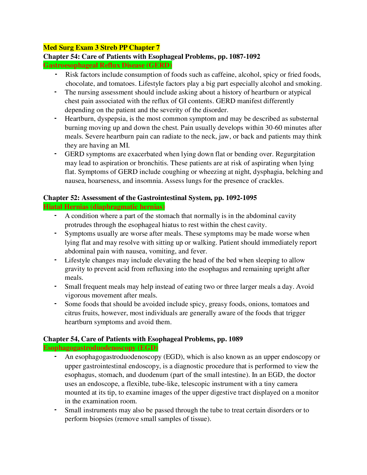
Med Surg Exam 3 Streb PP Chapter 7
Chapter 54: Care of Patients with Esophageal Problems, pp. 1087-1092
Gastroesophageal Reflux Disease (GERD)
Risk factors include consumption of foods such as caffeine, alcohol, spi
...
Med Surg Exam 3 Streb PP Chapter 7
Chapter 54: Care of Patients with Esophageal Problems, pp. 1087-1092
Gastroesophageal Reflux Disease (GERD)
Risk factors include consumption of foods such as caffeine, alcohol, spicy or fried foods,
chocolate, and tomatoes. Lifestyle factors play a big part especially alcohol and smoking.
The nursing assessment should include asking about a history of heartburn or atypical
chest pain associated with the reflux of GI contents. GERD manifest differently
depending on the patient and the severity of the disorder.
Heartburn, dyspepsia, is the most common symptom and may be described as substernal
burning moving up and down the chest. Pain usually develops within 30-60 minutes after
meals. Severe heartburn pain can radiate to the neck, jaw, or back and patients may think
they are having an MI.
GERD symptoms are exacerbated when lying down flat or bending over. Regurgitation
may lead to aspiration or bronchitis. These patients are at risk of aspirating when lying
flat. Symptoms of GERD include coughing or wheezing at night, dysphagia, belching and
nausea, hoarseness, and insomnia. Assess lungs for the presence of crackles.
Chapter 52: Assessment of the Gastrointestinal System, pp. 1092-1095
Hiatal Hernias (diaphragmatic hernias)
A condition where a part of the stomach that normally is in the abdominal cavity
protrudes through the esophageal hiatus to rest within the chest cavity.
Symptoms usually are worse after meals. These symptoms may be made worse when
lying flat and may resolve with sitting up or walking. Patient should immediately report
abdominal pain with nausea, vomiting, and fever.
Lifestyle changes may include elevating the head of the bed when sleeping to allow
gravity to prevent acid from refluxing into the esophagus and remaining upright after
meals.
Small frequent meals may help instead of eating two or three larger meals a day. Avoid
vigorous movement after meals.
Some foods that should be avoided include spicy, greasy foods, onions, tomatoes and
citrus fruits, however, most individuals are generally aware of the foods that trigger
heartburn symptoms and avoid them.
Chapter 54, Care of Patients with Esophageal Problems, pp. 1089
Esophagogastroduodenoscopy (EGD)
An esophagogastroduodenoscopy (EGD), which is also known as an upper endoscopy or
upper gastrointestinal endoscopy, is a diagnostic procedure that is performed to view the
esophagus, stomach, and duodenum (part of the small intestine). In an EGD, the doctor
uses an endoscope, a flexible, tube-like, telescopic instrument with a tiny camera
mounted at its tip, to examine images of the upper digestive tract displayed on a monitor
in the examination room.
Small instruments may also be passed through the tube to treat certain disorders or to
perform biopsies (remove small samples of tissue).
Certain medications (such as aspirin, anticoagulants and the anti-inflammatory drugs
called NSAIDs) should be discontinued at least five to seven days before an EGD to
reduce the risk of bleeding.
NPO - Patients will be asked not to eat or drink anything for at least 8 hours before the
procedure to ensure that the upper intestinal tract will be empty.
Before the procedure, patients may be given a moderate sedative and/or pain medication,
usually by intravenous injection.
Monitor gag reflex, the sedation will block the gag reflex to prevent aspiration.
Keep NPO until they get their gag reflex back. (1 to 2 hours before the anesthetic is out
the system).
Patient must have someone accompany them home after recovery.
Chapter 55: Care of Patients with Stomach Disorders, pp. 1103-1107
Gastritis
Gastritis occurs when the lining of the stomach known as the mucosa becomes inflamed
or swollen. When the stomach mucosa becomes inflamed edema, hemorrhage and erosion
of the mucosa occur.
Medical treatment for gastritis depends on the specific cause. Patients will be instructed
to stop taking irritating medications such as ASA and NSAIDS. Medications to decrease
the amount of hydrochloric acid in the stomach are usually prescribed. These would
include Antacids, H2 antagonists, and Proton pump inhibitors.
The patient with gastritis is at risk for deficient fluid volume. A nursing priority is to
access for the patient’s hydration status. This would include I&O, daily weights, & VS.
Chapter 55: Care of Patients with Stomach Disorders, pp. 1107-1115
Peptic Ulcer Disease (PUD)
Peptic ulcers are a break in the mucous lining of gastrointestinal tract from continued
contact with gastric juice. This results in inflammation. Pain that is worsened by the
ingestion of food.
Ulcers in the mucosa of GI tract occur from several different causes. Duodenal ulcers are
associated with a H. pylori infection.
Gastric ulcers may cause a dull, aching pain, often right after a meal; eating does not
relieve the pain and may even worsen it. Pain may also occur at late at night.
Other symptoms associated with PUD are nausea with or without vomiting, weight loss,
anorexia, belching and dyspepsia or indigestion. Patient may report a distended abdomen
that is painful.
Smoking contributes to the pathogenesis of peptic ulcer disease. Smoking causes an
acceleration of gastric emptying of liquids, promotes of duodenogastric reflux and causes
a reduction in mucosal blood flow. Patient should attend a smoking cessation course.
Chapter 55, Care of Patients with Stomach Disorders, pp. 1116-1119
Gastric Cancer
Stomach cancers tend to develop slowly over many years. Before a true cancer develops,
pre-cancerous changes often occur in the inner lining (mucosa) of the stomach. These
early changes rarely cause symptoms and therefore often go undetected.
The decline of stomach cancer has been linked to the frequent use of antibiotics to treat
infections. Antibiotics can kill the bacteria called Helicobacter pylori (H. pylori), which is
thought to be a major cause of stomach cancer.
Administer protein and vitamin supplements to foster wound repair and tissue building.
Eat small, frequent meals rather than three large meals. Reduce fluids with meals but take
them between meals. Stress the importance of long-term vitamin B12 injections after
gastrectomy to prevent surgically induced pernicious anemia.
Chapter 56, Care of Patients with Noninflammatory, pp. 1135-1137
Irritable Bowel Syndrome (IBS)
Irritable bowel syndrome (IBS) refers to a disorder that involves abdominal pain and
cramping, as well as changes in bowel movements
Risk factors include consuming a diet high in fats and gas producing foods. Consuming
carbonated beverages, caffeine and alcohol contribute to the development of IBS.
Smoking and stress are other related factors. Emotional experiences such as anxiety and
depression are also a factor as this affects the autonomic nervous system and its
innervation to the bowel.
Nursing care for the patient with IBS focuses on education and emotional support. Help
the patient implement lifestyle changes that reduce stress. Remind the patient about
regular exercise, discourage smoking while encouraging the need for regular physical
examinations.
Chapter 56, Care of Patients with Noninflammatory, pp. 1137-1139
Herniation
A weakness in the abdominal muscle wall through which a segment of the bowel or other
abdominal structure protrudes. Hernias can also penetrate through any other defect in the
abdominal wall, through the diaphragm, or through other structures in the abdominal
cavity.
The most significant factors contributing to increased intra-abdominal pressure are
obesity, pregnancy, and lifting heavy objects.
Indirect Inguinal Hernia is a sac formed from the peritoneum that contains a
portion of the intestine or omentum. The hernia pushes downward at an angle into
the inguinal canal. In males, indirect inguinal hernias can become large and often
descend into the scrotum.
Direct Inguinal Hernias, in contrast, pass through a weak point in the abdominal
wall.
Femoral Hernias protrudes through the femoral ring. A plug of fat in the femoral
canal enlarges and eventually pulls the peritoneum and often of the urinary
bladder into the sac.
Umbilical Hernias are congenital or acquired. Congenital umbilical hernias
appear in infancy. Acquired umbilical hernias directly result from increased intra-
abdominal pressure. They are most commonly seen in people who are obese.
Incisional, or Ventral Hernias occur at the site of a previous surgical incision.
These hernias result from inadequate healing of the incision, which is usually
caused by post-operative wound infections, inadequate NUTRITION, and obesity.
Chapter 56: Care of Patients with Noninflammatory Intestinal Disorders, pp. 1121-1126
Intestinal Obstruction
Intestinal obstruction is a partial or complete blockage of the bowel that results in the
failure of the intestinal contents to pass through. With obstruction, gas and fluid
accumulate proximal to and within obstructed segment causing bowel distention. A bowel
obstruction is divided into two basic categories: mechanical and non-mechanical.
Treatment of intestinal obstruction is directed toward relieving symptoms, managing fluid
and electrolyte imbalances, preventing complications, and treating the cause of the
obstruction.
Surgery may be needed to relieve the obstruction if gastric decompression does not
relieve the symptoms, or if there are signs of bowel necrosis. The type of surgery will
depend on the type and area of obstruction and may include intestinal resection with an
anastomosis or creation of an ileostomy or colostomy.
SMALL BOWEL:
If the small bowel obstruction is complete, the peristaltic waves become quite vigorous,
assuming reverse direction, propelling intestinal contents toward the mouth rather than
the rectum.
The patient vomits stomach contents first, then the bilious contents of the duodenum, and
finally the fecal contents of the ileum.
A distended abdomen, a bloated sensation, and altered bowel sounds may indicate a small
bowel obstruction. Patients with Ileostomy who develops distention and cramping should
apply warm, moist towels to abdomen or lightly massage abdomen.
LARGE BOWEL:
Constipation may be the only symptom for several days. Barium enema may be ordered
to reveal a distended, air-filled colon. Monitor the patient for bowel movement after a
barium enema.
Patients with large bowel obstructions may experience intermittent persistent lower
abdominal cramping. Severe pain may result from strangulation or bowel perforation.
Chapter 57, Care of Patients with Inflammatory Intestinal Disorders, pp. 1117-1119
Dumping Syndrome (postop) (No treatment, patient waits it out).
Rapid emptying of gastric contents into the small intestines. This results in a fluid shift
into the gut causing abdominal distention.
Observe for early manifestation of this syndrome, which typically occur within 30
minutes of eating.
Symptoms include vertigo, tachycardia, syncope, sweating, pallor, palpitation, and the
desire to lie down
Monitor patient for late dumping syndrome, which occurs 90 minutes to 3 hours after
eating, is caused by a released of an excessive amount of insulin. The insulin release
follows a rapid rise in the blood glucose level that results from the rapid entry of high
carbohydrate food into the jejunum.
Observe for manifestations including dizziness, lightheadedness, palpitations,
diaphoresis, and confusion.
Dumping syndrome is managed by nutrition changes that include decreasing the amount
of food taken at one time and eliminating liquids ingested with meals.
Teach small frequent feedings, no liquids before or with meals.
Teach to eat high protein, fat foods, low to moderate carbohydrate diet.
Avoid smoking and NSAIDS.
All we can do is to teach, there is nothing we a do for the patient.
Patient will need to lie down and wait it out.
Chapter 56: Care of Patients with Noninflammatory Intestinal Disorders: pp. 1130-1135
Colostomy
When are colostomies performed?
Colostomies are performed because of problems with the lower bowel. Some problems
can be corrected by temporarily diverting stool away from the bowel. This is when
temporary colostomies are used to keep stool out of the colon. If the colon becomes
diseased, as in the case of colon cancer, permanent colostomies are performed, and the
colon may be removed completely.
How does it initially look like?
Must be patent, must be functional, healthy and do not injury it (need to protect the
stoma).
It will be quite swollen from surgery at first but will shrink to its final size about 6 to 8
weeks after surgery.
What you are seeing is the inner lining of the intestinal wall folded under to form a
stoma.
It should nice beefy red, and moist. (if not red, pink and must be moist)
Should not be dry, pale, yellow, dusky, ash and the surrounding skin should not be dry
and shiny around the stoma (all indication something wrong with the circulation).
There should be no drainage except for the normal, if an ileostomy the drainage will be
dark green liquid drainage, and a colostomy once patient starts having bowel movement
there will be fecal material coming out into the pouch.
If the drainage is greenish, with pus, and a foul odor, should not happen, contact doctor.
What does a healthy stoma look like?
When you look at a stoma, you are looking at the lining (the mucosa) of the intestine
wall, which looks a lot like the inside lining of your cheek. The stoma will look pink to
red. It’s warm and moist and secretes small amounts of mucus.
What teaching would you do regarding diet and pouching/application of appliance?
The patient will receive only IV fluids for two to three days after a colectomy or
colostomy, to give the colon time to heal. After that, you can try clear liquids, such as
soup broth and juice, followed by easy-to-digest foods, such as toast and oatmeal.
You will be able to go back to your normal diet after this, but if you have a colostomy,
you may want to avoid certain foods that cause odors or gas, which can over-inflate the
colostomy bag and make it more difficult to manage.
There should be an ostomy nurse come in initially and do a plan of care, teaching and we
will do a lot of reinforcing.
Teach them what the stoma looks like and how to monitor for infection (increase of
drainage, the increase of redness around the stoma, warm to the touch and pain are signs
of infection and need to report it).
Need to monitor the drainage and that it is the correct drainage in the pouch, in the
ileostomy there will always be drainage from the small intestine like enzymes, fluids, and
partially digested food.
No thickening agents with an ileostomy, can cause an obstruction.
If there is no drainage does not take laxatives with an ileostomy or an enema.
You can have an enema with a colostomy.
Teach to place moist towel on abdominal to stimulate the peristalsis or lying down on the
side in a fetal position, and if cramping drink hot tea.
Do not do anything else to start peristalsis.
Teaching to avoid offending foods like cabbage, broccoli, beans or and foods that can
cause gas.
Application of their appliance or the wafer, every time they change the wafer, the stoma
needs to be remeasured until the stoma remains to their normal size and make sure it is
seal good, no moisture or skin exposure. (paste to seal, 1/8 -1/16 may want to draw a
stencil once normal size)
Do not allowed the bag to get more than 1/3 full or 1⁄2 full at the max, will need to be
empty.
[Show More]


.png)

.png)
.png)
.png)