HEMATOLOGIC DISORDERS: 7-10 questions
• Identify general nursing strategies associated with altered anemia
• Describe the morphology, pathophysiology, clinical manifestations, therapeutic management and nursing conside
...
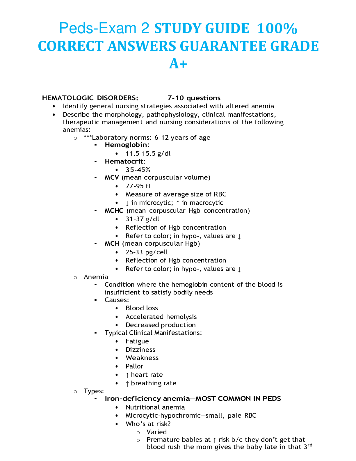
HEMATOLOGIC DISORDERS: 7-10 questions
• Identify general nursing strategies associated with altered anemia
• Describe the morphology, pathophysiology, clinical manifestations, therapeutic management and nursing considerations of the following anemias:
o ***Laboratory norms: 6-12 years of age
▪ Hemoglobin:
• 11.5-15.5 g/dl
▪ Hematocrit:
• 35-45%
▪ MCV (mean corpuscular volume)
• 77-95 fL
• Measure of average size of RBC
• ↓ in microcytic; ↑ in macrocytic
▪ MCHC (mean corpuscular Hgb concentration)
• 31–37 g/dl
• Reflection of Hgb concentration
• Refer to color; in hypo-, values are ↓
▪ MCH (mean corpuscular Hgb)
• 25–33 pg/cell
• Reflection of Hgb concentration
• Refer to color; in hypo-, values are ↓
o Anemia
▪ Condition where the hemoglobin content of the blood is insufficient to satisfy bodily needs
▪ Causes:
• Blood loss
• Accelerated hemolysis
• Decreased production
▪ Typical Clinical Manifestations:
• Fatigue
• Dizziness
• Weakness
• Pallor
• ↑ heart rate
• ↑ breathing rate
o Types:
▪ Iron-deficiency anemia—MOST COMMON IN PEDS
• Nutritional anemia
• Microcytic-hypochromic—small, pale RBC
• Who’s at risk?
o Varied
o Premature babies at ↑ risk b/c they don’t get that blood rush the mom gives the baby late in that 3rd trimester, b/c premies are born early—this is why premature infants need the iron supplements at 2-3 months, whereas term babies don’t need it until 4-6 months
o Multiples and twins at ↑ because only finite amount of blood available in that last rush of blood; so not all the babies get enough iron
o Breast-fed exclusively babies at ↑ risk—breast milk doesn’t have same iron content as fortified iron formula
o Kiddos with chronic blood loss—GI bleeders, hemophiliacs
o Kiddos with poor dietary intake; whole milk doesn’t have same iron concentration as iron fortified formula
• Pathophysiology:
o Body stores iron that is used for erythropoiesis
o Iron necessary mineral for erythropoiesis to occur
o Not enough iron to transport to the bone marrow to make RBCs
o Bone marrow still makes RBCs but they are very small and have a lower hemoglobin concentration
• Clinical Manifestations:
o Typical signs of anemia plus:
▪ Irritability—due to tissue hypoxia/O2 craving
▪ Pica (especially ice, dirt, chalk, powder or pure starch)—eating of substances that are not typical of kiddos to eat; however this could be hard to distinguish from this occurring normally in toddlerhood
▪ Poor muscle tone
▪ Growth retardation
▪ Headache
▪ Nail bed deformities called?? Spoon nail
▪ Tachycardic; possible murmurs
• Lab Values:
o Everything is ↓↓↓↓ b/c its microcytic-hypochromic
• Implementations:
o EDUCATION***
▪ Prevention*
▪ High iron foods*
• Foods that a toddler would eat; eggs, breads
• Whole milk takes up room in belly!!! So kiddo may not intake enough iron- rich food b/c they’re not hungry; remember whole milk does not have same iron concentration like fortified formula so if kiddo is drinking equal amounts of whole milk as he/she used to drink the formula he/she is not getting enough iron
▪ Iron supplements
• Best on an empty stomach
• Best with Vitamin C
• Don’t give with milk
• If taking efficiently they will have black/green tarry stools
• Lab values best indicator
• Use a straw to prevent staining of teeth
• Monitor for constipation & treat
▪ Beta-Thallasemia
• Hemolytic anemia; bone marrow issue is the underlying patho—blood cells are extremely unstable, RBCs lyse very easily
• Not producing correct beta chains of hgb—alpha and gamma attempt to take over and produce RBCs that are fragile and prone to lysing
• ***kiddos BM is not making right hemoglobin chains—they break down very quickly and easily
• Microcytic-hypochromic
• Defective synthesis of the protein component of hemoglobin
• Who’s at Risk
o Mediterranean descent—Italians and Greeks
• Pathophysiology:
o Hemoglobin synthesis is impaired
o Fragile RBC w/short life
o Severe anemia
• Types:
o Major
▪ Most severe
▪ Occurs in infancy
▪ If kiddo doesn’t receive freq. transfusions and treatments they rarely survive childhood
▪ Without treatment they die before age 7
o Intermediate
▪ Develop normally into adulthood
▪ Need to be treated
▪ Delayed puberty
o Minor
▪ Carry the trait, don’t have it
▪ Have normal life-span
• Clinical Manifestations:
* occurring b/c of BM involvement
o Frequent epistaxis*
o Osteoporosis*
o Pathologic fractures*
o Chronic CHF
o Myocardia fibrosis
o Murmurs
o Hepatosplenomegaly—b/c spleen has to work harder to clean up dysfunctional RBCs
o Diabetes mellitus
o Darkening of the skin—indication of iron toxicity that’s occurring
• Therapeutic Management:
o Frequent blood transfusion
▪ Subsequent iron chelation therapy
▪ ***Enough transfusions to maintain the Hgb greater than 9.5
▪ Monitor for iron overload
o Splenectomy
• Nursing Management:
o Promote compliance
o Assist w/ coping strategies
o Observe for complications of multiple transfusions
o Differentiate between Iron Deficiency Anemia & Thalassemia
IDA Thalassemia
Ferritin Levels
Serum iron
TIBC
MCHC
Hemoglobin
Electrophoresis
Ethnic Background
▪ Sickle-cell anemia
• Hemolytic anemia
• Autosomal recessive condition where normal hemoglobin is partially or completely replaced by the sickle-shaped, abnormal S Hgb
• Won’t see manifestations until about 6-12 months after birth b/c kiddo still had fetal hemoglobin circulating; fetal hemoglobin does not sickle!
• Who’s at Risk?
• Pathophysiology:
o Biggest trigger is hypoxia—stress, high altitudes, cold, not breathing well, excessive exercises, low body or environment temp, anesthesia—watch closely in OR, dehydration, infection, acidosis
o Hemoglobin S becomes elongated and rigid
o Cells clump together and obstruct capillary blood flow
o Ischemia and tissue infarction occur beyond the obstruction
o Hypoxia continues which leads to tissue infarcts— ischemia of tissue is extremely painful
• Crisis experienced:
o Vaso-occlusive crisis
▪ Norm/MC—typically non-life threatening; associated with a lot of pain
▪ Seriousness depends upon where occlusion occurs; ex: brain vs. hand
▪ Manifestations
• Severe pain #1
• Tissue engorgement
• Fever
o Sequestration crisis
▪ Life-threatening
▪ Blood pooling within spleen
▪ Occurs more often in younger kiddos—late in infancy 8-10 months of age up to 5 years of age
▪ Manifestations
• Profound anemia
• Hypovolemia
• Rupture of spleen possible
o Aplastic crisis
▪ Triggered by fragile RBC leading to lysing of RBCs
▪ Signals to bone marrow (BM) we need more
RBCs, leading to crowding out of bone marrow
▪ Crowding of BM prevents normal production of WBCs and platelets
▪ Pt. becomes anemic, thrombocytopenic &
neutropenic
• Complications:
o Acute chest syndrome
▪ Appears similar to pneumonia
▪ Pulmonary infiltrate present on CXR
▪ Are in severe respiratory distress
▪ Manifestations:
• Acute, severe onset of chest pain
• Fever—high temp
• Congested cough
• Wheezing
• Hypoxic
• Dyspnea
o Cerebral vascular accident
▪ Vaso-occlusive crisis occurs in brain
• Clinical Manifestations:
o Anemia
o Episodes of pain
o Hand-foot syndrome—swelling b/c of micro- vasculature gets occluded quickly
o Jaundice
o Frequent infections
o Stunted growth
o Vision problems—occlusion crisis within micro- vasculature in eyes
• Therapeutic Management:
o Rest
o Hydration
o Pain management/analgesics
o Oxygenation
o Electrolyte replacement
o Blood replacement—monitor iron levels
o Antibiotics—aggressively treat infections
• Describe the etiology, pathophysiology, clinical manifestations, and therapeutic management of idiopathic thrombocytopenic purpura (ITP)
o Disorder with increased destruction of platelets, despite normal platelet production
o Usually follows a viral infection
o Characterized by rapid destruction of platelets, despite normal platelet production occurring
o Who’s at Risk?
▪ Children who have had a viral infection past couple weeks
▪ 2-5 years, some say 2-10 years
o Types:
▪ Acute: (focus for PEDS)
• Occurs 1-3 weeks after viral infection: URI, measles, mumps, chicken pox
• Autoimmune response causes antibody to coat platelets that the spleen recognizes as foreign—when platelets circulate through the spleen they are destroyed
• Decreased platelet count—prone to bleeding
▪ Chronic:
• If it lasts more than 6 months—MC in adults
o Pathophysiology:
▪ Autoimmune response causes antibodies to coat platelets
▪ Antibody-coated platelets are seen as foreign
▪ Spleen sees them as foreign and destroys them
o Clinical Manifestations:
▪ Nosebleeds
▪ Oral bleeding—gums
▪ Purpura—big large, purple discoloration/bruising caused by bleeding under the skin
▪ Petechiae—small, pinpoint bruising
▪ Internal hemorrhaging
o Therapeutic Management:
▪ Use steroids and IVIG—anti-inflammatory and autoimmune treatment
▪ Anti-D antibody—causes a transient hemolytic anemia—trying to destroy all blood cells hoping to clear the antibody-coated platelets to essentially “start over new”
o Nursing Management:
▪ SAFETY
▪ Maintain kiddo’s safety while they are in active process of the disease
▪ BED REST
▪ Limitation in activity when platelet count is low
• Limited when platelet count is between 50-100 (50,000- 100,000)
▪ Avoid aspirin and ibuprofen products b/c of blood thinning aspects
• Discuss the etiology, Pathophysiology, clinical manifestations and management of disseminated intravascular coagulopathy (DIC)
o Abnormal simultaneous activation of body’s thrombin (clotting) mechanisms and fibrinolytic system
o “DEATH IS COMING” = DIC
o Secondary disease process meaning kiddo has something else going on to cause the kiddo to go into multi-system failure
▪ Shock, cancer, transfusion reactions, overwhelmingly septic, spinal and brain injuries, other immune disorders
o Who’s at Risk?
o Pathophysiology:
▪ Body’s clotting mechanisms are activated throughout the body instead of just at the site of injury
▪ Clotting factors depleted
▪ Clot dissolving mechanisms increased
▪ Clots being inappropriately placed which can lead to tissue infarction/ischemia—lead to organ damage
o Clinical Manifestations:
▪ BP and pulse
▪ Purpura, ecchymosis, petechiae
▪ Cyanosis
▪ “Oozing” to profound blood loss from every opening—eyes, nose, wounds, etc.
▪ GI bleeding
o Lab Values:
▪ platelet count
▪ serum fibrinogen
▪ Prolonged PT/PTT
▪ Fibrin degradation products—increased by-products of clot
▪ **D dimer—test for type of fibrinogen that is classic/indicative of DIC
▪ clotting factors
o Therapeutic Management:
▪ Treat underlying cause***
▪ If underlining cause can’t be treated—Cryoprecipitate, platelets and fresh frozen plasma infusions
▪ IV heparin to deal with the clots that are being thrown to avoid end-organ damage and tissue ischemia/infarction
o Nursing Management:
▪ Constant vigilance/ assessments
▪ Monitor bleeding
• Compare and contrast hemophilia and von Willebrand’s disease in terms of Pathophysiology, clinical manifestations, therapeutic management and nursing management
o Hemophilia
▪ Hereditary clotting factor disorders characterized by prolonged coagulation times
▪ Types:
• Hemophilia A (classic)
o MC; deficiency in factor VIII
• Hemophilia B (Christmas disease)
o Deficiency in factor IX
▪ Who’s at Risk?
• X-linked recessive—MC in men; 1/3 new onset occurs from gene mutation, so females can be affected too
▪ Pathophysiology:
• Not able to make thromboplastin—necessary for clotting
• Able to form a clot but clot is not stable, you will have re- bleeding
• Form platelet plugs at site of bleeding
• Clotting factor deficiency impairs coagulati
[Show More]