# RHYTHM ANALYSIS
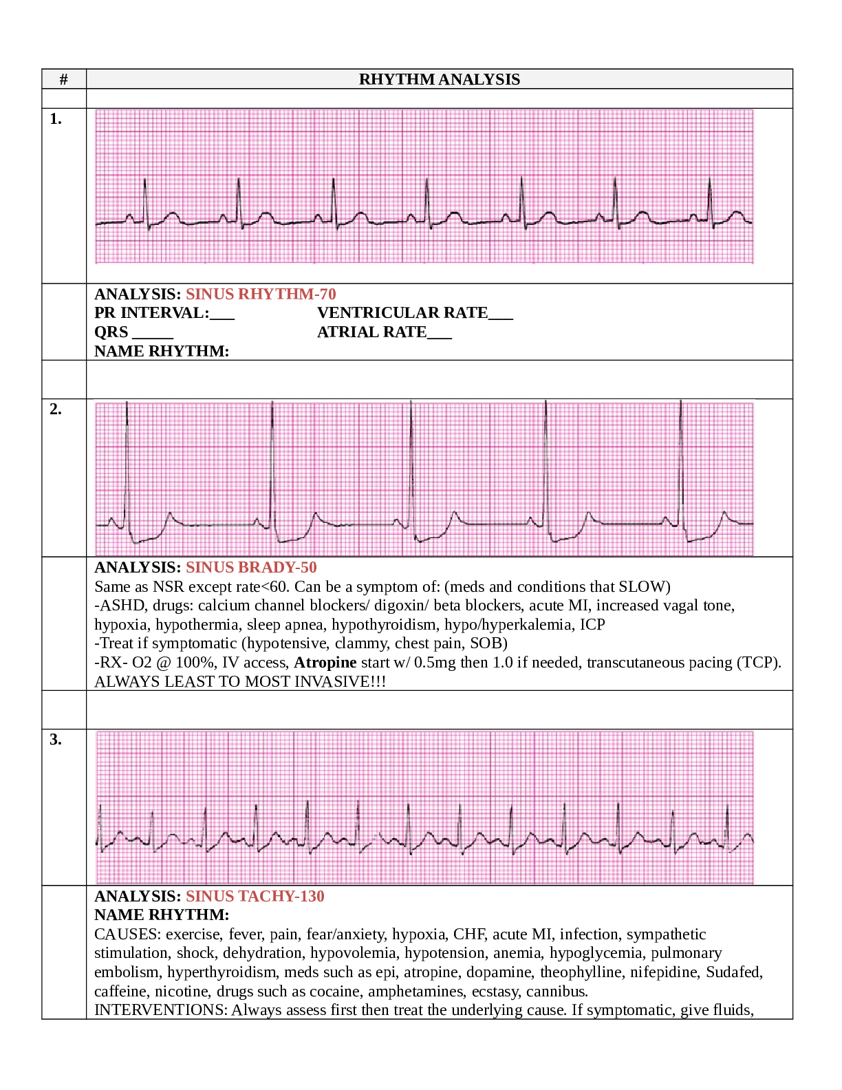
1.
ANALYSIS: SINUS RHYTHM-70
PR INTERVAL: VENTRICULAR RATE QRS ATRIAL RATE
NAME RHYTHM:
2.
ANALYSIS: SINUS BRADY-50
Same as NSR except rate350
Atria quivering-no discernable ‘
...
# RHYTHM ANALYSIS
1.
ANALYSIS: SINUS RHYTHM-70
PR INTERVAL: VENTRICULAR RATE QRS ATRIAL RATE
NAME RHYTHM:
2.
ANALYSIS: SINUS BRADY-50
Same as NSR except rate<60. Can be a symptom of: (meds and conditions that SLOW)
-ASHD, drugs: calcium channel blockers/ digoxin/ beta blockers, acute MI, increased vagal tone, hypoxia, hypothermia, sleep apnea, hypothyroidism, hypo/hyperkalemia, ICP
-Treat if symptomatic (hypotensive, clammy, chest pain, SOB)
-RX- O2 @ 100%, IV access, Atropine start w/ 0.5mg then 1.0 if needed, transcutaneous pacing (TCP). ALWAYS LEAST TO MOST INVASIVE!!!
3.
ANALYSIS: SINUS TACHY-130 NAME RHYTHM:
CAUSES: exercise, fever, pain, fear/anxiety, hypoxia, CHF, acute MI, infection, sympathetic stimulation, shock, dehydration, hypovolemia, hypotension, anemia, hypoglycemia, pulmonary embolism, hyperthyroidism, meds such as epi, atropine, dopamine, theophylline, nifepidine, Sudafed, caffeine, nicotine, drugs such as cocaine, amphetamines, ecstasy, cannibus.
INTERVENTIONS: Always assess first then treat the underlying cause. If symptomatic, give fluids,
stop related meds, relieve pain, fever, anxiety, vagal maneuver, if from MI give BB (lol’s), if no heart
failure and it doesn’t work…shock.
# RHYTHM ANALYSIS
4.
ANALYSIS: SINUS ARRYTHMIA-60 PR INTERVAL:
VENT RATE irregular, rate is 60-100, everything else normal---not enough to be a block. QRS ATRIAL RATE
NAME RHYTHM:
-rhythm fluctuates with respiration appear irregular, No treatment/not a problem
-Rate usually 60-100
5.
ANALYSIS: SINUS BLOCK-60
PR INTERVAL: VENTRICULAR RATE regular rhythm w/ one complex missing QRS ATRIAL RATE
NAME RHYTHM:
CAUSES: Acute MI, Meds (Digoxin, quinidine, procainamide, (antidysrhythmics), salicylates, CAD, myocarditis, CHF, carotid sinus sensitivity, increased vagal tone.
INTERVENTIONS: No treatment if asymptomatic, if due to meds…withhold, if happening too often give atropine (anticholinergic-speeds heart/fixes blocks drug toxicity), temp pacing or pacemaker if needed.
6.
ANALYSIS: SINUS PAUSE/ARREST-60 (2 SECOND PAUSE) more than one complex missing PR INTERVAL: VENTRICULAR RATE
QRS ATRIAL RATE
NAME RHYTHM: Sinus Arrest-regular rhythm with absence of PQRST (brief asystole!)
-Also called “sinus pause” or “SA arrest”
-Sinus impulses are not generated/look longer than one
-CAUSES: Hypoxia, Myocardial ischemia or infarction, Hyperkalemia, Digitalis toxicity, Reactions to medications: beta-blockers and calcium channel blockers, Increased vagal tone (Elvis)
TREATMENT: observation if asymptomatic, if symptomatic and frequent and longer than 3 s needs temporary pacing and/or permanent pacemaker, also review labs, dig, BB and CC blockers and stop meds.
# RHYTHM ANALYSIS
7.
ANALYSIS: ST-RATE 110 W/ UNIFOCAL PAC’S (3)
PR INTERVAL: VENTRICULAR RATE QRS ATRIAL RATE
NAME RHYTHM:
■ Premature Atrial Contraction (PAC) **looks like the P’s want to jump in front of the T’s
Atria fires an early impulse causing a premature complex
“noncompensatory” (incomplete) pause often follows a PAC
Represents the delay during which the SA node resets its rhythm for the next beat
CAUSES: emotional stress, CHF, ACS (acute coronary syndrome), mental/physical fatigue, dig tox, stimulants such as caffeine/tobacco/cocaine, hyperthyroid, electrolyte imbalance INTERVENTIONS: treat underlying cause and if needed anti-anxiety meds, BB/Ca+ blockers
8.
ANALYSIS: PARYOXYSMAL SVT/PSVT-150
PR INTERVAL: VENTRICULAR RATE QRS ATRIAL RATE
NAME RHYTHM:
SVT (supraventricular tachycardia), PSVT (P=PAROXYSMAL- SUDDEN START AND STOP)
• Typically 170 to 250 beats/min (rate is 170 or above) Three primary types of PSVT:
Atrial tachycardia-impulse begins in irritable spot in atrias, three or more PAC’s in a row, Rate
from 150-250
Atrioventricular nodal reentrant tachycardia (AVNRT)-originates in AV node
AV reentrant tachycardia (AVRT)-begins above vents but takes a route other than by AV node CAUSES: stimulants, infection, electrolyte imbalance, acute illness, MI
TREATMENT: if tachy lasts longer than 30 s and symptomatic give O2, iv access, vagal manouvers, Adenosine (except w asthma), CA blockers, BB’s, Amiodarone, synched cardioversion and/or radiofrequency ablation if no response.
9
a .
ANALYSIS: ATRIAL FLUTTER-VENTRICULAR RATE-70, ATRIAL RATE 300 PR INTERVAL: VENTRICULAR RATE
QRS ATRIAL RATE NAME RHYTHM:
■ Atrial Flutter
Started by a PAC
Atrial impulse from irritable focus Atrial rate 250-350
Flutter waves- “Sawtooth Pattern” LOOKS LIKE LITTLE TEETH AT TOP
■ Rarely occurs in patients without cardiac disease Flutter Type 1 250-350 BPM
Flutter TYPE II 350-450 BPM and can develop into atrial fibrillation
■ TREATMENT: Vagal Stimulation, Ca blocker-Cardizem/diltiazem, BB’s (to regulate/slow rhythm), cardioversion if needed.
# RHYTHM ANALYSIS
10
ANALYSIS: ATRIAL FIBRILLATION/QUIVER
PR INTERVAL: VENTRICULAR RATE QRS ATRIAL RATE >350
Atria quivering-no discernable ‘P’ waves Atrial rate > 350
Ventricular rate varies Irregularly irregular R-R
Baseline appears wavy-QUIVERS WITH R’S
CAUSES: Rheumatic heart disease, Coronary artery disease, Hypertension, Mitral or tricuspid valve disease, Congestive heart failure, Acute or chronic pulmonary disease, Enhanced vagal tone, Enhanced sympathetic tone, Hypokalemia, Hyperthyroidism, Pericarditis, Pulmonary embolism, Cardiomyopathy, Hypoxia, Drugs or intoxicants, Alcohol, Carbon monoxide
INTERVENTIONS:
If cardiac function is normal:
Calcium channel blockers (Diltizem/Cardizem) Beta-blockers (except w pulmonary/heart failure)
If cardiac function is impaired: Digoxin
Amiodarone
In the presence of severe symptoms such as hypotension, shock or heart failure (anticoags before cardioversion if AFIB longer than 48H:
Cardioversion
Cath Ablation (w syncope and WPW syndrome or if cardioversion contraindicated)
11
ANALYSIS: ST RATE 110 WITH ONE PJC
PR INTERVAL: VENTRICULAR RATE QRS ATRIAL RATE
NAME RHYTHM:
■ Premature Junctional Complex (PJC) Result of irritable focus at AV junction The QRS will usually measure 0.10 second or less
A P wave may or may not be present with a PJC
Often followed by a noncompensatory (incomplete) pause Usually no treatment and asymptomatic if occur occasionally
12
ANALYSIS: JUNCTIONAL ESCAPE RHYTHM 60
PR INTERVAL: VENTRICULAR RATE QRS ATRIAL RATE
NAME RHYTHM:
■ Junctional Escape Rhythm
Due to conduction disturbance above AV junction- protective mechanism
Rate 40-60
# RHYTHM ANALYSIS
13
ANALYSIS: ACCELERATED JUNCTIONAL RHYTHM-80 with no P’s PR INTERVAL: VENTRICULAR RATE
QRS ATRIAL RATE NAME RHYTHM:
Due to conduction disturbance above AV junction- protective mechanism.
RATE IS 61-100 (Normal AV junction rate 40-60). Enhanced automaticity of Bundle of His
CAUSES:
1. Digoxin Toxicity
2. Acute MI
3. Cardiac Surgery
4. Rheumatic Fever
5. COPD
6. Hypokalemia TREATMENT:
1. Asymptomatic-monitor
2. Due to dig tox-stop giving (Digibind)
14
ANALYSIS: JUNCTIONAL TACHYCARDIA-110 (over 100) PR INTERVAL: VENTRICULAR RATE QRS ATRIAL RATE
NAME RHYTHM:
Junctional Tachycardia: Junctional-starts at bundle of his. Increased automaticity.
Causes:
1. Acute Coronary Syndrome (Ischemia)
2. Acute MI
3. Congestive Heart Failure
4. Theophilline
5. Digitalis toxicity
Treatment: Stable but symptomatic:
1. O2
2. IV access
3. Vagal maneuvers (to stabilize)
4. Adenosine (to stabilize-super fast)
5. If due to Digoxin Toxicity give Digibind
6. If due to Theophilline stop or slow the infusion
7. If nothing works and continues to decline, synchronized cardiogenic shock.
15
ANALYSIS: VENTRICULAR BIGEMINY
PR INTERVAL: VENTRICULAR RATE QRS ATRIAL RATE
NAME RHYTHM:
# RHYTHM ANALYSIS
16
ANALYSIS: SR-100 WITH 2 UNIFOCAL PVCs
PR INTERVAL: VENTRICULAR RATE QRS ATRIAL RATE
NAME RHYTHM:
17
ANALYSIS: ST-130 WITH MULTIFOCAL PVC’S (3)
PR INTERVAL: VENTRICULAR RATE QRS ATRIAL RATE
NAME RHYTHM:
18
ANALYSIS:
PR INTERVAL: VENTRICULAR RATE QRS ATRIAL RATE
NAME RHYTHM:
# RHYTHM ANALYSIS
19
ANALYSIS:
PR INTERVAL: VENTRICULAR RATE QRS ATRIAL RATE
NAME RHYTHM:
20
ANALYSIS: VENTRICULAR TACHYCARDIA
PR INTERVAL: VENTRICULAR RATE QRS ATRIAL RATE
NAME RHYTHM:
21
ANALYSIS: TORSADE DE POINTES **SPINDLE-polymorphic VTACH** CAUSES:
1. Acute Coronary Syndrome
2. Cardiomyopathy
3. Antidepressent overdose
4. Dig Toxicity
5. Valvular Heart Disease
6. Cocaine abuse
7. Mitral Valve Prolapse
8. Acid base imbalance
9. Trauma (cardiac procedure/accident)
10. Electrolyte imbalance (hypo/hyperkalemia, hypomagnesium)
Treatment:
stable but symptomatic (Amiodarone) or MAGNESIUM SULFATE for HYPOMAGNESIUM
# RHYTHM ANALYSIS
22
COURSE (QUIVERING/FIBRILLATION)
FINE
ANALYSIS: VENTRICULAR FIBRILLATION
RATE CAN NOT BE DETERMINED, NO PATTERN, NO DISCERNABLE P’S, PRI OR QRS.
CAUSES:
1. Increased CNS stimulation
2. Vagal Stimulation
3. Electrolyte imbalance
4. Anti-arrhythmics
5. Electrocution
6. Hypertrophy
7. Acute Coronary Syndrome
8. Heart Failure
9. Arrhythmias ***Acute MI- #1 cause of death arrhythmias like VFIB
TREATMENT: (Patient will be unresponsive/apnic/pulseless and no meds can help VFIB)
1. Check the patient’s pulse to make sure its not artifact. ALWAYS CHECK THE PATIENT FIRST!!!
2. Immediately start CPR, call the code, get the crash card and defibrillate (cardiogenic shock).
3. Once a rhythm is established meds can be given to stabilize.
4. ONLY If shock does not work try Epinephrine, Vasopressin, Procainamide, Amiodarone or Lidocaine.
23
ANALYSIS: ASYSTOLE W P WAVES
PR INTERVAL: VENTRICULAR RATE QRS ATRIAL RATE
NAME RHYTHM:
24 RHYTHM ANALYSIS
ANALYSIS: PULSELESS ELECTRICAL ACTIVITY (PEA) PR INTERVAL: VENTRICULAR RATE QRS ATRIAL RATE
NAME RHYTHM:
25
ANALYSIS: FIRST DEGREE AV BLOCK
Everything is normal except the PRI is longer than 0.20
PR INTERVAL: LONGER THAN 0.20 VENTRICULAR RATE-60 rhythm is regular QRS ATRIAL RATE
NAME RHYTHM: SINUS RHYTHM 60 BPM WITH FIRST DEGREE AV BLOCK CAUSES:
1. Athletes (normal)
2. Sleep (brady-normal)
3. Ischemia or injury to the AV node or junction
4. Medications
5. RHD
6. Hyperkalemia
7. Acute MI
8. Increased vagal tone
MEDS THAT CAN CAUSE FIRST DEGREE AV BLOCK: (ABCDPQ)
1. Quinidine
2. Procainamide
3. Beta-blockers
4. Ca+ blockers
5. Digitalis
6. Amiodarone
TREATMENT-Monitor closely- no intervention needed.
HOW TO INTERPRET AV BLOCKS:
1. Look at the PRI
a. Is it constant? YES Is there a P for every QRS? YES--1ST DEGREE AV BLOCK
b. Constant with MORE than 1 P for every QRS-2ND DEGREE MOBITZ TYPE II
2. If the PRI is NOT constant---evaluate if the R to R’s are regular
a. Constant-3rd DEGREE BLOCK
b. If they vary-2nd DEGREE MOBITZ I (TYPE I) WENCKEBACH (Neither PRI or R to R’s are constant)
3. Anterior wall MI-wide QRS-greater than 0.10 (0.12-0.14 is already considered wide, more than
2.5 tiny boxes)
4. Inferior wall MI-narrow QRS-less than 0.10
HOW TO DESCRIBE BLOCKS:
• PRI -type of AV block
• Level/Location-width of QRS complex and in 3rd degree the rate of the escape rhythm
26
ANALYSIS: 2ND DEGREE AV BLOCK, TYPE I (WENCKEBACH, MOBITZ I) (GOING, GOING, GONE W/ ONE QRS MISSING)
PRI Inconsistent, R to R’s inconsistent VENTRICULAR RATE-60
QRS- > 0.10 CAUSES:
1. Increased parasympathetic tone
2. ischemia to right coronary artery (Inferior wall MI)
3. Acute MI
Conduction starts above AV Node (the bundle of his)
TREATMENT: ASYMPTOMATIC-MONITOR SYMPTOMATIC:
1. Atropine
2. Temporary Pacing
3. Inferior Wall MI (ischemia to right coronary artery)-monitor for increased AV block for 48-72 hours…after that it should disappear.
2 TO 1; TWO ARE IRREGULAR BEATS TO ONE MISSING….
27 RHYTHM ANALYSIS
ANALYSIS: 2ND DEGREE AV BLOCK, TYPE II- (MOBITZ II) VENTR.RATE-30
**(the husband and the wife are together, he leaves and comes back)**
PRI is Constant (when there’s a QRS) with MORE than 1 P for every QRS-2ND DEGREE MOBITZ TYPE II
PR INTERVAL: Constant VENTRICULAR RATE-IRREGULAR QRS More P’s than QRS’s ATRIAL RATE-REGULAR
NAME RHYTHM: MORE P’S THAN QRS-P’S WILL BE REGULAR W/ MISSING QRS’S
How do you know its anterior (inferior/top…anterior/front) wall MI (left coronary artery affected? Conduction starts below AV Node (bundle branches-which makes QRS wider >0.10)
• Wide QRS-anterior wall MI (left coronary artery)
CAUSES:
1. Anterior MI (ischemia to left coronary artery-ischemia to bundle branches)
2. Acute myocarditis
TREATMENT:
1. Due to the slow rate very rarely are they asymptomatic. Decreased cardiac output.
2. Prepare for pacing/permanent pacemaker
3. DO NOT USE ATROPINE!!! Will further decrease conduction to SA Node.
28
ANALYSIS: 3RD DEGREE BLOCK
(R to R’s are regular and the PRI is inconsistent)
PR INTERVAL: irregular VENTRICULAR RATE-regular-30 QRS -regular ATRIAL RATE
CAUSES:
1. Inferior wall MI-Narrow QRS < 0.10 (above bundle of his-progresses from 1st degree or 2nd degree type I block)
2. THIS STRIP IS: Anterior wall MI-QRS is wide > 0.10 (ischemia to right or left bundle branch block-escape pacemaker/ventricular) usually starts as 2nd Degree AV block type II and progresses to III.
3. Acute ischemia
TREATMENT:
1. Atropine
2. PERMANENT PACEMAKER
29
ANALYSIS: ATRIAL PACER-80 (spikes before the P’s) NOT for an AV block because it doesn’t pace ventricles.
Notes for pacemakers:
• First letter indicates the chamber paced (O-none, A-right atria, V-right ventrical, D-dual/both
• Second letter is the chamber sensed (OAVD)
• Third letter-response to sensing (fixed-discharged all the time. Demand pacemaker-only
discharges when rate drops below preset rate) (O-None/fixed, T-triggers pacing, I-Inhibits pacing, D-dual-triggers and inhibits)
30 RHYTHM ANALYSIS
ANALYSIS: VENTRICULAR PACER-75
(pacer stimulates spike followed by a wide QRS resembling a ventricular ectopic beat)
Pacer is placed in right ventricle
31
ANALYSIS: AV SEQUENTIAL PACEMAKER/DDD (dual pacemaker) PR INTERVAL: VENTRICULAR RATE
QRS ATRIAL RATE NAME RHYTHM:
32 RHYTHM ANALYSIS
ANALYSIS: FAILURE TO CAPTURE
Visible spikes NOT followed by P’s or QRS’s. Some are captured others are not. CAUSES:
1. Battery failure
2. Broken lead
3. Displaced lead wire (commom)-turn the patient on side.
4. Perforation of myocardium by lead
5. Edema or scar tissue (fibrosis) formation on lead
6. Pacemaker set too low (common)
7. Antiarrythmic meds (too much)
8. Electro’s imbalances
TREATMENT: (remember-always least to most invasive)
1. Reposition the patient
2. Increase setting
3. Change the battery
4. Surgically replacing or reposition the lead
33
ANALYSIS: PACEMAKER-FAILURE TO SENSE
Not recognizing spontaneous myocardial depolarization pacemaker spikes that follow too closely behind the RIF earlier R on T pheonomenon and can lead to I or FK patient complains of palpitations or skipped beats.
TREATMENT: (remember-always least to most invasive)
1. Reposition the patient
2. Increase setting
3. Change the battery
4. Surgically replacing or reposition the lead
KEY
■ SA node: rate 60-100
■ AV node/junction: rate 40-60
■ Bundle of HIS: 40-60 continues into right and left bundle branch
■ Purkinje Fibers: 20-40 located in ventricle
■ All rhythms coming from above the ventricles are have narrow QRS< .10
■ All rhythms coming from below the ventricles have wide QRS > .10
■ Heart rate 60-100 bpm
■ Rhythm regular
■ P wave before every QRS
■ PR interval .12-.20 seconds (3-5 tiny boxes)
■ QRS complex < .10 seconds (2.5 or less tiny boxes)
■ 1 small box= .04 seconds/ 1mm in height
■ 1 large box= .20 seconds/ 5 mm in height
■ 15 boxes= 3 seconds
■ 30 boxes= 6 seconds [used to measure rate]
TEST #1
1) SINUS RHYTHM-70
2) SINUS BRADYCARDIA-50
3) SINUS TACHYCARDIA-130
4) SINUS ARRHYTHMIA-60
5) SINUS BLOCK-60
6) SINUS ARREST-60 (2 SEC PAUSE APPROX)
7) ST-RATE 110 WITH UNIFOCAL PACs (3)
8) PAROXSYSMAL SVT or (PSVT)-150
9) ATRIAL FLUTTER-VENT. RATE-70 ATRIAL RATE (300)
TEST #2
10) ATRIAL FIBRILLATION -90
11) 11- ST-RATE 110- WITH ONE PJC
12) 12- JUNCTIONAL ESCAPE RHYTHM-50
13) 13- ACCELERATED JUNCTIONAL RHYTHM-80
14) 14- JUNCTIONAL TACHYCARDIA-110
15) 15- VENTRICULAR BIGEMINY-100
16) 16- SR-100 WITH 2 UNIFOCAL PVCs
17) ST-130 WITH MULTIFOCAL PVCs (3)
18) IDIOVENTRICULAR RHYTHM-30 TEST #3
19) IDIOVENTRICULAR RHYTHM-ACCELERATED-50
20) VENTRICULAR TACHYCARDIA (180)
21) TORSADE DE POINTES-(POLYMORPHIC V. TACH)
22) VENTRICULAR FIBRILLATION-COARSE AND FINE
23) ASYSTOLE (HAS P WAVES)
24) ANSWER: PULSELESS ELECTRICAL ACTIVITY (PEA) TEST #4
25) FIRST DEGREE AV BLOCK- PR: 0.28 RATE-60
26) 2ND DEGREE AV BLOCK, TYPE I (MOBITZ I/ WENCKEBACH) RATE-50
27) 2ND DEGREE AV BLOCK, TYPE II- (MOBITZ II) VENTR.RATE-30
28) 3RD DEGREE AV BLOCK (COMPLETE) – ATRIAL RATE-70, VENT. RATE-30
29) ATRIAL PACER-80
30) VENTRICULAR PACER-75
31) A-V PACER-60
32) PACER SPIKES-FAILURE TO CAPTURE
33) PACER-FAILURE TO SENSE
[Show More]