Student
Access Code (located on the lid of your lab kit):
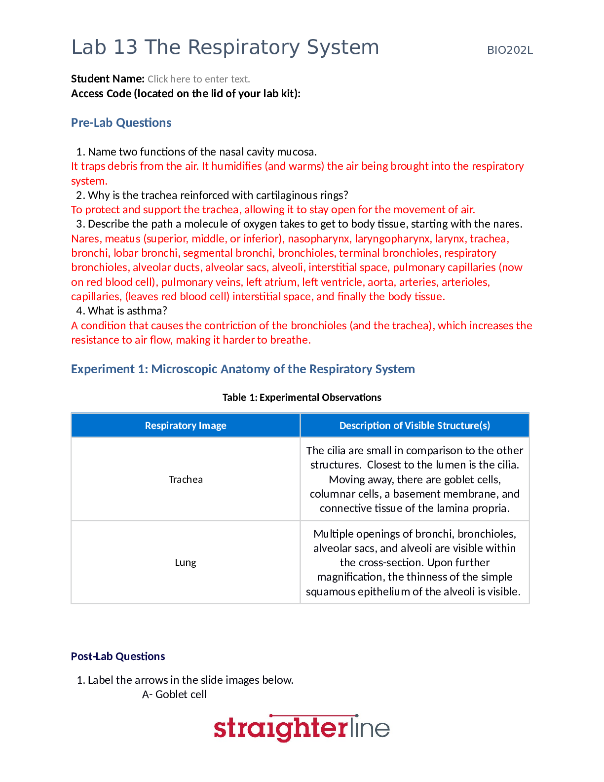
Pre-Lab Questions
1. What is the function of the ureter?
To transport urine from the kidney to the bladder.
2. Compare and contrast the male and fe
...
Student
Access Code (located on the lid of your lab kit):
Pre-Lab Questions
1. What is the function of the ureter?
To transport urine from the kidney to the bladder.
2. Compare and contrast the male and female urethra.
In both females and males, the urethra transports urine from the bladder to the outside of the body. In females the urethra is about 3-4cm long, straight, and goes straight from the neck of the bladder to the vulva. In males the urethra is about 20cm long and curves, going from the neck of the bladder through the prostate gland and then the penis. In males the urethra also transports semen to the outside of the body. The urethra in females is more prone to infections due to its shorter size.
3. Describe the two layers of the kidneys.
The two layers are the medulla and the cortex. The medulla is the inner layer and darker, and the cortex is the outer layer and lighter. The medulla contains renal pyramids which are cone-shaped structures. The bases of these renal pyramids are called medullary rays and extend into the cortex. The tips of the renal pyramids are called renal papillae and point towards the renal sinus. Projections from the cortex, called renal columns, are present between the renal pyramids. The main functional unit of the kidneys are nephrons. Nephrons contain a renal corpuscle, a proximal convoluted tubule, a loop of Henle, and a distal convoluted tubule. There are two types of nephrons: juxtamedullary nephrons and cortical nephrons. Cortical nephrons are entirely housed within the cortex, and the renal corpuscles of cortical nephrons (the initial portion of the nephron) are distributed throughout the entire cortex.
Juxtamedullary nephrons have loops of Henle that extend into the deep parts of the medulla, but the rest of the juxtamedullary nephrons are within the cortex. The kidneys filter blood, which first interact with nephrons in the renal corpuscles within the cortex. Urine is produced by the function of the kidneys, and this is collected within the medulla in collecting ducts.
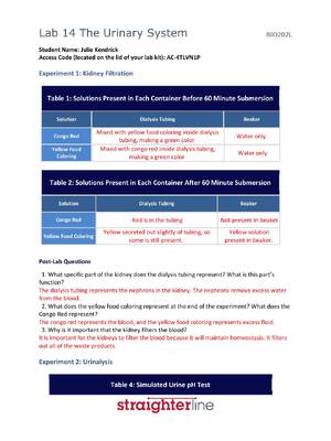
Experiment 1: Kidney Filtration
Table 1: Solutions Present in Each Container Before 60 Minutes Submersion
Solution Dialysis Tubing Beaker
Congo Red Present Not Present
Yellow Food Coloring Present Not Present
Table 2: Solutions Present in Each Container After 60 Minutes Submersion
Solution Dialysis Tubing Beaker
Congo Red Present Not Present
Yellow Food Coloring Not Present Present
Post-Lab Questions
1. What specific part of the kidney does the dialysis tubing represent? What is this part’s function?
The nephron. At first I was going to say the filtration membrane of the renal corpuscle (which is made up of the capillary endothelium, basement membrane, and the podocytes of the Bowman capsule). This is because the lab instructions stated that the solution initially placed into the dialysis tubing containing the yellow food coloring, Congo Red, and water represents the blood entering the kidney through the renal artery, which means it represents blood that has yet to be filtered. And when placed in the beaker of water, the yellow food coloring filtered out, and the Congo Red remained within the dialysis tubing. This would seemingly represent the filtration that occurs within the renal corpuscle, more specifically, by the filtration membrane. However, I noticed that while the yellow food coloring diffused out of the dialysis tubing, water had diffused into the dialysis tubing, seen by an increase in fluid volume within the tubing. This would represent the reabsorption of water, which does not take place within the renal corpuscle. Therefore, while the renal corpuscle is part of the nephron, it is more accurate to say that the dialysis tubing represents the nephron as a whole. The nephron is the main functional unit of the kidney. Its function is to filter the blood and regulate the fluid composition of the body in doing so. Essentially, nephrons function to sort substances from blood, secreting the bad ones, and keeping the good/necessary ones.
2. What does the yellow food coloring represent at the end of the experiment? What does the Congo Red represent?
The yellow food coloring represents the urine at the end of the experiment. The Congo Red represents the filtered blood (heading out of the kidney eventually through the renal vein).
3. Why is it important that the kidney filters the blood?
So that homeostasis can be maintained by excreting toxins, waste products, and excess materials, regulating blood volume/pressure, and regulating the solute concentrations within the body.
Experiment 2: Urinalysis
Table 3: Urine Test
Test Normal Abnormal
pH
4.5 - 7.5
Acidic Urine (below 4.5) - Diabetes, starvation, dehydration, respiratory acidosis.
Alkaline Urine (above 7.5) - Kidney disease, kidney failure, urinary tract infection, respiratory alkalosis.
Glucose None Glucose present (red or green color after test); diabetes mellitus.
Protein None Protein present (violet color after test); kidney disease.
Yeast
None Yeast present (bubbles form after test); yeast infection in urinary tract.
Ketones
Little or None Large amount of ketones present (sweet smell of urine); starvation, prolonged vomiting, diabetes, hyperthyroidism, and other metabolic disorders.
Table 4: Simulated Urine pH Test
Simulated Urine Sample pH
A 8
B 4
C 7
D 6
Table 5: Simulated Urine Glucose Test
Simulated Urine Sample Color Before Hot Water Bath Color After Hot Water Bath
A Blue Blue
Simulated Urine Sample Color Before Hot Water Bath Color After Hot Water Bath
B Blue Red
C Blue Orange
D Blue Blue
Table 6: Simulated Urine Protein Test
Simulated Urine Sample Color Before Biuret Solution Color After Biuret Solution
A Clear Purple
B Clear Blue
C Clear Purple
D Clear-yellow Blue
Table 7: Simulated Urine Yeast Test
Simulated Urine Sample Bubbles Before Hydrogen Peroxide? Bubbles After Hydrogen Peroxide?
A Absent Absent
B Absent Absent
C Absent Absent
D Absent Absent
Table 8: Simulated Urine Ketone Test
Simulated Urine Sample Odor Observation
A No odor
B No odor
C No odor
D No odor
Fill in Tables 9 through 12. Refer to Table 3 to determine if each result was normal or abnormal. If abnormal, include the data which indicates this (e.g., a pH of 3.2 means that glucose is present). Using the test results from each of the urine samples, diagnose the condition(s), if any, that each of the sample patients is experiencing.
Table 9: Sample A
Test Test Results
pH Abnormal (pH of 8 = alkaline urine)
Glucose Normal
Protein Abnormal (violet color = protein present)
Yeast Normal
Ketones Normal
Table 10: Sample B
Test Test Results
pH Abnormal (pH of 4 = acidic urine)
Glucose Abnormal (red color = glucose present)
Protein Normal
Yeast Normal
Ketones Normal
Table 11: Sample C
Test Test Results
pH Normal
Glucose Abnormal (orange color = glucose/reducing sugar present)
Protein Abnormal (violet color = protein present)
Yeast Normal
Ketones Normal
Table 12: Sample D
Test Test Results
pH Normal
Glucose Normal
Protein Normal
Yeast Normal
Ketones Normal
Post-Lab Questions
1. Using the results from each urine sample, along with Table 3, diagnose the condition(s), if any, that each sample patient is experiencing.
Table 9 Diagnosis: Kidney disease Table 10 Diagnosis: Diabetes mellitus
Table 11 Diagnosis: Kidney disease due to diabetes mellitus Table 12 Diagnosis: No condition
2. If you were a doctor and a patient’s urinalysis came back with high level of glucose, ketones and an acidic pH, what diagnosis would you immediately look into?
Diabetes mellitus
3. If you were a doctor and a patient’s urinalysis came back with an alkaline pH and high levels of protein, what diagnosis would you immediately look into?
Kidney disease
4. What other conditions can urine be used to test for?
Ketoacidosis, starvation, dehydration, liver disease, kidney stones, cancer (bladder), respiratory alkalosis, respiratory acidosis, kidney failure, hyperthyroidism, yeast infection, urinary tract infection, and more.
Experiment 3: Virtual Model – The Urinary System
Insert screenshot of the prostate:
Insert screenshot of the renal medulla:
Insert screenshot of the renal pelvis:
Post-Lab Questions
1. Which component of the urinary system is more lateral: the left kidney or the urinary bladder? Left kidney.
4. Which component of the urinary system is a funnel shaped cavity formed by the union of calices, ending in the ureter.
The renal pelvis.
5. What is the most superior component of the urinary system? Suprarenal/adrenal glands.
6. Where is the prostatic sinus in relation to the prostate? It is medial to the prostate.
Experiment 4: Fetal Pig Dissection of the Urinary System
1. Identify the labeled components of the kidney in the image below.
A – Renal pyramid
B - Calyx
C – Renal cortex
D – Renal artery
E – Renal vein
F - Ureter
7. What is the function of the urinary bladder? It stores urine until it is excreted.
8. What is the function of the adrenal glands?
They produce essential hormones. The functions of which include regulating fluid retention and composition.
9. Would you think the kidneys are highly vascularized? Why or why not? Yes. Kidneys filter blood, and therefore must be vascularized by nature.
10. Explain in detail the process by which urine is made.
Blood flows into the kidney through the renal artery. After branching down through multiple different arteries, the blood reaches a renal corpuscle within the cortex of the kidney. Here the blood is filtered through a structure of intertwining capillaries called a glomerulus. The resulting fluid is called filtrate and enters the Bowman capsule, which is the second part of the renal corpuscle. The filtrate then travels to and through the renal tubule (which contains the proximal convoluted tubule, the loop of Henle, and the distal convoluted tubule). Within the renal tubule the filtrate is altered as solutes, water, and other molecules are reabsorbed from the filtrate into the interstitial fluid and subsequently back in into the blood. Additionally, solutes, waste products, water, and other molecules are secreted from the blood into the interstitial fluid and then into the filtrate. After the renal tubule the filtrate travels to a collecting duct where it is further altered via reabsorption and secretion of certain molecules before it concentrated to urine and exits the collecting ducts.
Insert photo of your pig’s dissected kidney with your name clearly visible in the background:
[Show More]